Decubitus ulcer
Synonym(s)
Decubitus pressure sore pressure ulcer ischemic ulcer bed sore
Definition
Localized area of devitalized tissue secondary to vascular occlusion from prolonged external pressure against an internal body prominence, such as the sacrum or heel
Pathogenesis
Microcirculatory occlusion as pressures rise above capillary filling pressure, resulting in ischemia, causing inflammation and tissue anoxia, leading to cell death, tissue necrosis, and ulceration; paralysis result in muscle and soft tissue atrophy, decreasing the bulk over which bony prominences are supported; sensory loss, malnutrition, hypo-proteinemia, and anemia can be contributing factors in prolonged healing 929b124j time
Clinical manifestation
Stage 1: intact skin with signs of impending ulceration, with blanching erythema from reactive hyperemia
Stage 2: partial-thickness loss of skin involving epidermis and some dermis; sometimes presenting as an abrasion, blister, or superficial ulceration
Stage 3: full-thickness loss of skin with extension into subcutaneous tissue but not through the underlying fascia
Stage 4: full-thickness loss of skin and subcutaneous tissue and extension into muscle, bone, tendon, or joint capsule
Differential diagnosis
Pyoderma gangrenosum;
squamous cell carcinoma;
factitial ulcer (artificially produced; produced unintentionally)
burn;
contact dermatitis;
bullous pemphigoid;
spider bite;
stasis ulcer;
vasculitis
Therapy
Reduction or elimination of the source of external pressure, with frequent turning, protective pads, special mattresses, etc
stage 2: hydrocolloid dressings;
stages 3 and 4: wet dressings; silver sulfadiazine cream; hydrogels; xerogels; daily whirlpool use
Controversies about the decubitus ulcer
The decubitus ulcer is one of the most maligned diagnoses made in contemporary medicine . The nomenclature is straightforward, but there are those health care providers who like to spend time adjusting terms . The morphology and location should provide no controversy, but these characteristics often create a closed mind on the part of the practitioner . The differential diagnosis should also not present difficulties; however, other types of ulcers are not even considered . Assessments can be interesting, but their use may be questionable . Because the available therapies and preventative measures often are empirics or not proved by double-blind studies, irrational measures come into vogue
Nomenclature
The decubitus ulcer has existed for what seems like an eternity. The word ''decubitus'' is derived from ''decumbere'' (to lie down). The traditional term, bedsore, known in the nineteenth century and even until recent years, suggests that only bed-ridden patients might develop this ulceration. Neither term limits the scope of the disease . Unfortunately, within the past two decades there has been a movement to call these defects of the epidermis, dermis, and subcutaneous tissue pressure ulcers or sores . This conveys the notion that sustained pressure is the only factor and disregards friction, shear force, moisture, temperature elevation,
sensory impairment, or oxygen deprivation in its pathogenesis. So litigious has American society become, that some observers prefer to call these cutaneous defects breakdowns, or as has been facetiously proposed, torsion stress, to deflect the incendiary notion of the so-called ''pressure sore'' . Although the term pressure ulcer was once accepted, now the term decubitus ulcer is preferred.
To elaborate how simplistic the term ''pressure ulcer'' has become, one of us (JAW) manned a scientific exhibit at a national meeting where the booth was titled ''Pressure Ulcer.'' A passing delegate commented to her colleague that she always knew about pressure at work being the cause of ulcer formation. On further questioning as to what she was referring, she replied, ''duodenal and gastric ulcers.'' Needless to say, there is no relationship.
Morphology
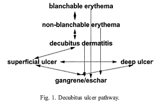
The decubitus ulcer can be described very readily in its various manifestations. Early on, there are blanchable and nonblanchable erythemas. These are followed by decubitus dermatitis, which encompasses these signs, plus the option of bullae and scaling.
When the skin breaks, there may be an ulcer, either superficial or deep. There also may be eschar or gangrene formation. The condition might be complicated by superimposed bacterial infection and evolving necrosis . The decubitus ulcer pathway shown in Fig. 1 illustrates this concept.
Periodically, staging of the ulcer has been suggested. Stage 1 is nonblanchable erythema; stage 2 has loss of the epidermis or dermis; stage 3 is a fullthickness loss of skin with destruction of subcutaneous fat; and stage 4 has involvement of the bone, tendon, or joint capsule
Why staging presents problems
Stage 1 cannot be determined in patients with pigmented skin except indirectly by feeling for warmth, edema, and induration. Blanchable erythema is ignored even though there is histologic evidence of damage . Secondly, explaining the various stages to neophytes is difficult. Lastly, staging also conveys an orderly progression of damage to the epidermis, extending through the dermis, and finally reaching the underlying tissues.
Unfortunately, staging is considered as a static process, whereas it is actually a fluid situation that is constantly evolving . The decubitus ulcer pathway illustrates this concept. One part of the ulcer might be labeled stage 1, where another portion should properly be defined as stage 4. An ulcer might be viewed as a stage 2 ulcer, until the underlying damage becomes more evident (the volcano effect). Unless the eschar or gangrene material is debrided, staging of that ulcer cannot be accomplished
The value of staging
Staging can suggest a therapeutic approach. It is an indicator of severity of damage to the integument, and it can be a predictor of the length of time of the healing process. By facilitating communication among health care providers, it has great potential for the development of studies on therapy, prevention, and determination of incidence and prevalence . Unfortunately, in today's litigious society, lawyers have used staging to determine the so-called ''neglect'' on the part of the health providers or a determination of the amount of monetary compensation that should be awarded to families
Location
The decubitus ulcer has traditionally been considered an ulcer appearing over bony prominences, such as the sacrum, ischial tuberosities, and heels. It has been shown that such an ulcer can also develop in unusual locations, such as the helix or the palm
The decubitus ulcer theoretically could develop on any part of the body where there is sustained pressure under the right circumstances.
Conversely, just because an ulcer is present over the sacrum, other types of ulceration should not be excluded. There is a differential diagnosis for leg ulcers , and so there should be for the decubitus ulcer. Deep mycotic infections, various vasculitides, abscess formations, and malignancies must be excluded by history, physical examination, biopsy, or bacteriologic culture.
Diagnosis
Are cultures of wounds useful for demonstrating infections in ulcers?
Bacteria colonize all decubitus ulcers. Swab cultures merely recover a sample of the flora on the ulcer surface. To discover the etiology of an infection in an ulcer, culture fluid should be obtained by aspiration, a forcefully applied swab, or a biopsy of the ulcer tissue . A swab, taken once over lightly, may not reveal the actual pathogens . An ancillary tool for identifying an anaerobic infection is aWood's light examination of the ulcer . These are the bacteria that are most often associated with a fatal outcome
The role of debridement in the diagnosis of decubitus ulcers
When a wound is covered with necrotic slough, the morphology is obscured. Not only can an underlying malignancy be masked, but pathogens could be allowed to proliferate . Although debridement is another diagnostic modality, taken to excess it can destroy any semblance of healing . Eliminating the necrotic debris may be accomplished medically, surgically, and physically. Occasionally, it is beneficial to allow the dead tissue to separate spontaneously.
Assessments
Varieties of assessment scales have been introduced to determine the potential vulnerability of the patient to develop decubitus ulcers. These include the Braden and Norton scales, several being developed by nurses , and the Norman Scale, created by a physician (personal communication 2003). Such scales often include grading for the physical condition, mental make-up, activity that can be performed, mobility, fecal and urinary incontinence, and nutritional status. The total of the grades then suggests the status of the patient.
Although it is important to know where one is so that one can move forward, sometimes too much emphasis is placed on these tools. They can only reveal a static situation for the criteria that they include
Treatment and prevention
Treatment and prevention of the decubitus ulcer are often so intermingled that a modality is either criticized or extolled without justification. A case at point is the subject of nutrition. Malnourished patients are more prone to the development of decubitus ulcers, but there are no satisfactory studies to prove that correcting the protein depleted state leads to rapid healing . In addition, there are no data to confirm that inserting a feeding tube to improve the patient's nutritional status makes a difference in the healing process
Are all decubitus ulcers preventable?
Skin integrity is dependent on the function of all the other organs. The health of the skin relies on other organs for provision of nutrients, hydration, sensation, and immune function. When other organs are failing, should not the skin do likewise?
The decubitus ulcer must be accepted as being a legitimate result of a combination of environmental, biochemical, and physiologic factors. When the heart fails, the health care provider is not accused of neglect. The skin, then, can also fail. There are limits to prevention even with excellent care
Is massage useful in treatment or prevention?
Massage is often used, especially by laypersons, with the mistaken impression that it stimulates the circulation. It is reminiscent of the old nursing procedure of applying balsam of Peru ''to harden the skin.''
In actuality, such manipulation causes friction and shear injury, leading to deep tissue destruction . This is especially true in the elderly where there is age-related flattening of the dermoepidermal junction.
Are all pressure-relieving devices equally effective?
There are two types of support surfaces: static and dynamic. The static support surfaces include air, foam, and water mattress overlays. The dynamic support surfaces encompass the alternating air overlay, the low air loss bed, and the air-fluidized bed. The static support surfaces are appropriate for a patient who has an ulcer but who is at risk for developing additional ulcers and can assume a variety of positions without lying on the ulcer. The dynamic support surfaces are appropriate for a patient who cannot assume a variety of positions without laying on the ulcer, bottoms out when laying on a static support surface, or does not heal after 2 to 4 weeks of optimal care
Donut-type devices are not recommended because they cause further ischemia of the area, often enlarging the ulcer . Similarly, sheepskins do not relieve pressure, although they diminish friction and shear force damage. Egg crate mattresses may increase comfort, but they are not thick enough to relieve pressure areas.
What makes turning the patient every 2 hours so magical?
The decubitus ulcer represents a defect created when the vessels are occluded because of excessive pressure. There are many contributory factors ranging from depleted oxygenation, decreased wound healing capability, superimposed infection, and overall health status. Moving the patient by keeping the patient off of bony prominences may help in slowing down the development of decubitus ulcers and assist in the wound healing process.
The development of the 2-hour rotation is unclear. It may have started at the Stoke Mandeville Hospital, England, during the early years of WorldWar II. Spinal cord-injured patients were surviving longer because of the introduction of antimicrobial therapy, but they were developing horrendous ulcers because of plaster casts. The medical director, Sir Ludwig Guttman, introduced ''log rolling'' and had a team of orderlies whose job was to turn the patients. It was said that it took 2 hours to see every patient in the hospital, and hence the 2-hour rotation
Another possibility for the introduction of the 2-hour interval can be traced to a handbook for home health care, written during World War I. The author recommended. ''If the patient is at all in a condition to be moved, he should be turned to his side and allowed to remain in that position for a few hours.'' The concept of a 2-hour interval was used for changing the diaper (nappies)
We must move, as we sit, sleep, or walk. Each person has different requirements, and no one has ever proved how often or how much someone needs to move, although there have been interesting experiments . This is not to say that patients should not be moved, if they are incapable of doing so themselves, but there is no proved time interval. The authors have no objection to the arbitrary order of ''turn every 2 hours,'' but one should remember that it is just a time span of convenience. No known catastrophe
occurs if the intervals are shorter or longer.
Summary
Knowledge of the decubitus ulcer has made great progress in recent decades. No longer is the diagnosis of a bedsore, as it was in the iron lung days, almost a death warrant. One cannot cause to heal a patient who is in organ failure, nor can one necessarily prevent ulcers in a new spinal cord-injured patient
Reflecting on the common cold, one realizes that prevention is limited to covering the face when sneezing, and treatment is symptomatic. There is no way of eradicating the virus of the common cold, nor is there an appropriate treatment. The decubitus ulcer should be conceived of in a similar vein, until understanding is more complete and specific treatments are available.
|
