Metabolic and Hereditary Disorders
Acid-Base
Disorders
- In analyzing acid-base
disorders, several points should be kept in mind:
- Determination of pH and
blood gases should be performed on arterial
blood. Venous blood is useless for judging oxygenation but offers an
estimate acid-base status.
- Blood specimens should
be packed in ice immediately; delay of even a few minutes causes
erroneous results, especially if WBC is high.
- Determination of
electrolytes, pH, and blood gases ideally should be performed on blood
specimens obtained simultaneously, because the acid-base situation is
very labile.
- Repeated determinations
may often be indicated because of the development of complications, the
effect of therapy, and other factors.
- Acid-base disorders are
often mixed rather than in the pure form usually described in textbooks.
- These mixed disorders
may represent simultaneously occurring diseases, complications
superimposed on the primary condition, or the effect of treatment.
- Changes in chronic forms
may be notably different from those in the acute forms.
- For judging hypoxemia,
one must also know the patient's Hb or Hct and whether the patient was
breathing room air or oxygen when the specimen was drawn.
- Arterial blood gas
values cannot be interpreted without clinical information about the
patient.
- Renal compensation for a
respiratory disturbance is slower (37 days) but more successful than
respiratory compensation for a metabolic disturbance but cannot completely
compensate for pCO >65 mm Hg unless
another stimulus for HCO
retention is present. Respiratory mechanism responds quickly but can only
eliminate sufficient CO to balance the most mild
metabolic acidosis.
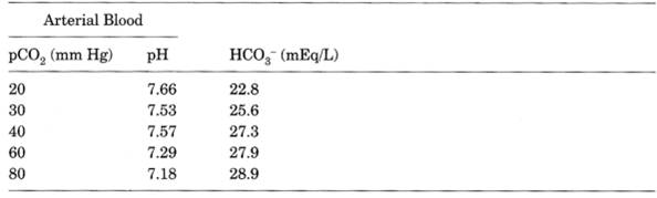
- Most laboratories measure
pH and pCO directly and calculate
HCO using the
Henderson-Hasselbalch equation:
Arterial pH = 6.1 + log [(HCO ) ÷ (0.03 × pCO
where 6.1 is the dissociation constant for CO in aqueous solution and 0.03 is a constant
for the solubility of CO in plasma at
37°C.
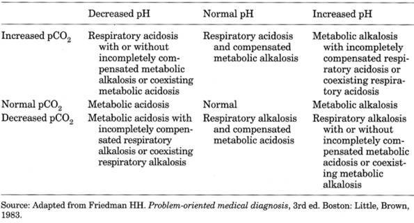
- A
normal pH does not ensure the absence of an acid-base disturbance if the
pCO is
not known
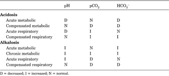
- An abnormal HCO means a metabolic rather
than a respiratory problem; decreased HCO indicates metabolic
acidosis, and increased HCO
indicates metabolic alkalosis. Respiratory acidosis is associated with a
pCO of >45 mm Hg, and respiratory alkalosis is
associated with a pCO of <35 mm Hg. Thus
mixed metabolic and respiratory acidosis is characterized by low pH, low
HCO , and high pCO .
Mixed metabolic and respiratory alkalosis is characterized by high pH,
high HCO ,
and low pCO
- See Tables
12-1, , and .
- In severe metabolic acidosis, respiratory compensation is limited
by inability to hyperventilate pCO to less
than ~15 mm Hg; beyond that, small increments of H ion produce disastrous changes in pH and prognosis; thus patients
with lung disorders (e.g., COPD, neuromuscular weakness) are very
vulnerable because they cannot compensate by hyperventilation. In
metabolic alkalosis, respiratory compensation is limited
P.490
by CO retention,
which rarely causes pCO levels >5060
mm Hg (because increased CO and hypoxemia
stimulate respiration very strongly); thus pH is not returned to normal
|
|
|
Table 12-1. Metabolic and Respiratory
Acid-Base Changes in Blood
|
- Base excess is a value
that hypothetically corrects pH to 7.40 by first adjusting pCO
to 40 mm Hg, thereby allowing comparison of resultant HCO with normal value at
that pH (24 mEq/L). Base excess can be calculated from determined values
for pH and HCO
by the following formula:
- Base excess (mEq/L) = HCO + 10(7.40 pH) 24
- Negative base excess indicates
depletion of HCO .
Does not distinguish primary from compensatory derangement.
- See Tables
12-1, , , and
; section on metabolic and respiratory
acid-base changes in blood.
Pearls
- Pulmonary embolus: Mild to moderate respiratory alkalosis is
present unless sudden death occurs. The degree of hypoxia often correlates
with the
size and extent of the pulmonary embolus. pO
of >90 mm Hg when patient breathes room air virtually excludes a lung
problem.
- Acute pulmonary edema:
Hypoxemia is usual. CO is not increased unless
the situation is grave.
- Asthma: Hypoxia occurs
even during a mild episode and increases as the attack becomes worse. As
hyperventilation occurs, the pCO falls (usually <35 mm
Hg); a normal pCO (>40 mm Hg) implies
impending respiratory failure; increased pCO
in a patient with true asthma (not bronchitis or emphysema) indicates
impending disaster and the need to consider intubation and ventilation
assistance.
- COPD (bronchitis and emphysema): May show two patternspink puffers
with mild hypoxia and normal pH and pCO and blue bloaters with
hypoxia and increased pCO ; normal pH suggests
compensation, and decreased pH suggests decompensation.
- Neurologic and neuromuscular disorders (e.g., drug overdose,
Guillain-Barré syndrome, myasthenia gravis, trauma, succinylcholine administration): Acute alveolar
hypoventilation causes uncompensated respiratory acidosis with high pCO ,
low pH, and normal HCO .
Acidosis appears before significant hypoxemia, and rising CO
indicates rapid deterioration and need for mechanical assistance.
- Sepsis: Unexplained respiratory alkalosis may be the earliest sign
of sepsis. It may progress to cause metabolic acidosis, and the mixed
picture may produce a normal pH; low HCO is useful to recognize
this situation. With deterioration and worsening of metabolic acidosis,
the pH falls.
P.491
|
|
|
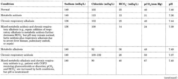
Table 12-2. Illustrative Serum Values in
Acid-Base Disturbances
|
P.492
|
|
|
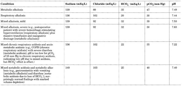
Table 12-2. (continued)
|
P.493
|
|
|
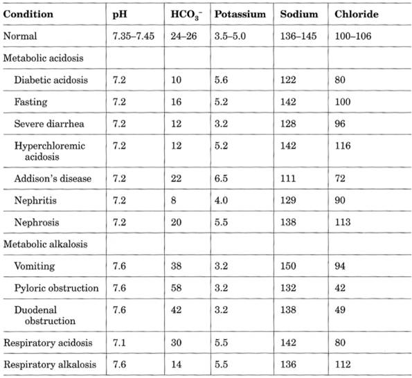
Table 12-3. Illustrative Serum
Electrolyte Values in Various Conditions
|
- Salicylate poisoning: Characteristically, poor correlation is seen
between serum salicylate level and presence or degree of acidemia (because
as pH drops from 7.4 to 7.2, the proportion of nonionized to ionized salicylate
doubles and the nonionized form leaves the serum and is sequestered in the
brain and other organs, where it interferes with function at a cellular
level without changing blood levels of glucose, etc.). In adults
salicylate poisoning typically causes respiratory alkalosis, but in children
this progresses rapidly to mixed respiratory alkalosismetabolic acidosis
and then to metabolic acidosis (in adults, metabolic acidosis is said to
be a rare and a near-terminal event).
- Isopropyl (rubbing) alcohol poisoning: Produces enough circulating
acetone to produce a positive nitroprusside test (it therefore may be
mistaken for diabetic ketoacidosis; thus insulin should not be given until
the blood
glucose is known). In the absence of a history, positive serum ketone test
associated with normal AG, normal serum HCO , and normal blood
glucose suggests rubbing alcohol intoxication.
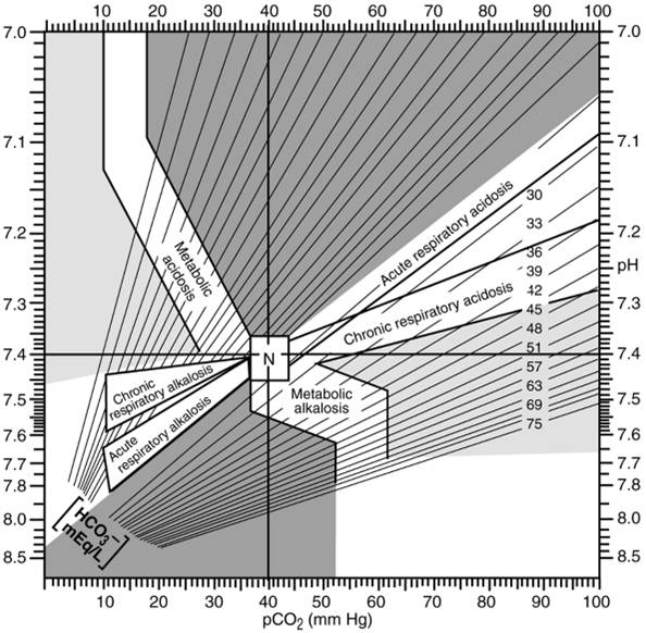
- Acid-base maps (Fig. 12-1) are a graphic solution of the
Henderson-Hasselbalch equation that predicts the HCO value for each set of
pH/pCO coordinates. They also
allow a check of the consistency of arterial blood gas and some chemical
analyzer determinations, because the chemical analyzer determines the
total CO content, of which 95% is
HCO . These maps contain
bands that show the 95% probability range of values for each disorder. If
the pH/pCO coordinate is outside
the 95% confidence band, then the patient has at least two acid-base
disturbances. These maps are of particular use when one of the acid-base
disturbances is not suspected clinically. If the coordinates lie within a
band, however, there is no guarantee of a simple acid-base disturbance.
P.494
|
|
|
Table 12-4. Upper Limits of Arterial
Blood 16216e48q pH and HCO Concentrations (Expected for Blood pCO Values)
|
Acid-Base
Disturbances, Mixed
- (Must
always be interpreted with clinical data and other laboratory findings)
- See Table
12-2.
Respiratory
Acidosis with Metabolic Acidosis
- Examples: Acute pulmonary
edema, cardiopulmonary arrest (lactic acidosis due to tissue anoxia and CO
retention due to alveolar hypoventilation)
- Acidemia may be extreme
with
- pH <7.0 (H
>100 mEq/L).
- HCO
<26 mEq/L. Failure of HCO3 to increase ≥3 mEq/L for each 10 mm
Hg rise in pCO suggests metabolic
acidosis with respiratory acidosis.
- Mild
metabolic acidosis superimposed on chronic hypercapnia causing partial
suppression of HCO may be indistinguishable from adaptation to
hypercapnia alone
Metabolic
Acidosis with Respiratory Alkalosis
- Examples: Rapid
correction of severe metabolic acidosis, salicylate intoxication,
septicemia due to gram-negative organisms, initial respiratory alkalosis
with subsequent development of metabolic acidosis.
- Primary metabolic acidosis with primary respiratory
alkalosis with an increased AG is characteristic of salicylate
intoxication in absence of uremia and diabetic ketoacidosis
|
|
|
Table 12-5. Summary of Pure and Mixed
Acid-Base Disorders
|
|
|
|
Fig. 12-1. Acid-base map. The values
demarcated for each disorder represent a 95% probability range for each pure disorder (N = normal). Coordinates lying outside
these zones suggest mixed acid-base disorders. (Adapted from
Goldberg M, et
al. Computer-based instruction and diagnosis of acid-base disorders. JAMA
Copyright 1973 American Medical
Association.)
|
- pH may be normal or
decreased.
- Hypocapnia remains
inappropriate to decreased HCO
for several hours or more.
Respiratory
Acidosis with Metabolic Alkalosis
- Examples: Chronic
pulmonary disease with CO retention in which
patient develops metabolic alkalosis due to administration of diuretics,
severe vomiting, or sudden improvement in ventilation (posthypercapnic
metabolic alkalosis)
- Decreased or absent urine chloride indicates that
chloride-responsive metabolic alkalosis is a part of the picture.
- In clinical setting of respiratory acidosis but with normal blood
pH and/or HCO
higher than predicted, complicating metabolic alkalosis may be present.
Respiratory
Alkalosis with Metabolic Alkalosis
- Examples: Hepatic
insufficiency with hyperventilation plus administration of diuretics or
severe vomiting; metabolic alkalosis with stimulation of ventilation
(e.g., sepsis, pulmonary embolism, mechanical ventilation) that causes
respiratory alkalosis
P.496
- Marked alkalemia with decreased pCO
and increased HCO
is diagnostic.
Acute
and Chronic Respiratory Acidosis
- Examples: Chronic
hypercapnia with acute deterioration of pulmonary function causing further
rise of pCO
- May be suspected when HCO
in intermediate range between acute and chronic respiratory acidosis
(similar findings in chronic respiratory acidosis with superimposed
metabolic acidosis or acute respiratory acidosis with superimposed
metabolic alkalosis)
Coexistence
of Metabolic Acidoses of Hyperchloremic Type and Increased AG Type
- Examples: Uremia and
proximal renal tubular acidosis, lactic acidosis with diarrhea, excessive
administration of sodium chloride to patient with organic acidosis
- May be suspected when plasma HCO level is lower than is
explained by the increase in anions (e.g., AG = 16 mEq/L and HCO = 5 mEq/L)
Coexistence
of Metabolic Alkalosis and Metabolic Acidosis
- Examples: Vomiting
causing alkalosis plus bicarbonate-losing diarrhea causing acidosis
- May be suggested by acid-base values that are too normal for
clinical picture
Acidosis,
Lactic
- Indicates acute
hypoperfusion and tissue hypoxia.
- Should be considered in any metabolic acidosis with increased AG
(>15 mEq/L).
- Diagnosis is confirmed by exclusion of other causes of metabolic acidosis and serum
lactate ≥5 mEq/L (upper limit of normal = 1.6 for plasma and 1.4 for
whole blood). Considerable variation in literature in limits of serum
lactate and pH to define lactic acidosis.
- Exclusion of other causes by
- Normal serum creatinine
and BUN. (Increased acetoacetic acid [but not
beta-hydroxybutyric acid] causes false increase of creatinine by
colorimetric assay.)
- Osmolar gap <10
mOsm/L.
- Negative nitroprusside
reaction. (Nitroprusside test for ketoacidosis
measures acetoacetic acid but not beta-hydroxybutyric acid; thus blood
ketone test may be negative in diabetic ketoacidosis.)
- Urine negative for
calcium oxalate crystals.
- No known ingestion of
toxic substances.
- Laboratory findings due
to underlying diseases (e.g., diabetes mellitus, renal insufficiency,
etc.)
- Laboratory tests for
monitoring therapy
- Arterial pH, pCO , HCO , serum electrolytes, every 12 hrs until patient is stable
- Urine electrolytes every
6 hrs
- Associated or
compensatory metabolic or respiratory disturbances (e.g., hyperventilation
or respiratory alkalosis may result in normal pH)
Due To
- Type A due to clinically
apparent tissue hypoxia, e.g., acute hemorrhage, severe anemia, shock,
asphyxia; marathon running, seizures
- Type B without clinically
apparent tissue hypoxia due to
- Common disorders (e.g.,
diabetes mellitus, uremia, liver disease, infections, malignancies,
alkaloses).
- Drugs and toxins (e.g.,
ethanol, methanol, ethylene glycol, salicylates, metformin).
- Hereditary enzyme
defects (e.g., methylmalonicaciduria, propionicaciduria, defects of fatty
acid oxidation, pyruvate-dehydrogenase deficiency, pyruvate-carboxylase
deficiency, multiple carboxylase deficiency, glycogen storage disease
type I).
- Others (e.g.,
short-bowel syndrome).
- With a typical clinical
picture (acute onset after nausea and vomiting, altered state of
consciousness, hyperventilation, high mortality)
P.497
- Decreased serum
bicarbonate.
- Low serum pH, usually
6.987.25.
- Increased serum potassium,
often 67 mEq/L.
- Serum chloride normal or
low with increased AG.
- WBC is increased
(occasionally to leukemoid levels).
- Increased serum uric
acid is frequent (up to 25 mg/dL in lactic acidosis).
- Increased serum phosphorus. Phosphorus/creatinine ratio >3
indicates lactic acidosis either alone or as a component of other
metabolic acidosis.
- Increased serum AST, LD,
and phosphorus.
- See Table
12-3.
Acidosis,
Metabolic
With Increased Anion Gap (AG >15 mEq/L)
- Lactic acidosismost
common cause of metabolic acidosis with increased AG (frequently >25
mEq/L) (see previous section)
- Renal failure (AG <25
mEq/L)
- Ketoacidosis
- Diabetes mellitus (AG
frequently >25 mEq/L)
- Associated with alcohol
abuse (AG frequently 2025 mEq/L)
- Starvation (AG usually
510 mEq/L)
- Drug effects
- Salicylate poisoning (AG
frequently 510 mEq/L; higher in children)
- Methanol poisoning (AG
frequently >20 mEq/L)
- Ethylene glycol
poisoning (AG frequently >20 mEq/L)
- Paraldehyde treatment
(AG frequently >20 mEq/L)
With Normal
Anion Gap
- (Hyperchloremic
acidosis)
- Decreased serum potassium
- Renal tubular acidosis
- Acquired (e.g., drugs, hypercalcemia)
- Inherited (e.g.,
cystinosis, Wilson's disease)
- Carbonic anhydrase
inhibitors (e.g., acetazolamide, mafenide)
- Increased loss of
alkaline body fluids (e.g., diarrhea, loss of pancreatic or biliary
fluids)
- Ureteral diversion
(e.g., ileal bladder or ureter, ureterosigmoidostomy)
- Normal or increased serum
potassium
- Hydronephrosis
- Early renal failure
- Administration of HCl
(e.g., ammonium chloride)
- Hypoadrenalism (diffuse, zona glomerulosa, or hyporeninemia)
- Renal aldosterone
resistance
- Sulfur toxicity
- In lactic acidosis the increase in AG is usually
greater than the decrease in HCO , in contrast to diabetic ketoacidosis in
which the increase in AG is identical to the decrease in HCO
Laboratory Findings
- Serum pH is decreased
(<7.3).
- Total plasma CO
content is decreased; value <15 mEq/L almost certainly rules out
respiratory alkalosis.
- Serum potassium is
frequently increased; it is decreased in renal tubular acidosis, diarrhea,
or carbonic anhydrase inhibition.
- Azotemia suggests
metabolic acidosis due to renal failure.
- Urine is strongly acid
(pH = 4.55.2) if renal function is normal.
- In evaluating acid-base
disorders, calculate the AG (see below).
Acidosis,
Respiratory
Laboratory findings differ in acute and
chronic conditions.
P.498
Acute
- Due to decreased alveolar
ventilation impairing CO excretion
- Cardiopulmonary (e.g.,
pneumonia, pneumothorax, pulmonary edema, foreign-body aspiration,
laryngospasm, bronchospasm, mechanical ventilation, cardiac arrest)
- CNS depression (e.g.,
general anesthesia, drug effects, brain injury, infection)
- Neuromuscular conditions
(e.g., Guillain-Barré syndrome, hypokalemia, myasthenic crisis)
- Acidosis is severe (pH 7.057.10) but HCO concentration is only
2930 mEq/L.
- Severe mixed acidosis is
common in cardiac arrest when respiratory and circulatory failure cause
marked respiratory acidosis and severe lactic acidosis.
Chronic
- Due to chronic
obstructive or restrictive conditions
- Nerve disease (e.g.,
poliomyelitis)
- Muscle disease (e.g.,
myopathy)
- CNS disorder (e.g.,
brain tumor)
- Restriction of thorax
(e.g., musculoskeletal disorders, scleroderma, pickwickian syndrome)
- Pulmonary disease (e.g.,
prolonged pneumonia, primary alveolar hypoventilation)
- Acidosis is not usually
severe.
- Beware of commonly
occurring mixed acid-base disturbances
- Chronic respiratory
acidosis with superimposed acute hypercapnia resulting from acute
infection, such as bronchitis or pneumonia.
- Superimposed metabolic
alkalosis (e.g., due to diuretics or vomiting) may exacerbate the
hypercapnia.
Alkalosis,
Metabolic
Due To
- Loss of acid
- Vomiting, gastric
suction, gastrocolic fistula
- Diarrhea in
mucoviscidosis (rarely)
- Villous adenoma of colon
- Aciduria secondary to
potassium depletion
- Excess of base due to
- Administration of
absorbable antacids (e.g., sodium bicarbonate; milk-alkali syndrome)
- Administration of salts
of weak acids (e.g., sodium lactate, sodium or potassium citrate)
- Some vegetarian diets
- Potassium depletion
(causing sodium and H to enter cells)
- Gastrointestinal loss
(e.g., chronic diarrhea)
- Lack of potassium intake
(e.g., anorexia nervosa, administration of IV fluids without potassium
supplements for treatment of vomiting or postoperatively)
- Diuresis (e.g.,
mercurials, thiazides, osmotic diuresis)
- Extracellular volume
depletion and chloride depletion
- All forms of
mineralocorticoid excess (e.g., primary aldosteronism, Cushing's
syndrome, administration of steroids, ingestion of large amounts of
licorice)
- Glycogen deposition
- Chronic alkalosis
- Potassium-losing
nephropathy
- Hypoproteinemia per se
may cause a nonrespiratory alkalosis. Decreased albumin of 1 gm/dL causes
an average increase in standard bicarbonate of 3.4 mEq/L, an apparent base
excess of +3.7 mEq/L, and a decrease in AG of ~3 mEq/L.1
Laboratory Findings
- Serum pH is increased
(>7.60 in severe alkalemia).
- Total plasma CO
is increased (bicarbonate >30 mEq/L).
P.499
- pCO
is normal or slightly increased.
- Serum pH and bicarbonate
are above those predicted by the pCO (by nomogram or Table 12-4).
- Hypokalemia is an almost
constant feature and is the chief danger in metabolic alkalosis.
- Decreased serum chloride
is relatively lower than sodium.
- BUN may be increased.
- Urine pH is >7.0
(≤7.9) if potassium depletion is not severe and concomitant sodium
deficiency (e.g., vomiting) is not present. With severe hypokalemia
(<2.0 mEq/L), urine may be acid in presence of systemic alkalosis.
- When the urine chloride is low (<10 mEq/L) and the patient
responds to chloride treatment, the cause is more likely loss of gastric juice,
diuretic therapy, or rapid relief of chronic hypercapnia. Chloride
replacement is completed when urine chloride remains >40 mEq/L. When
the urine chloride is high (>20 mEq/L) and the patient does not respond
to sodium chloride treatment, the cause is more likely hyperadrenalism or
severe potassium deficiency.
- See Table
12-4.
Alkalosis,
Respiratory
(Decreased pCO of <38 mm Hg)
Due To
- Hyperventilation
- CNS disorders (e.g.,
infection, tumor, trauma, cerebrovascular accident [CVA])
- Salicylate intoxication
- Fever
- Bacteremia due to
gram-negative organisms
- Liver disease
- Pulmonary disease (e.g.,
pneumonia, pulmonary emboli, asthma)
- Mechanical
overventilation
- Congestive heart failure
- Hypoxia (e.g., decreased
barometric pressure, ventilation-perfusion imbalance)
- Anxiety-hyperventilation
Laboratory
Findings
- Acute hypocapniausually
only a modest decrease in plasma HCO
concentrations and marked alkalosis
- Chronic
hypocapniausually only a slight alkaline pH (not usually >7.55)
Anion
Gap Classification
(Calculated as
Na [Cl + HCO ]; typically normal = 816 mEq/L; if K is included, normal = 1020
mEq/L; reference interval varies considerably depending on instrumentation.)
Use
- Identification of cause
of metabolic acidosis
- Supplement to laboratory
quality control along with its components
Increased
In
- Increased unmeasured anions
- Organic (e.g., lactic
acidosis, ketoacidosis)
- Inorganic (e.g.,
administration of phosphate, sulfate)
- Protein (e.g., transient
hyperalbuminemia)
- Exogenous (e.g., salicylate, formate, nitrate, penicillin,
carbenicillin)
- Not completely
identified (e.g., hyperosmolar hyperglycemic nonketotic coma, uremia,
poisoning by ethylene glycol, methanol, salicylates)
- Artifactual
- Falsely increased serum
sodium
- Falsely decreased serum
chloride or bicarbonate
P.500
- Decreased unmeasured
cations (e.g., hypokalemia, hypocalcemia, hypomagnesemia)
- When AG >1214 mEq/L, diabetic ketoacidosis is
the most common cause, uremic acidosis is the second most common cause,
and drug ingestion (e.g., salicylates, methyl alcohol, ethylene glycol,
ethyl alcohol) is the third most common cause; lactic acidosis should
always be considered when these three causes are ruled out
Decreased
In
- Decreased unmeasured anion (e.g., hypoalbuminemia is probably most
common cause of decreased AG)
- Artifactual
- Hyperchloremia in
bromide intoxication (if chloride determination by colorimetric method)
- Hyponatremia due to
viscous serum
- False decrease in serum
sodium; false increase in serum chloride or HCO
- Increased unmeasured cations
- Hyperkalemia,
hypercalcemia, hypermagnesemia
- Increased proteins in
multiple myeloma, paraproteinemias, polyclonal gammopathies (these
abnormal proteins are positively charged and lower the AG)
- Increased lithium,
tris(hydroxymethyl)aminomethane buffer (tromethamine)
- AG >30 mEq/L almost always indicates organic
acidosis even in presence of uremia. AG of 2029 mEq/L occurs in absence
of identified organic acidosis in 25% of patients
- AG is
rarely >23 mEq/L in chronic renal failure
- Simultaneous
changes in ions may cancel each other out, leaving AG unchanged (e.g.,
increased chloride and decreased HCO
- AG may
provide a clue to the presence of a mixed rather than simple acid-base
disturbance
Nutritional
Deficiencies
Deficiency,
Copper
Nutritional
Copper Deficiency
- Found in patients on
parenteral nutrition and in neonates and premature infants and children
recovering from severe protein-calorie malnutrition fed iron-fortified
milk formula with cane sugar and cottonseed oil.
- Anemia not responsive to
iron and vitamins
- Leukopenia with WBC
<5000/cu mm and neutropenia (<1500/cu mm)
- Copper administration corrects neutropenia in 3 wks and anemia responds
with reticulocytosis.
- Decreased copper and
ceruloplasmin in plasma and decreased hepatic copper confirm diagnosis.
Kinky-Hair
Syndrome
- (X-linked
recessive error of copper metabolism causing accumulation of excess copper
in a low-molecular-weight protein; syndrome of neonatal hypothermia,
feeding difficulties, and sometimes prolonged jaundice; at 23 mos,
seizures and progressive change of hair from normal to steel woollike
texture with light color; striking facial appearance, increasing mental
deterioration, infections, failure to thrive, death in early infancy;
changes in elastica interna of arteries)
- Decreased copper in serum and liver; normal in RBCs
- Increased copper in
amniotic fluid, cultured fibroblasts, and amniotic cells
- Decreased serum
ceruloplasmin
Serum
Copper Also Decreased In
- Nephrosis (ceruloplasmin
lost in urine)
- Wilson's disease
- Acute leukemia in
remission
- Some iron deficiency
anemias of childhood (that require copper as well as iron therapy)
- Kwashiorkor
P.501
- ACTH and corticosteroid
use
Serum
Copper Increased In
- Anemias
- PA
- Megaloblastic anemia of
pregnancy
- Iron-deficiency anemia
- Aplastic anemia
- Leukemia, acute and
chronic
- Infection, acute and
chronic
- Malignant lymphoma
- Biliary cirrhosis
- Hemochromatosis
- Collagen diseases
(including SLE, RA, acute rheumatic fever, GN)
- Hypothyroidism
- Hyperthyroidism
- Frequently associated
with increased CRP
- Ingestion of oral
contraceptives and estrogens
- Pregnancy
Deficiency,
Niacin (Pellagra)
- Whole blood niacin level <24 µmol/L
- Decreased excretion of niacin metabolites (nicotinamide) in 6- or
24-hr urine sample
- Plasma tryptophan level markedly decreased
Deficiency,
Riboflavin
- Decreased riboflavin level in plasma, RBCs, WBCs
- RBC glutathione reductase activity coefficient is ≥1.20.
Deficiency,
Thiamine (Beriberi)
- Increased blood pyruvic
acid level
- Decreased thiamine level in blood and urine; becomes normal within 24 hrs after
therapy begins (thus baseline levels should be established first).
- RBC transketolase <8 U
(baseline) and addition of thiamine pyrophosphate causes >20% increase.
- Laboratory findings due
to complications (e.g., heart failure)
- Laboratory findings due
to underlying conditions (e.g., chronic diarrhea, inadequate intake,
alcoholism)
Deficiency,
Vitamin A
- Decreased plasma level of vitamin A
- Elevated carotenoids may
cause false low values for vitamin A.
- Laboratory findings due
to preceding conditions (e.g., malabsorption, alcoholism, restricted diet)
Deficiency,
Vitamin B (Pyridoxine)
- Decreased pyridoxic acid in urine
- Decreased serum levels of vitamin B
Deficiency,
Vitamin B and Folic Acid
See Table 11-11.
Deficiency,
Vitamin C (Scurvy)
- Plasma level of ascorbic acid is decreasedusually 0 in frank scurvy. (Normal =
0.51.5 mg/dL, but lower level does not prove diagnosis.) Ascorbic acid in
buffy coat (WBC) is decreasedusually absent in clinical scurvy. (Normal is 30 mg/dL.)
- Tyrosyl compounds are
present in urine (detected by Millon's reagent) in patients with scurvy
but are absent in normal persons after protein meal or administration of
tyrosine.
P.502
- Serum ALP is decreased;
serum calcium and phosphorus are normal.
- Rumpel-Leede test is
positive.
- Microscopic hematuria is
present in one-third of patients.
- Stool may be positive for
occult blood.
- Laboratory findings due
to associated deficiencies (e.g., anemia due to folic acid deficiency)
Deficiency
(Or Excess), Vitamin D
See Rickets, and
discussion of excess
1,25-Dihydroxy-vitamin
D
- Formed from
25-hydroxy-vitamin D by kidney, placenta, granulomas
- Use
- Differential diagnosis of
hypocalcemic disorders
- Monitoring of patients
with renal osteodystrophy
- Increased
In
- Hyperparathyroidism
- Chronic granulomatous
disorders
- Hypercalcemia associated
with lymphoma
- Decreased
In
- Severe vitamin D
deficiency
- Hypercalcemia of
malignancy (except lymphoma)
- Tumor-induced
osteomalacia
- Hypoparathyroidism
- Pseudohypoparathyroidism
- Renal osteodystrophy
- Type I vitamin
Dresistant rickets
25-Hydroxy-vitamin
D
- Use
- Evaluation of vitamin D
intoxication or deficiency
- Increased
In
- Vitamin D intoxication
(distinguishes this from other causes of hypercalcemia)
- Decreased
In
- Rickets
- Osteomalacia
- Secondary
hyperparathyroidism
- Malabsorption of vitamin
D (e.g., severe liver disease, cholestasis)
- Diseases that increase
vitamin D metabolism (e.g., tuberculosis, sarcoidosis, primary
hyperparathyroidism)
Deficiency,
Vitamin E
- Plasma tocopherol <0.4 mg/dL in adults; <0.15 mg/dL in infants
aged 1 mo.
- Laboratory findings due
to underlying conditions (e.g., malabsorption in adults; diet high in
polyunsaturated fatty acids in premature infants)
Deficiency,
Vitamin K
Deficiency,
Zinc
Due To
- Acrodermatitis
enteropathica (rare autosomal recessive disease of infancy due to block in
intestinal absorption of zinc)
- Inadequate nutrition
(e.g., parenteral alimentation)
- Excessive requirements
- Decreased absorption or
availability
- Increased losses
P.503
- Iatrogenic causes
- Plasma zinc levels do not
always reflect nutritional status.
- Measurement of zinc in
hair may be helpful.
- Findings of decreased or
very excessive urinary zinc excretion may be helpful.
- Plasma, RBC, or WBC zinc
levels are insensitive markers for zinc status.
- Plasma concentrations
- Normal range = 70120
µg/dL
- Moderate depletion =
4060 µg/dL
- Severe depletion = 20
µg/dL
Dehydration,
Hypertonic
Due To
- Loss of water in excess
of electrolyte loss (e.g., gastroenteritis with diarrhea,
hyperventilation, high fever, diabetes insipidus)
- Excessive intake of
high-solute mixtures (e.g., accidental ingestion, iatrogenic infusion)
- Increased serum sodium to >150 mEq/L
- Metabolic acidosis is almost always present.
- Increased blood glucose
is common, often >200 mg/dL.
- BUN is increased, often ≥60 mg/dL.
- Serum osmolality is increased
- Hypocalcemia is common
and may persist if calcium is not administered.
- Urine is concentrated
with specific gravity usually >1.020.
- Other laboratory findings
of dehydration
- Rehydration
with return of serum sodium to normal should not be completed in <48
hrs because of risk of permanent CNS damage
Dehydration,
Hypotonic
- (Usually
in children with vomiting and diarrhea treated with oral replacement of
tap water)
- Decreased serum sodium, usually <135 mEq/L
- Other laboratory findings
of dehydration
- Urine pH is >7.0
(≤7.9) if potassium depletion is not severe and concomitant sodium
deficiency (e.g., vomiting) is not present.
- When urine chloride is low (<1020 mEq/L) and the patient
responds to sodium chloride treatment, the cause is more likely loss of gastric juice,
diuretic therapy, or relief of chronic hypercapnia.
- When the urine chloride is high (>1020 mEq/L) and the patient
does not respond to sodium chloride treatment, the cause is more likely
hyperadrenalism or severe pulmonary deficiency.
Infant
Who Fails To Thrive, Laboratory Evaluation
- Initial tests
- Pathologic examination
of placenta
- CBC (anemia,
hemoglobinopathy)
- Urinereducing
substances, ferric chloride test, pH, specific gravity, microscopic
examination, colony count and culture
- Stooloccult blood, ova
and parasites, pH
- Serumsodium, potassium,
chloride, bicarbonate, creatinine, calcium
- More detailed tests
- Sweat chloride and
sodium (see section on cystic fibrosis,)
- Serum TSH and T (hypothyroidism)
- Serum and urine amino
acids (aminoacidurias)
- Rectal biopsy
- Serologic tests for
congenital infection (rubella, CMV infection, toxoplasmosis, syphilis)
- Duodenal enzyme
measurements
- Chromosomal studies
(trisomy D, E)
- Premature
infants (shortened gestation period) should be differentiated from infants
whose weight is below that expected for gestational age
P.504
Some
Causes of Failure to Thrive
|
|
% of Cases
|
|
Inadequate caloric intake
|
|
|
|
Maternal deprivation (e.g.,
caloric restriction, child abuse, emotional disorders)
|
|
|
Congenital abnormalities
(e.g., cleft lip or palate, tracheoesophageal fistula, esophageal webs,
macroglossia, achalasia)
|
|
|
Acquired abnormalities (e.g.,
esophageal stricture, subdural hematoma, hypoxia, diabetes insipidus)
|
|
|
Decreased intestinal function
|
|
|
|
Abnormal digestion, e.g.,
|
|
|
Cystic
fibrosis
|
|
|
|
Trypsin
deficiency
|
|
|
Mono-
and disaccharidase deficiencies
|
|
|
Abnormal absorption, e.g.,
|
|
|
Celiac
syndrome
|
|
|
|
Gastroenteritis
|
|
|
Biliary
atresia
|
|
|
Megacolon
|
|
|
Giardiasis
|
|
|
Protein-losing
enteropathy
|
|
|
Increased utilization of calories
|
|
|
Infant of narcotic-addicted
mother
|
|
|
Prolonged fever (e.g.,
chronic infections)
|
|
|
Excessive crying
|
|
|
Congenital heart disease
|
|
|
Renal loss of calories
|
|
|
Aminoaciduria, e.g.,
|
|
|
Maple
syrup disease
|
|
|
|
Methylmalonicacidemia
|
|
|
|
Chronic renal disease, e.g.,
|
|
|
Renal
tubular acidosis
|
|
|
Pyelonephritis
|
|
|
Polycystic
disease
|
|
|
Congenital/acquired
nephritis
|
|
|
Congenital
nephrosis
|
|
|
Nephrogenic
diabetes insipidus
|
|
|
Other
|
|
|
Anemia
|
|
|
Fetal-maternal
transfusion
|
|
|
Hemoglobinopathies
|
|
|
Iron
deficiency
|
|
|
Hypercalcemia
|
|
|
Hyperparathyroidism
|
|
|
Vitamin
A or D intoxication
|
|
|
Idiopathic
|
|
|
Endocrine
|
|
|
Hypothyroidism
|
|
|
|
Hypoadrenalism
|
|
|
Hyposomatotropism
|
|
|
Congenital
hyperthyroidism
|
|
|
Metabolic
|
|
|
Glycogen
storage disease
|
|
|
|
Galactosemia
|
|
|
Hypophosphatasia
|
|
|
Mucopolysaccharidosis
|
|
|
Rickets
|
|
|
CNS
lesions
|
|
|
Subdural
hematoma
|
|
|
|
Intracerebral
hemorrhage
|
|
|
Tumors
|
|
|
Unknown
|
|
|
- Iatrogenic causes
- Plasma zinc levels do not
always reflect nutritional status.
- Measurement of zinc in
hair may be helpful.
- Findings of decreased or
very excessive urinary zinc excretion may be helpful.
- Plasma, RBC, or WBC zinc
levels are insensitive markers for zinc status.
- Plasma concentrations
- Normal range = 70-120
µg/dL
- Moderate depletion =
40-60 µg/dL
- Severe depletion = µg/dL
P.505
Intrauterine
Growth Retardation
(Low-birth-weight
infants who are mature by gestational age)
Due To
- Chronic hypertension,
especially with renal involvement and proteinuria
- Chronic renal disease
- Severe, long-standing
diabetes mellitus
- Preeclampsia and
eclampsia with underlying chronic vascular disease
- Hypoxia, e.g.,
- Cyanotic heart disease
- Pregnancy at high
altitudes
- Hemoglobinopathies,
especially sickle cell disease
- Maternal protein-calorie
malnutrition
- Placental conditions
- Extensive infarction
- Parabiotic transfusion
syndrome
- Hemangioma of placenta
or cord
- Abnormal cord insertion
- Fetal factors
- Chromosomal
abnormalities, especially trisomies of D group and chromosome 18
- Malformations of GI
tract that interfere with swallowing
- Chronic intrauterine
infections (e.g., rubella, CMV and herpesvirus infection, syphilis,
toxoplasmosis)
- Unexplained
- No specific diagnostic laboratory tests are available.
Malnutrition,
Protein-Calorie
Adult
Malnutrition and Kwashiorkor
- (Occur
in patients with inadequate protein intake in presence of low caloric
intake or normal caloric intake and increased catabolism [e.g., trauma,
severe burns, respiratory or renal failure, nonmalignant GI tract
disease]; may develop quickly. Major loss of protein from visceral
compartments may impair organ function.)
- Decreased serum albumin
(2.13.0 mg/dL in moderate deficiencies, <2.1 mg/dL in severe
deficiencies, 2.83.4 mg/dL in mild deficiencies) is a poor marker.
- Decreased serum
prealbumin (transthyretin) is more sensitive than albumin due to shorter
half-life (normal range = 1836 mg/dL; severe malnutrition is <10.7
mg/dL; moderate malnutrition = 10.716 mg/dL; patient is likely to benefit
from early therapy). With therapy, increases >1 mg/dL daily. Other
proteins with short half-lives that have been suggested as markers are
retinol-binding protein and fibronectin. Effective in monitoring growth
rate in preterm infants. Also decreased in impaired liver function (e.g.,
hepatitis, cirrhosis, obstructive jaundice) and some types of amyloidosis.
- Decreased serum
transferrin (150200 mg/dL in mild, 100150 mg/dL in moderate, <100
mg/dL in severe deficiencies) or TIBC. Increase in transferrin due to
inflammation decreases diagnostic utility. Direct measurement is preferred
because calculation is affected by iron metabolism and laboratory
variability. Poor sensitivity in this condition.
- All serum complement
components except C4 and sometimes C5 are decreased.
- Decreased total
lymphocyte count evidencing diminished immunologic resistance.
(20003500/cu mm is normal; <1500/cu mm is indication for further
assessment; 8001200/cu mm is moderate; <800/cu mm is severe; should
always be interpreted with total WBC count.)
- Diminished delayed
hypersensitivity reaction (measured by skin testing)
- Normal anthropometric
measurements (e.g., creatinine-height index, triceps skinfold, arm
circumference measurements)
- Clinically, may show
pitting edema, ascites, enlarged liver, diarrhea.
- These laboratory
tests all have low sensitivity and specificity or are not easily
obtainable.
P.506
Marasmus
- (Chronic
deficiency in total energy intake as in wasting illnesses [e.g., cancer]
with protein loss from somatic compartment without necessary losses in
visceral component)
- Normal serum protein
levels
- Impaired immune function
- Clinically, patient shows
severe wasting of skeletal muscle and fat; edema is distinctively absent.
May progress to marasmic kwashiorkor.
- Laboratory findings due
to underlying diseases (e.g., cancer) or complications (e.g., infection)
Monitoring
of Nutritional Therapy
- Weekly 24-hr urine
nitrogen excretion reflects degree of hypermetabolism and correction of
deficits.
- Increase of serum
prealbumin and retinol-binding proteins by 1 mg/dL/day indicates good
response. Measure 23 times/wk. May precede improvement in albumin levels
by 710 days.
- Somatomedin C has also
been suggested for monitoring.
- Fluid and electrolyte
levels should be corrected.
Nutritional
Factors In Young Children, Laboratory Indicators
- ProteinBUN <6 mg/dL
or urine <8 mg/gm of creatinine suggests recent low protein intake
- Serum albumin <3.2
gm/dL suggests low protein intake, but this is a rather insensitive,
nonspecific indicator of protein status.
- Iron
- Vitamin Aserum carotene
<40 µg/dL suggests low intake of carotene. Serum vitamin A <20 µg/dL
suggests low stores of vitamin A or may indicate failure of retinol
transport out of liver into circulation.
- Ascorbic acidserum
ascorbate <0.3 mg/dL suggests recent low intake. Whole blood ascorbate
<0.3 mg/dL indicates low intake and reduction in body pool of ascorbic
acid. Leukocyte ascorbic acid <20 mg/dL suggests poor nutritional
status.
- Riboflavin<250 µg/gm
of creatinine in urine suggests low recent intake of riboflavin.
- Glutathione
reductaseflavin adenine dinucleotide effect expressed as ratio of
>1.2:1 suggests poor nutritional status.
- Thiamine<125 µg/gm of
creatinine in urine suggests low intake of thiamine.
Transketolasethiamine pyrophosphate effect expressed as a ratio of
>1.5:1 suggests poor nutritional status.
- Folateserum folate <6
µg/dL suggests low intake. RBC folate <20 µg/dL or increased excretion
of formiminoglutamic acid in urine after histidine load suggests poor
nutritional status.
- Iodine<50 µg/gm of
creatinine in urine suggests recent low intake of iodine.
- Calcium, phosphorus,
ALPrickets
Total
Parenteral Nutrition (Tpn), Metabolic Complications
- Decreasing serum
prealbumin (transthyretin) level after 2 wks of TPN indicates poor
prognosis, but increasing or unchanged level indicates anabolism and
protein replenishment and suggests probable survival.
- Serum cholesterol
decreases rapidly during first 2 days, then remains at low level. Apo A decreases 3050% after long-term TPN but apo B
is usually unchanged.
- Hyperglycemia (which may
cause osmotic diuresis and hyperosmolarity) or hypoglycemia
- Serum electrolytes are
usually unchanged but sodium may decrease slightly and potassium may
increase slightly after fifth day. Changes depend on solution composition
and infusion rate. Frequent monitoring is indicated.
- Ketosis develops if
insufficient calories or low glucose concentration; may indicate onset of
infection.
- Hyperosmolarity due to
TPN infusion
- Lactic or hyperchloremic
metabolic acidosis develops in some patients.
P.507
- Serum creatinine and
creatinine clearance are not significantly changed.
- Serum uric acid decreases
markedly after 217 day of TPN and returns to pretreatment level 37 days
after cessation of TPN.
- Abnormal plasma amino
acid levels
- Deficiency of essential
fatty acids (on fat-free TPN), zinc, or copper
- Transiently increased
serum AST (34×), ALT (37×), ALP (2×), and GGT.
- Direct bilirubin and LD
normal or slightly increased. Improve 1 wk after cessation of TPN and
return to normal in 14 mos.
- Serum folate falls 50% if
not supplemented.
- 67% of children show
eosinophilia (>140/cu mm) after 9 days of TPN.
- Laboratory findings of
sepsis (e.g., Candida) due to infection of
catheter.
Some
Guidelines for Monitoring Patients on TPN
- Twice weekly: chemistry
profile, electrolytes, transthyretin
- Weekly: CBC, urinalysis,
chemistry and acid-base profiles, iron, zinc, copper, magnesium,
triglycerides, ammonia
- Every 2 wks: folate,
Vitamin B
- Baseline: all of the
above tests
- Unstable clinical
condition may require testing daily or more often.
Nutritional
Dwarfism
- Serum proteins, amino
acids, and BUN are usually normal.
- Anemia is not prominent.
- Laboratory changes due to
underlying condition (e.g., intestinal malabsorption, chronic vomiting,
congenital heart disease, chronic infections, chronic renal insufficiency)
Vitamin Reference Ranges (Blood)
|
Limited utility because blood levels
may not reflect tissue stores.
|
|
Vitamin A
|
|
Retinol
|
3601200 µg/L
|
|
|
<20 µg/dL indicates low intake and
tissue stores
|
|
|
2036 µg/dL indeterminate
|
|
Retinyl esters
|
≤1.0 µg/dL
|
|
Carotene
|
48200 µg/dL
|
|
Vitamin C (ascorbic acid)
|
0.22.0 mg/dL
|
|
|
<0.2 mg/dL represents deficiency
|
|
Vitamin D
|
Indirect estimate by measuring serum
ALP, calcium, and phosphorus
|
|
Total
25-hydroxy-vitamin D
|
1442 ng/mL (winter)
|
|
|
1580 ng/mL (summer)
|
|
1,25-dihydroxy-vitamin
D
|
1560 pg/mL
|
|
Vitamin E (alpha-tocopherol)
|
|
|
Children
|
3.015.0 µg/mL
|
|
Adults
|
5.517.0 µg/mL
|
|
Deficiency
|
<3.0 µg/mL
|
|
Excess
|
>40 µg/mL
|
|
Vitamin B (thiamine)
|
5.37.9 µg/dL
|
|
Vitamin B (riboflavin)
|
3.713.7 µg/dL
|
|
Vitamin B (cobalamin)
|
|
|
Low
|
<150 pg/mL
|
|
Normal
|
190900 pg/mL
|
|
Unsaturated vitamin B binding capacity
|
8701800 pg/mL
|
|
Folate, serum
|
≥3.5 ng/mL
|
|
RBC
|
|
|
<1 yr
|
74995 ng/mL
|
|
111 yrs
|
96362 ng/mL
|
|
≥12 yrs
|
180600 ng/mL
|
|
P.508
Prenatal
Screening and Diagnosis2, , ,
(See also Chapter 14,
Obstetrical Monitoring of Fetus and Placenta.)
Use
- General risk factors
- Maternal age ≥35
yrs at delivery
- Abnormal maternal serum
AFP, hCG, or unconjugated estriol
- Ethnic risk factors
- Sickle cell anemia
(presence of sickling; confirmed by Hb electrophoresis)
- Tay-Sachs disease
(decreased serum hexosaminidase A)
- Alpha- and
beta-thalassemia (decreased MCV; confirmed by Hb electrophoresis)
- Specific risk factors
- Rubella, toxoplasmosis,
or CMV infection
- Maternal disorder, e.g.,
diabetes mellitus, PKU
- Teratogen exposure,
e.g., radiation, alcohol, isotretinoin, anticonvulsants, lithium
- Previous stillbirth or
neonatal death
- Previous child with
chromosomal abnormality or structural defect
- Inherited disorders,
e.g., cystic fibrosis, metabolic disorders, sex-linked recessive
disorders
- Either parent with
balanced translocation or structural abnormality
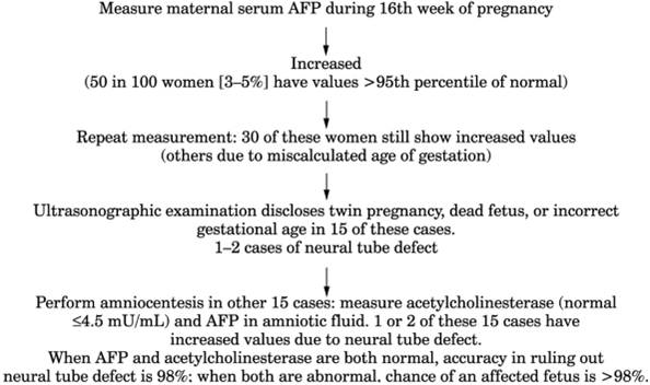
Maternal
Serum Sampling
- See Table
12-6 and Fig. 12-2.
- AFP is increased 4×
normal in open neural tube, 7× normal in anencephaly, and in ventral wall
defects; associated with exposed fetal-membrane and blood-vessel surfaces.
- Maximum serum AFP
concentration is between 1618 wks, but sampling should not be done before
14 or after 20 wks. If both serum and amniotic fluid show increased
levels, contamination of amniotic fluid with fetal or maternal blood is
ruled out by assay for fetal Hb and acetylcholinesterase. If only maternal
serum AFP is increased without demonstrable defect, pregnancy is at
increased risk (e.g., premature delivery, low-birth-weight baby, or fetal
death).
- Decreased AFP and
unconjugated estriol in trisomy 21 (Down syndrome) and 18 hCG significantly
increased in trisomy 21
Amniocentesis
- Generally done between 8
and 12 wks of gestation. Risk of fetal loss is ~0.5%.
- Cell culture takes 57
days; activity similar to that in fibroblasts.
Use
- Can detect intermediary
metabolites of some inborn errors, especially organic acid disorders.
- AFP is increased ~20× in
anencephaly, 7× in open neural tube, and in ventral wall defects
associated with exposed fetal-membrane and blood-vessel surfaces. See
preceding paragraph.
Chorionic
Villus Sampling
- Generally done between 8
and 12 wks of gestation; sometimes as early as 67 wks. Risk of fetal loss
is 0.52%.
- Contamination with
maternal decidua must be avoided for accurate diagnosis based on fetal
chromosomes, enzyme assay, or DNA analysis.
P.509
|

|
|
Table 12-6. Serum Markers in Detection of
Various Prenatal Conditions
|
- In some patient
populations, a negative culture for Neisseria
gonorrhoeae or HSV may be required.
- Associated with ~7% fetal
loss similar to amniocentesis (spontaneous rate ~4.5%).
- False-positive in 2% of
cases compared with 0.3% of cases in amniocentesis.
- Most prenatal diagnoses
of enzyme defects are now made using this assay.
Indications
- Chromosomal examination
- Previous child with
chromosomal trisomy
- Mother carrier of
X-linked disorder (to determine fetal sex)
- Parent carrier of chromosomal
translocation
- Maternal age >35 yrs
- Restriction enzyme assay
- Hemoglobinopathy (e.g.,
thalassemia)
- Lesch-Nyhan syndrome
|
|
|
Fig. 12-2. Algorithm for
alpha-fetoprotein (AFP) testing in pregnancy (detects virtually all cases of
anencephaly and 80% of cases of open spina bifida with very few
false-positives).
|
- P.510
-
- Alpha -antitrypsin deficiency
- PKU
- Metabolic assay, e.g.,
- Adenosine deaminase
deficiency
- Adrenoleukodystrophy
- Argininosuccinicaciduria
- Citrullinemia
- Cystinosis
- Fabry's disease
- Fanconi's anemia
- Farber's disease
- Gaucher's disease
- GM gangliosidosis
- GM gangliosidosis (Tay-Sachs disease)
- Homocystinuria
- Krabbe's disease
- Lesch-Nyhan syndrome
- Maple syrup urine
disease
- Menkes' syndrome
- Metachromatic
leukodystrophy
- Methylmalonicaciduria
- Mucolipidosis II (I-cell
disease)
- Mucopolysaccharidosis
(Ia, II, III, IV)
- Multiple sulfatase
deficiency
- Niemann-Pick disease
- Pompe's disease
- Wolman's disease
- Zellweger syndrome
Fetal
Blood Sampling
Generally done at ~15th week but usually
also successful between 18th and 23rd wks. Check for maternal serum
contamination by determining hCG concentration. Additional risk to fetus of 2%.
Use
- Prenatal diagnosis of
- RBC isoimmunization,
e.g., Rh, minor antigens
- Alloimmune or autoimmune
thrombocytopenia
- Hemoglobinopathies
(e.g., thalassemias, sickle cell disorders, spherocytosis, enzyme
deficiencies [e.g., G-6-PD])
- Coagulation defects
(e.g., factor VIII and IX hemophilias and fetal sex, other factor
deficiencies, von Willebrand's disease)
- Immune-deficiency
disorders (e.g., SCID, Wiskott-Aldrich syndrome, ataxia-telangiectasia,
chronic granulomatous disease, homozygous C3 deficiency, Chédiak-Higashi
syndrome)
- Intrauterine infections
(detection of specific IgM and increased total IgM, increased WBC and
eosinophil count, decreased platelet count, various blood chemistries)
(e.g., rubella, toxoplasmosis, varicella, CMV, and parvovirus B19
infection)
- Chromosomal disorders
(e.g., mosaicism, fragile X syndrome)
- Metabolic and
cytogenetic disorders (e.g., PKU, Alpha -antitrypsin deficiency, cystic fibrosis, Duchenne's muscular
dystrophy)
- Other conditions (e.g.,
familial hypercholesterolemia, hyperphenylalaninemia,
adrenoleukodystrophy)
- Fetal acid-base balance
and metabolic state
Fetal
Biopsy
Use
- Liver biopsy for
diagnosis of deficiency of long-chain 3-hydroxyacylcoenzyme A (CoA)
dehydrogenase, ornithine transcarbamylase deficiency, atypical PKU due to
deficiency
P.511
of glutamyl transpeptidase cyclohydrolase I, type I primary hyperoxaluria,
glycogen storage disease type I.
- Skin biopsy (e.g., for
certain genetic disorders such as epidermolysis bullosa)
- Muscle biopsy for
Duchenne's muscular dystrophy
Ultrasonography
and Echocardiography
Use
- To guide sampling process
- To verify gestational age
- Karyotyping is done if
malformations are found because one-third of these fetuses have a
chromosomal disorder.
- May be abnormal in
trisomy 13, 18, 21, 45, X, and in triploidy.
- ~50% of major heart,
kidney, and bladder abnormalities not detected by maternal serum AFP
screening.
Karyotype
Analysis
Use
Determine status of chromosomes X, Y, 21, 18,
13
Molecular
Diagnosis
Use
Direct detection of gene deletions and
mutations and linkage analysis using cultured amniocytes or chorionic villi can
make some diagnoses even when gene products are not present (e.g., adult
polycystic kidney disease, sickle cell disease, alpha-thalassemia, cystic
fibrosis, Gaucher's disease, Duchenne's muscular dystrophy, fragile X syndrome,
factor VIII and factor IX deficiencies).
Isolation
Of Fetal Cells In Maternal Blood
(Usual ratio =
1:10001:5000)
Use
Still an investigational procedure but
would allow diagnosis by flow cytometry and PCR. PCR can demonstrate Y
chromosome in women carrying male fetuses.
Newborn
Screening
Chromosome
Analysis (Karyotyping)
Use
- Suspected autosomal
syndromes, e.g.,
- Down syndrome
(mongolism)
- Trisomy E, 18
- Trisomy D, 13
- Cri du chat syndrome
- Suspected sex-chromosome
syndromes, e.g.,
- Klinefelter's syndrome,
XXY, XXXY
- Turner's syndrome, XO
- Superfemale XXX, XXXX
- Supermale XYY
- Funny-looking kid
syndromes, especially with multiple anomalies including mental
retardation and low birth weight
- Possible myelogenous
leukemia to demonstrate Ph chromosome
- Ambiguous genitalia
- Infertility (some
patients)
P.512
- Repeated miscarriages
- Primary amenorrhea or
oligomenorrhea
- Mental retardation with
sex anomalies
- Hypogonadism
- Delayed puberty
- Abnormal development at
puberty
- Disturbances of somatic
growth
Inherited
Disorders That Can Be Identified By Molecular Genetics
- Adult polycystic disease
- Achondroplasia
- Alpha -antitrypsin
deficiency
- Canavan's disease
- Charcot-Marie-Tooth
disease
- Congenital adrenal
hyperplasia
- Cystic fibrosis
- Duchenne's and Becker's
muscular dystrophies
- Familial adenomatous
polyposis
- Familial
hypercholesterolemia
- Fragile X syndrome
- Galactosemia
- Gaucher's disease
- Hemophilia A and B
- Huntington's disease
- Marfan syndrome
- Mitochondrial disorders
- Myotonic dystrophy
- Neurofibromatosis types 1
and 2
- Ornithine
transcarbamoylase deficiency
- PKU
- Spinal muscular atrophy
- Spinocerebellar ataxia
- Sickle cell disease
- Tay-Sachs disease
- Alpha- and
beta-thalassemia
Metabolic
Conditions (Inherited), Classification
(Deficient enzyme is shown in parentheses.)
|
Disorders of carbohydrate metabolism
|
|
|
Diabetes mellitus
|
|
|
Pentosuria
|
|
|
Fructose
|
|
|
Fructosuria
(aldolase B)*
|
|
|
Fructose-1,6-bisphosphatase
deficiency*
|
|
|
Lactose
|
|
|
Familial
lactose intolerance
|
|
|
Galactose
|
|
|
Galactosemia
(galactose 1-phosphate uridyltransferase)*
|
PD
|
|
Galactokinase
deficiency
|
PD
|
|
Glycogen storage
diseases*
|
PD for some
|
|
Disorders of amino acid metabolism
|
|
|
Phenylalanine
|
|
|
PKU
(phenylalanine hydroxylase)
|
PD
|
|
Methionine
|
|
|
Homocysteinuria
(cystathionine synthase)
|
PDP.513
|
|
Tyrosine
|
|
|
Tyrosinemia I
(fumarylacetoacetate hydrolase)*
|
PD
|
|
Tyrosinemia II
(tyrosine aminotransferase)
|
|
|
Valine, leucine, isoleucine
|
|
|
Maple syrup
urine disease (branched-chain ketoacid dehydrogenase)*
|
PD
|
|
Glycine
|
|
|
Nonketotic
hyperglycinemia (glycine cleavage system)*
|
PD
|
|
Lysine
|
|
|
Hyperlysinemia
(aminoadipic semialdehyde synthase)
|
|
|
Proline
|
|
|
Hyperprolinemia
I (proline oxidase)
|
|
|
Hyperprolinemia
II (pyrroline-5-carboxylate dehydrogenase)
|
|
|
Hyperimidodipeptiduria
(prolidase)
|
|
|
Urea cycle disorders
|
|
|
Citrullinemia
(argininosuccinic acid synthetase)*
|
PD
|
|
Argininemia (arginase)
|
PD
|
|
Argininosuccinicaciduria
(argininosuccinate lyase)*
|
PD
|
|
Ornithine
carbamoyltransferase deficiency*
|
PD
|
|
N-acetylglutamate
synthetase deficiency
|
|
|
Carbamyl phosphate
synthetase deficiency*
|
|
|
Organic acidurias
|
|
|
Propionate and methylmalonate
metabolism
|
|
|
Propionicacidemia
(propionylCoA carboxylase)*
|
PD
|
|
Methylmalonicacidemia
(methylmalonylCoA mutase, adenosylcobalamin synthesis)*
|
PD
|
|
Multiple
carboxylase deficiency (holocarboxylase synthetase, biotinidase)
|
|
|
Pyruvate and lactate
metabolism
|
|
|
LD deficiency
|
|
|
Pyruvate
dehydrogenase deficiency
|
|
|
Pyruvate
carboxylase deficiency*
|
PD
|
|
Phosphoenolpyruvate
carboxykinase deficiency*
|
|
|
Branched-chain organic
acidemias
|
|
|
Isovalericacidemia
(isovalerylCoA dehydrogenase)*
|
PD
|
|
Mevalonicaciduria
(mevalonate)
|
PD
|
|
Other organic acid
disorders
|
|
|
Alkaptonuria
(homogentisic acid oxidase)
|
|
|
Hyperoxaluria
type I, glycolicaciduria (alanine-glyoxylate aminotransferase)
|
|
|
Hyperoxaluria
type II, glycericaciduria (glyceric dehydrogenase)
|
|
|
Glycerol kinase
deficiency
|
|
|
Canavan's
disease (aspartoacylase)
|
|
|
Lysosomal enzyme defects
|
|
|
Mucopolysaccharidoses
|
PD
|
|
Mucolipidosis
II and III (uridine diphosphateN-acetyl-glucosaminelysosomal
enzyme N-acetylglucosaminyl-L-phosphotransferase)
|
PD
|
|
Glycoproteinoses
|
|
|
Alpha-
and beta-mannosidosis (alpha- and beta-mannosidase)
|
PD
|
|
Sialidosis
types I, II (neuraminidase)
|
PD
|
|
Fucosidosis
(alpha-fucosidase)
|
PD
|
|
GM gangliosidoses
|
|
|
Tay-Sachs
disease (hexosaminidase A)
|
PD
|
|
Sandhoff's
disease (hexosaminidase A, B)
|
PD
|
|
GM activator deficiency
|
|
|
Other lysosomal storage
disorders
|
|
|
Metachromatic
leukodystrophy (arylsulfatase A)
|
PD
|
|
Multiple
sulfatase deficiency (multiple lysosomal sulfatases)
|
PD
|
|
Niemann-Pick
disease (sphingomyelinase)*
|
PD
|
|
Farber's
disease (ceramidase)
|
PD
|
|
Gaucher's
disease (cerebroside beta-glucosidase)*
|
PDP.514
|
|
Pompe's disease
(glycogen storage disease type II) (alpha-1,4-glucosidase deficiency)
|
PD
|
|
Krabbe's
disease (galactocerebrosidase)*
|
PD
|
|
Fabry's disease
(alpha-galactosidase)
|
PD
|
|
GM gangliosidosis (beta-galactosidase)*
|
PD
|
|
Wolman's
disease (acid lipase)*
|
PD
|
|
Cholesteryl
ester storage disease (acid lipase)
|
PD
|
|
Mucolipidosis
type IV
|
|
|
Peroxisomal disorders
|
|
|
Acatalasia (catalase)
|
|
|
Refsum's disease (phytanic
acid hydroxylase)
|
PD
|
|
Zellweger syndrome
(peroxisome biogenesis)*
|
PD
|
|
Purine and pyrimidine
metabolism disorders
|
|
|
Lesch-Nyhan
syndrome (hypoxanthine phosphoribosyltransferase)
|
PD
|
|
Oroticaciduria
(uridine 5'-monophosphate synthase)
|
|
|
Xanthinuria
(xanthine oxidase)
|
|
|
Disorders of metal
metabolism
|
|
|
Wilson's
disease
|
|
|
Hemochromatosis
|
|
|
Menkes' syndrome
|
PD
|
|
Disorders of lipid
metabolism see Table 12-7)
|
|
|
Disorders of heme proteins
|
|
|
Porphyrinurias
|
PD for some
|
|
Bilirubin
metabolism
|
|
|
Crigler-Najjar
syndromes I and II (uridine diphosphateglucuronyl transferase)
|
|
|
Gilbert's
syndrome (uridine diphosphateglucuronyl transferase)
|
|
|
Dubin-Johnson
syndrome
|
|
|
Rotor's
syndrome
|
|
|
Membrane transport
disorders
|
|
|
Cystinuria
|
|
|
Hartnup disease
|
|
|
Cystinosis
|
PD
|
|
Hypophosphatemic
rickets
|
|
|
Disorders of serum enzymes
|
|
|
Hypophosphatasia
(ALP)
|
PD
|
|
Hyperphosphatasia
|
|
|
Alpha -antitrypsin deficiency
|
|
|
Disorders of plasma
proteins
|
|
|
Analbuminemia
|
|
|
Agammaglobulinemia
|
|
|
Atransferrinemia
|
|
|
Disorders of blood
|
|
|
Coagulation
diseases (e.g., hemophilias)
|
PD
|
|
RBC G-6-PD
deficiency
|
PD
|
|
Hemoglobinopathies
and thalassemias
|
PD
|
|
Hereditary
spherocytosis
|
PD
|
|
Hereditary
nonspherocytic hemolytic anemia
|
PD
|
|
Others
|
|
|
Congenital
adrenal hyperplasia
|
|
|
Menkes'
syndrome
|
|
|
PD = Prenatal diagnosis is
possible.
|
|
|
* May
present in neonate.
|
|
Newborn Screening For Metabolic
Disorders
Indications
- Screen for disorders that
are asymptomatic until irreversible damage has occurred and for which
effective treatment exists.
- Population prevalence
sufficient to limit false-positive and false-negative results.
- High cost/benefit ratio
- Adequate follow-up to
assure appropriate treatment
P.515
Interpretation
- PKU
- Neonatal hypothyroidism
(see Fig. 13-5)
- Galactosemia
- Maple syrup urine disease
- Homocystinuria
- Biotinidase deficiency
(one cause of multiple carboxylase deficiency; incidence ~1 in 40,000;
ketoacidosis and organic aciduria can develop late)
- Sickle cell disease
- Congenital adrenal
hyperplasia
- Cystic fibrosis
- Toxoplasmosis
Nuclear
Sexing
- Epithelial cells from
buccal smear (or vaginal smear, etc.) are stained with cresyl violet and
examined microscopically.
- A dense body (Barr body)
on the nuclear membrane represents one of the X chromosomes and occurs in
3060% of female somatic cells. The maximum number of Barr bodies is one
less than the number of X chromosomes.
- If <10% of the cells
contain Barr bodies in a patient with female genitalia, karyotyping should
be done to delineate probable chromosomal abnormalities.
- A normal count does not
rule out chromosomal abnormalities.
- Two Barr bodies may be
found in
- 47 XXX female
- 48 XXXY male
(Klinefelter's syndrome)
- 49 XXXYY male
(Klinefelter's syndrome)
- Three Barr bodies may be
found in
- 49 XXXXY male
(Klinefelter's syndrome)
Sex
Chromosome In Leukocytes
- Presence of a drumstick
nuclear appendage in ~3% of leukocytes in normal females indicates the
presence of two X chromosomes in the karyotype. It is not found in males.
- It is absent in the XO
type of Turner's syndrome.
- In Klinefelter's syndrome
(XXY) the presence of drumsticks shows a lower incidence than the presence
of the extra Barr body. (Mean lobe counts of
neutrophils are also decreased.)
- Incidence of drumsticks
is decreased and mean lobe counts are lower in trisomy 21 as well.
- Double drumsticks are
exceedingly rare and impractical for diagnostic use.
Tests
of Lipid Metabolism
- See Chapter
5, Coronary Heart Disease.
- Blood lipid tests should
not be performed during stress or acute illness, e.g., recent myocardial
infarction, stroke, pregnancy, trauma, weight loss, use of certain drugs; should not be performed on hospitalized patients until 23
mos after illness.
- Abnormal lipid test
results should always be confirmed with a new specimen, preferably 1 wk
later, before beginning or changing therapy.
- Keeping tourniquet in
place longer than 3 mins may cause 5% variation in lipid values.
Apolipoproteins,
Serum
(Protein
component of lipoprotein that regulates their metabolism; each of four major
groups consists of a family of two or more immunologically distinct proteins.)
Use
- Assess risk of CHD
- Classify hyperlipidemias
P.516
- Apo A is the major
protein of HDL; Apo A-I and A-II
constitute 90% of total HDL protein in ratio of 3:1.
- Apo B is the major
protein in LDL; important in regulating cholesterol synthesis and
metabolism. Decreased by severe illness and abetalipoproteinemia.
- Apo C-I, C-II, and C-III are
associated with all lipoproteins except LDL; C-II is important in
triglyceride metabolism.
- Serum apo A-I and B
levels are more highly correlated with severity and extent of coronary
artery disease (CAD) than total cholesterol and triglycerides.
- Ratio of apo A-I to apo B
shows greater sensitivity and specificity for CAD than LDL/HDL cholesterol
ratio or HDL cholesterol/triglyceride ratio or any of the individual
components.7
- Because apo B is the only
protein in LDL and apo A-I is the major protein constituent of HDL and
VLDL, the ratio of apo B to apo A-I reflects the ratio of LDL to HDL and
may be a better discriminator of CAD than the individual components, but
data on apolipoproteins are still limited.
Cholesterol,
HDL (High-Density Lipoprotein), Serum
Intraindividual variation may be
~3.612.4%.
Use
- Assessment of risk for
CAD
- Diagnosis of various
lipoproteinemias (see below)
Increased
In
- (>60
mg/dL is negative risk factor for CAD)
- Vigorous exercise
- Increased clearance of
triglyceride (VLDL)
- Moderate consumption of
alcohol
- Insulin treatment
- Oral estrogen use
- Familial lipid disorders
with protection against atherosclerosis (illustrates importance of
measuring HDL to evaluate hypercholesterolemia)
- Hyperalphalipoproteinemia
(HDL excess)
- 1 in 20 adults with mild
increased total cholesterol levels (240300 mg/dL) secondary to increased
HDL (>70 mg/dL)
- LDL not increased
- Triglycerides are normal.
- Inherited as simple
autosomal dominant trait in families with longevity or may be caused by
alcoholism, extensive exposure to chlorinated hydrocarbon pesticides,
exogenous estrogen supplementation.
- Hypobetalipoproteinemia
Decreased
In
- (<32
mg/dL in men, <38 mg/dL in women)
- Is
inversely related to risk of CAD. For every 1 mg/dL decrease in HDL, risk
for CAD increases by 23%
- Secondary causes
- Stress and recent
illness (e.g., AMI, stroke, surgery, trauma)
- Starvation; nonfasting
sample is 510% lower.
- Obesity
- Lack of exercise
- Cigarette smoking
- Diabetes mellitus
- Hypo- and
hyperthyroidism
P.517
- Acute and chronic liver
disease
- Nephrosis
- Uremia
- Various chronic anemias
and myeloproliferative disorders
- Use of certain drugs
(e.g., anabolic steroids, progestins, antihypertensive beta-blockers,
thiazides, neomycin, phenothiazines)
- Genetic disorders
- Familial
hypertriglyceridemia.
- Familial
hypoalphalipoproteinemiacommon autosomal dominant condition with
premature CAD and stroke. One-third of patients with premature CAD may
have this disorder.
- HDL <10th percentile
(<30 mg/dL in men and <38 mg/dL in women of middle age).
- Homozygous Tangier
disease.
- Familial
lecithin-cholesterol acetyltransferase deficiency and fish eye disease.
- Nonneuropathic
Niemann-Pick disease.
- HDL deficiency with
planar xanthomas.
- Apo A-I and apo C-III
deficiency variant I and variant IIrare genetic conditions associated
with premature CAD and marked HDL deficiency.
Cholesterol,
LDL (Low-Density Lipoprotein), Serum
Use
Assess risk and decide treatment for CAD.
Increased
In
- (Is
directly related to risk of CAD)
- Familial
hypercholesterolemia
- Familial combined
hyperlipidemia
- Diabetes mellitus
- Hypothyroidism
- Nephrotic syndrome
- Chronic renal failure
- Diet high in cholesterol
and total and saturated fat
- Pregnancy
- Multiple myeloma,
dysgammaglobulinemia
- Porphyria
- Pregnancy
- Wolman's disease
- Cholesteryl ester storage
disease
- Anorexia nervosa
- Use of certain drugs
(e.g., anabolic steroids, antihypertensive beta-blockers, progestins,
carbamazepine)
Decreased
In
- Severe illness
- Abetalipoproteinemia
- Oral estrogen use
- LDL is measured by
ultracentrifugation and by analysis after antibody separation from HDL and
VLDL.
- LDL can be estimated by
the following formula (Friedewald equation):
- LDL = total cholesterol
(HDL cholesterol) (VLDL).
- VLDL = triglycerides/5.
- Formula underestimates
LDL (e.g., in chronic alcoholism), is unsuitable for monitoring,
misclassifies 1540% of patients when triglycerides = 200400 mg/dL, and
fails if fasting triglycerides are >400 mg/dL. Not reliable if type
III dyslipidemia is suspected or chylomicrons are present.
- Some laboratories also
report various ratios.
- Total cholesterol/HDL
ratio
|
Low risk
|
|
|
Average
risk
|
P.518
|
|
Moderate
risk
|
|
|
High risk
|
>11.0
|
|
Cholesterol
(Total), Serum
Use
- Monitoring for increased
risk factor for CAD
- Screening for primary and
secondary hyperlipidemias
- Monitoring of treatment
for hyperlipidemias
Interferences
- Note
effect of illness, intraindividual variation, position, season, drug use,
etc., when these values are used to diagnose and treat hyperlipidemias
- Intraindividual variation
may be 410% for serum total cholesterol. Repeat cholesterol values should
be within 30 mg/dL. Coefficient of variation should be <3%.
- Cholesterol values are up
to 8% higher in winter than in summer, 5% lower if patient bled when
sitting than when standing, and 1015% different when recumbent than when
standing.
- Cholesterol values of
EDTA plasma can be multiplied by 1.03 to make them comparable to serum
values.
- Serum cholesterol and HDL
can be nonfasting.
Increased
In
- Hyperlipoproteinemias
(see Table 12-7)
- Hyperalphalipoproteinemia
- Cholesteryl ester storage
disease
- Biliary obstruction
- Stone, carcinoma, etc.,
of duct
- Cholangiolitic cirrhosis
- Biliary cirrhosis
- Cholestasis
- von Gierke's disease
- Hypothyroidism
- Nephrosis (due to chronic
nephritis, renal vein thrombosis, amyloidosis, SLE, periarteritis,
diabetic glomerulosclerosis)
- Pancreatic disease
- Diabetes mellitus
- Total pancreatectomy
- Chronic pancreatitis
(some patients)
- Pregnancy
- Drug use (e.g.,
progestins, anabolic steroids, corticosteroids, some diuretics)
- Methodologic
interference (Zlatkis-Zak reaction) (e.g., bromides, iodides,
chlorpromazine, corticosteroids, viomycin, vitamin C, vitamin A)
- 10% of patients on
long-term levodopa therapy
- Hepatotoxic effect
(e.g., phenytoin sodium)
- Hormonal effect (e.g.,
corticosteroids, birth control pills, amiodarone)
- Total fasting that
induces ketosis leads to a rapid increase.
- Secondary
causes should always be ruled out
Decreased
In
- Severe liver cell damage
(due to chemicals, drugs, hepatitis)
- Hyperthyroidism
- Malnutrition (e.g.,
starvation, neoplasms, uremia, malabsorption in steatorrhea)
- Myeloproliferative
diseases
- Chronic anemia
- PA in relapse
- Hemolytic anemias
- Marked hypochromic
anemia
- Cortisone and ACTH
therapy
- Hypobeta- and
abetalipoproteinemia
- Tangier disease
P.519
- Infection
- Inflammation
- Drug use
- Hepatotoxic effect
(e.g., allopurinol, tetracyclines, erythromycin, isoniazid, monoamine
oxidase inhibitors)
- Synthesis inhibition
(e.g., androgens, chlorpropamide, clomiphene, phenformin)
- Diminished synthesis
(probable mechanism) (e.g., clofibrate)
- Other mechanisms (e.g.,
azathioprine, kanamycin, neomycin, oral estrogens, cholestyramine,
cortisone and ACTH therapy)
- Methodologic
interference (Zlatkis-Zak reaction) (e.g., thiouracil, nitrates)
Cholesterol
Decision Levels
See Fig. 12-3 and .
|
|
Cholesterol
(in mg/dL)
|
|
|
LDL
|
HDL
|
Total
|
LDL/HDL Ratio
|
|
Desirable level/low risk
|
<130
|
>60
|
<200
|
|
|
Borderline level/moderate risk
|
|
|
|
|
|
Elevated level/high risk
|
|
<35
|
|
>6.0
|
|
Chylomicrons,
Serum
Increased
In
- Lipoprotein lipase
deficiency (autosomal recessive disorder or due to deficient cofactor for
lipoprotein lipase) presenting in children with pancreatitis, xanthomas,
hepatosplenomegaly
- Apo C-II deficiency (rare
autosomal recessive disorder due to absence of or defective apo C-II).
Accumulation of VLDL and chylomicrons increases risk of pancreatitis.
- Type V
hyperlipoproteinemia
Lipoprotein
Electrophoresis
Use
- Identify rare familial
disorders (e.g., types I, III, V hyperlipidemias) to anticipate problems
in children. Shows a specific abnormal pattern in <2% of Americans
(usually type II, IV).
- May be indicated if
- Serum triglycerides are
>300 mg/dL.
- Fasting serum is
lipemic.
- Significant
hyperglycemia, impaired glucose tolerance, or glycosuria.
- Increased serum uric
acid.
- Strong family history of
premature CHD.
- Clinical evidence of CHD
or atherosclerosis in patient aged <40.
- If lipoprotein
electrophoresis is abnormal, tests should be performed to rule out secondary
hyperlipidemias (see below).
Lipoproteins,
Serum
Decreased
In
- Abetalipoproteinemia
(Bassen-Kornzweig syndrome)
- Tangier disease
- Hypobetalipoproteinemia
Increased
In
- Hyperbetalipoproteinemia
- Hyperalphalipoproteinemia
P.520
P.521
|

|
|
Table 12-7. Comparison of Classic Types
of Hyperlipoproteinemia
|
P.522
|
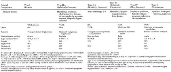
|
|
Table 12-7. (continued)
|
Triglycerides,
Serum
(80% in VLDL,
15% in LDL)
Classification8
|
Normal
|
<200 mg/dL
|
|
Borderline high
|
200400 mg/dL
|
|
High
|
4001000 mg/dL
|
|
Very high
|
>1000 mg/dL
|
|
P.523
Interferences
Diurnal variation causes triglycerides to
be lowest in the morning and highest around noon. Intraindividual variation in
serum triglycerides is 1240%; analytical variation is 510%.
Increased
In
- Genetic hyperlipidemias
(e.g., lipoprotein lipase deficiency, apo C-II deficiency, familial
hypertriglyceridemia, dysbetalipoproteinemia, cholesteryl ester storage
disease, Wolman's disease, von Gierke's disease)
- Secondary hyperlipidemias
- Gout
- Pancreatitis
- Acute illness (e.g., AMI
[rises to peak in 3 wks and increase may persist for 1 yr]; cold, flu)
P.524
|
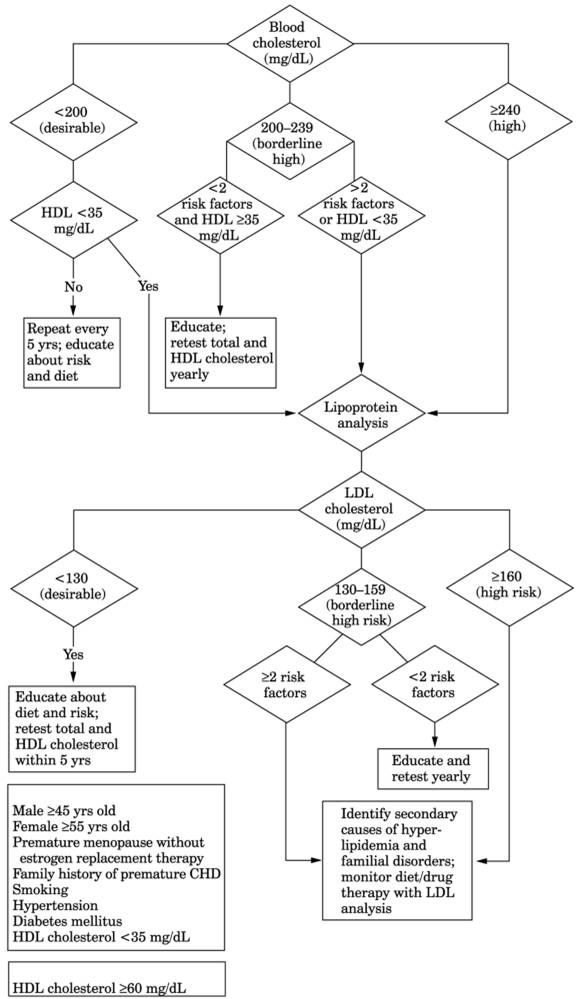
|
|
Fig. 12-3. Algorithm of recommended
testing and treatment of increased serum total and high-density lipoprotein
(HDL) cholesterol in adults without evidence of coronary heart disease (CHD).
Measure serum total cholesterol, HDL cholesterol, and triglycerides after 12-
to 14-hr fast. Average results of two or three tests; if difference of
≥30 mg/dL, repeat tests 18 wks apart and average results of three
tests. Use total cholesterol for initial case finding and classification and
monitoring of diet therapy. Do not use age- or sex-specific cholesterol values
as decision levels. Always rule out secondary and familial causes. (LDL =
low-density lipoprotein.) (Adapted from
Adult
Treatment Panel II. Report of the Expert Panel on Detection, Evaluation, and
Treatment of High Blood Cholesterol in Adults. National Cholesterol Education
Program. Bethesda, MD: National Heart, Lung, and Blood
Institute, National Institutes of Health, Sep 1993. NIH publication 93-3095.
|
P.525
- Drug use (e.g., thiazide
diuretics, anabolic steroids, cholestyramine, corticosteroids, amiodarone,
interferon)
- Pregnancy
- Concentrations associated
with certain disorders
- <250 mg/dL: not
associated with any disease state.
- 250500 mg/dL:
associated with peripheral vascular disease; may be a marker for patients
with genetic forms of hyperlipoproteinemias who need specific therapy.
- >500 mg/dL:
associated with high risk of pancreatitis.
- >1000 mg/dL:
associated with hyperlipidemia, especially type I or type V; substantial
risk of pancreatitis.
- >5000 mg/dL:
associated with eruptive xanthoma, corneal arcus, lipemia retinalis,
enlarged liver and spleen.
Decreased
In
- Abetalipoproteinemia
- Malnutrition
- Dietary change (within 3
wks)
- Recent weight loss
- Vigorous exercise
(transient)
- Drugs (e.g., ascorbic
acid, clofibrate, phenformin, asparaginase, metformin, progestins,
aminosalicylic acid)
- Total
and HDL cholesterol levels are similar when fasting or nonfasting but
triglycerides should be measured after 1214 hrs of fasting. Serum levels
are 35% higher than plasma levels
- Triglyceride
levels are not a strong predictor of atherosclerosis or CAD and may not be
an independent risk factor. Triglyceride levels are inversely related to
HDL cholesterol levels
Disorders
of Lipid Metabolism
Acid
Lipase Deficiencies
- (Inability
to hydrolyze lysosomal triglycerides and cholesteryl esters due to acid
lipase deficiency)
- Decreased acid lipase in leukocytes or cultured fibroblasts.
- Increased serum
triglycerides, LDL cholesterol, and cholesteryl esters.
Wolman's
Disease
- (Rare
autosomal recessive deficiency of lysosomal acid lipase activity causing
accumulation of cholesterol and triglycerides throughout body tissues and
death within first 6 mos)
- Prominent anemia develops
by 6 wks of age.
- Peripheral blood smear
shows prominent vacuolation (in nucleus and cytoplasm) of leukocytes.
- Characteristic foam cells in bone marrow resemble those in
Niemann-Pick disease.
P.526
|
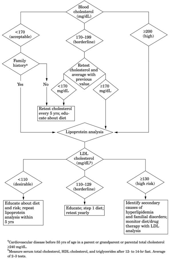
|
|
Fig. 12-4. Algorithm of recommended
testing and treatment of increased serum cholesterol in children and
adolescents. (HDL = high-density lipoprotein; LDL = low-density lipoprotein.)
(Adapted from Report of the Expert Panel on Detection, Evaluation, and
Treatment of High Blood Cholesterol in Children and Adolescents. National
Cholesterol Education Program. Bethesda,
MD: National Institutes of
Health, Sep 1991. NIH publication 91-2732.)
|
P.527
- Abnormal accumulation of cholesteryl esters and triglycerides in tissue biopsy (e.g.,
liver) establishes the diagnosis; cirrhosis may also be present.
- Assay shows absent acid lipase activity in many tissues, including leukocytes and
cultured fibroblasts. Heterozygotes have enzyme activity of ~50% of normal
in leukocytes or cultured fibroblasts.
- Prenatal diagnosis by demonstrating enzyme deficiency in cultured amniocytes.
- Laboratory findings due
to organ involvement
- Abnormal liver function
tests (due to lipid accumulation)
- Malabsorption
- Decreased adrenal
cortical function (diffuse calcification on CT scan)
Cholesteryl
Ester Storage Disease
(Rare inherited
deficiency of lysosomal acid lipase; milder than Wolman's disease)
- Pattern similar to that
of type II hyperlipidemia
- Increased LDL and
decreased HDL cholesterol
- Accelerated
cardiovascular disease; absent xanthomas; enlarged liver and spleen
Primary
Hyperlipidemias
See Table 12-7.
Severe
Hypertriglyceridemia (Type I) (Familial Hyperchylomicronemia Syndrome)
- (Rare
autosomal recessive trait due to deficiency of lipoprotein lipase [LPL] or
apo C-II or circulating inhibitor of LPL; marked heterogeneity in
causative molecular defects)
- Persistent very high
triglycerides (>1000 mg/dL) with marked increase in VLDL and
chylomicrons. Responds to marked dietary fat restriction.
- Patients with apo C-II
deficiency cannot activate LPL in vitro. Deficiency of apo C-II is shown
by isoelectric focusing or two-dimensional gel electrophoresis of plasma.
- Associated with recurrent
pancreatitis rather than CAD.
- Laboratory changes due to
fatty liver (increased serum transaminase)
Familial
Hypercholesterolemia (Type II)
- (Autosomal
dominant disorder)
- LDL receptors in fibroblasts or mononuclear blood cells are absent in homozygous
patients and 50% of normal levels in heterozygous patients (test performed
at specialized labs).
- Homozygousvery rare
condition (1 per million) in which serum cholesterol is very high (e.g.,
6001000 mg/dL) with corresponding increase (68× normal) in LDL. Both
parents are heterozygous. Clinical manifestations of increased total
cholesterol (xanthomata, corneal arcus, CAD that causes death, usually at
<30 yrs).
- Neonatal diagnosis requires finding increased LDL cholesterol in
cord blood;
serum total cholesterol is unreliable. Because of marked variation in
serum total cholesterol levels during first year of life, diagnosis should
be deferred until 1 yr of age.
- Prenatal diagnosis of homozygous fetus can be made by estimation
of binding
sites on fibroblasts cultured from amniotic fluids; useful when both
parents are heterozygous.
- Heterozygousincreased
serum total cholesterol (300500 mg/dL) and LDL (23× normal) with similar
change in a parent or first-degree serum triglycerides and VLDL
are normal in 90% and slightly increased in 10% of these cases. Gene
frequency is 1 in 500 in general population, but 5% in survivors of AMI
who are <60 yrs. Premature CAD, tendinous xanthomas, and corneal arcus
are often present.
- Plasma triglycerides are
normal in type II-A but increased in type II-B. This is not the most
common cause of phenotype II-A.
Polygenic
Hypercholesterolemia (Type II-A)
- Persistent total cholesterol
elevation (>240 mg/dL) and increased LDL without familial
hypercholesterolemia or familial combined hypercholesterolemia.
- Premature CAD occurs
later in life than with familial combined hyperlipidemia.
- Xanthomas are rare.
P.528
Familial
Combined Hyperlipidemia (Types II-B, IV, V)
- (Occurs
in 0.5% of general population and 15% of survivors of AMI <60 yrs old)
- Any combination of
increased LDL and VLDL and chylomicrons may be found; HDL is often low;
different family members may have increased serum total cholesterol or
triglycerides or both.
- Premature CAD occurs
later in life (>30 yrs of age) than with familial hypercholesterolemia.
- Xanthomas are rare.
- Patients are often
overweight.
Familial
Dysbetalipoproteinemia (Type III)
- (Occurs
in 1 in 5000 in the population.)
- Abnormality of apo E with
excess of abnormal lipoprotein (beta mobilityVLDL); total cholesterol
>300 mg/dL plus triglycerides >400 mg/dL should suggest this
diagnosis. VLDL cholesterol/triglyceride ratio = 0.3.
- Diagnosis by combination of ultracentrifugation and isoelectric
focusing that shows abnormal apo E pattern.
- Tuberous and tendinous
xanthomas and palmar and plantar xanthomatous streaks are present.
- Atherosclerosis is more
common in peripheral than in coronary arteries.
Familial
Hypertriglyceridemia (Type IV)
- (Autosomal
dominant condition present in 1% of general population and 5% of survivors
of AMI aged <60 yrs)
- Elevated triglycerides
(usually 200500 mg/dL) and VLDL with normal LDL and decreased HDL.
- Distinction from familial
combined hyperlipidemia is made only by extensive family screening.
Abetalipoproteinemia
(Bassen-Kornzweig Syndrome)
- (Extremely
rare autosomal recessive disorder; should be ruled out in children with
fat malabsorption, steatorrhea, failure to thrive, neurologic symptoms,
pigmented retinopathy, acanthocytosis)
- Marked decrease in serum triglycerides (<30 mg/dL) with little
increase after ingestion of fat, and in total cholesterol (2050 mg/dL)
- Chylomicrons, LDL, VLDL, and apo B are absent; HDL may be lower
than in normal persons.
- Plasma lipids are normal
in heterozygotes.
- Acanthocytes may be 5090% of RBCs and are characteristic.
- Decreased RBC life span
causes anemia that may vary from severe hemolytic anemia to mild
compensated anemia.
- Low serum levels of
carotene and other fat-soluble vitamins.
- Biopsy of small intestine shows characteristic lipid
vacuolization; not pathognomonic (occasionally seen in celiac disease,
tropical sprue, juvenile nutritional megaloblastic anemia).
- Negative sweat test
distinguishes this disorder from cystic fibrosis.
- Arteriosclerosis is
absent.
- A variant is
normotriglyceridemic abetalipoproteinemia in which patient can secrete apo
B-48 but not apo B-100, which results in normal postprandial triglyceride
values but marked hypocholesterolemia; associated with mental retardation
and vitamin E deficiency.
Hypobetalipoproteinemia
- (Autosomal
codominant disorder with increased longevity and lower incidence of
atherosclerosis; at least one parent shows decreased beta-lipoprotein)
- Marked decrease in LDL
and LDL/HDL ratio.
- Homozygous patients have
decreased serum cholesterol (<60 mg/dL) and triglycerides and
undetectable or trace amounts of chylomicrons, VLDL, and LDL.
- Heterozygotes are
asymptomatic and have serum total cholesterol, LDL, and apo B values of
50% of normal (consistent with codominant disorder). May also be caused by
P.529
malabsorption of fats, infection, anemia, hepatic necrosis, hyperthyroidism,
AMI, acute trauma.
L-Carnitine
Deficiency
- (Very
rare metabolic disorder of fatty acid metabolism [beta oxidation])
- Two types
- Myopathic: Deficiency
limited to muscle; normal levels in plasma and other tissues.
Myoglobinuria in older children or young adults. Biopsy shows lipid
deposits. Tissue homogenates do not support normal rates of beta
oxidation of long-chain fatty acids unless L-carnitine is added. Serum
carnitine is normal or slightly decreased.
- Systemic: More acute
clinical picture, presents earlier in life; may mimic Reye's syndrome.
- L-carnitine depleted in blood and all tissues.
- Tissue contains marked decreased activity of medium-chain acyl-CoA dehydrogenase.
- Hepatic encephalopathy
- Hypoglycemia without
ketosis
- Hyperammonemia may be
present.
- Serum uric acid may be
increased.
- Laboratory findings due
to cardiomyopathy
Due To
- Dietary deficiency
- Low renal reabsorption
(e.g., Fanconi's syndrome)
- Inborn deficiency of
medium-chain acyl-CoA dehydrogenase
- Valproic acid therapy
(inducing excretion of valprolycarnitine in urine)
- Excessive loss of free
carnitine in urine due to failure of carnitine transport across cells of
renal tubule, muscle, and fibroblasts
- Organic acidurias (e.g.,
methylmalonicaciduria, propionicacidemia)
- Other conditions (e.g.,
maternal deficiency, prematurity)
Lecithin-Cholesterol
Acyltransferase Deficiency (Familial)
- (Rare
autosomal recessive disorder of adults. Corneal opacities lead to
blindness.)
- Serum total cholesterol is normal but cholesteryl esters are
virtually absent. Plasma free cholesterol is extremely increased. HDL is
low.
- Anemia with large RBCs
that are frequently target cells
- Proteinuria
Lipodystrophy
(Total), Congenital
- (Rare
autosomal recessive disorder characterized by absence of fat in skin and
viscera, possibly due to deficiency in number or quality of insulin
receptors)
- No neonatal laboratory
abnormalities
- Later in life: marked
insulin resistance, glucose intolerance, development of diabetes mellitus
(although ketosis is unusual), increased serum triglycerides develop
- Laboratory findings due
to fatty liver, cirrhosis, acanthosis nigricans
- Similar syndromes of
leprechaunism, acquired and partial lipodystrophies
Tangier
Disease
- (Rare
autosomal recessive disorder causing defect in metabolism of apo A in
which a marked decrease [heterozygous] or absence [homozygous] of HDL is
seen)
- Plasma levels of apo A-I
and A-II are extremely low. In homozygotes, HDL is usually <10 mg/dL
and apo A-I is usually <5 mg/dL. In heterozygotes, HDL and apo A-I are
~50% of normal.
- Prebeta-lipoprotein is
absent.
- Serum total cholesterol
(<100 mg/dL), LDL cholesterol, and phospholipid are decreased;
triglycerides are normal or increased (100250 mg/dL).
- Deposits of cholesteryl
esters in RE cells cause enlarged liver, spleen, and lymph nodes, enlarged
orange tonsils, small orange-brown spots in rectal mucosa; premature CAD,
mild corneal opacification, and neuropathy may be present in homozygous
type.
P.530
Secondary
Hyperlipidemias
Due To
(Many are combined hyperlipidemias)
- Diabetes mellitus***
- Increased VLDL with
increased serum triglycerides, low HDL cholesterol; LDL cholesterol may
be normal or mildly increased. (Higher triglyceride values correlate with
hyperglycemia and poorer control of diabetes; reduced by insulin therapy)
- Hypothyroidism
- Increased LDL and total
cholesterol. Test for hypothyroidism whenever LDL
cholesterol is >190 mg/dL. Rapidly becomes normal with
treatment.
- Serum cholesterol is not
always increased.
- Nephrotic syndrome***
- Increased serum total
cholesterol and LDL cholesterol are usual.
- Increased VLDL and therefore
increased serum triglycerides may also occur.
- Other renal disorders
(chronic uremia, hemodialysis, after transplantation)
- Increased triglycerides
and total cholesterol and low HDL cholesterol may occur.
- Hepatic glycogenoses
- Increased serum
lipoprotein is common in any of the forms, but the pattern cannot be used
to differentiate the type of glycogen storage disease.
- Predominant increase in
VLDL in glucose-6-phosphatase deficiency.
- Predominant increase in
LDL in debrancher and phosphorylase deficiencies.
- Obstructive liver disease***
- Increased serum total
cholesterol is common until liver failure develops.
- Resistant to
conventional drug therapy. The type of lipoproteinemia is variable.
- In intrahepatic biliary
atresia, there is often increase in lipoprotein X with marked increase in
serum total cholesterol and even more marked increase in serum
phospholipids.
- Chronic alcoholism
- Marked increase in VLDL
producing type IV or V patterns
- Hyperlipoproteinemia of
affluence (dietary)***
- Pregnancy***
- Drugs
- Estrogens, steroids,
beta-blockers***
- Diuretics, cyclosporine
Metabolic
Errors Associated With Hyperammonemia In Children
- Defects in urea
cyclesevere hyperammonemia with respiratory alkalosis, e.g.,
- Arginosuccinate
synthetase deficiency
- Arginosuccinate lysase
deficiency
- Arginase deficiency
- Citrullinemia
- Ornithine
transcarbamylase deficiency
- N-Acetylglutamate
synthetase deficiency
- Carbamoyl phosphate
synthetase deficiency
- Organic acid defectsmild
to moderate hyperammonemia (≤500 mg/dL), e.g.,
- Methylmalonicacidemia****
- Isovalericacidemia****
- Multiple carboxylase
deficiency****
- Propionicacidemia****
- Glutaricaciduria type II
- Ketothiolase deficiency
- Hyperornithinemia
- Transient hyperammonemia
of newborn
- Fatty acid oxidation
defect
P.531
- Plasma
ammonia should be determined in any neonate with unexplained neurologic
deterioration or any patient with unexplained encephalopathy or episodic
lethargy and vomiting
Metabolic
Errors Causing Acidosis
- Amino acid disorders
- Maple syrup urine
disease
- Hypervalinemia
- Hyperleucineisoleucinemia
- Organic acid defects
- Isovalericacidemia
- Propionicacidemia
- Methylmalonicacidemia
- Glutaricacidemia
- Combined carboxylase
deficiency
- 3-Hydroxy-3-methylglutaricacidemia
- 2-Methyl-3-hydroxybutyricacidemia
- Acyl CoA dehydrogenase
deficiencies
- Glycogen storage diseases
Disorders
of Amino Acid Metabolism
See Fig. 12-5, Table 12-9.
Aminoaciduria,
Secondary
Due To
- Inherited
(Generalized)
- Cystinosis
- Fanconi's syndrome
(idiopathic)
- Fructose intolerance
- Galactosemia
- GSD Type I (rare)
- Lactose intolerance
- Lowe's syndrome
- Tyrosinosis
- Wilson's disease
Not
Inherited (all generalized except as indicated by
- Connective tissue
diseases
- Drugs (e.g., deficiency
of vitamins B , C, D; outdated tetracycline,
salicylates, steroids, toxic heavy metals)
- Endocrine (e.g.,
hyperparathyroidism, hyperthyroidism , neurosecretory tumors )
- Kidney disorders (e.g.,
nephrotic syndrome, renal transplant reaction)
- Liver necrosis
- Newborns (normal)
- Others
Occurs
in
- Severe liver disease
- Renal tubular damage due
to
- Lysol
- Heavy metals
- Maleic acid
P.532
|
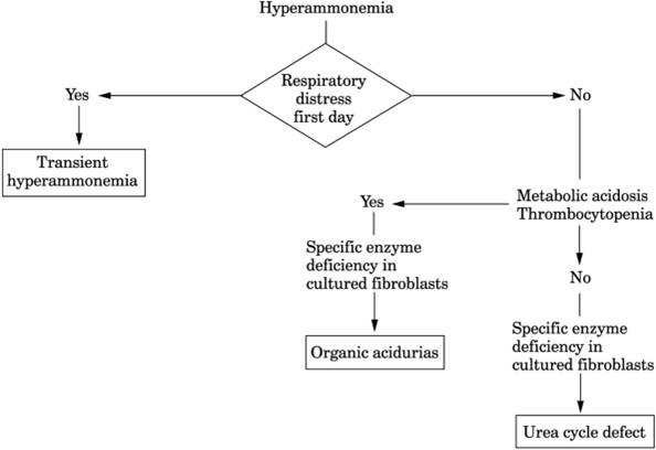
|
|
FIG. 12-5. Algorithm for neonatal
hyperammonemia.
|
- Burns
- Galactosemia
- Wilson's disease
- Scurvy
- Rickets
- Fanconi's syndrome
(e.g., outdated tetracycline, multiple myeloma, inherited)
- Neoplasms
- Cystathionine excretion
in neuroblastoma of adrenal gland
- Ethanolamine excretion
in primary hepatoma
Argininosuccinicaciduria
- (Autosomal
recessive deficiency of argininosuccinase; brittle hair, absence of
metabolic acidosis, and neurologic changes)
- Fasting blood ammonia is normal but level may be markedly
increased after eating.
- Argininosuccinic acid is increased in plasma and urine; may also be increased in
CSF.
- Because of block in urea
cycle, plasma arginine may be decreased and citrulline increased.
- Urine orotic acid is
increased.
- Serum ALP may be
increased.
- Heterozygous carriers show increased argininosuccinic acid in urine and decreased
argininosuccinase in RBCs.
- Prenatal diagnosis by assay of enzyme in cultured amniocytes (Mycoplasma
contamination may cause a false-negative result) or assay of amniotic
fluid for argininosuccinic acid
- Neonatal type is usually
fatal in infancy. Late-onset type may present at any age triggered by
intercurrent infection or stress.
Beta-Aminoisobutyricaciduria
- (Familial
recessive benign disorder of thymine metabolism)
- Increased beta-aminoisobutyric acid in urine (50200 mg/24 hrs)
- May
also occur in leukemia due to increased breakdown of nucleic acids
P.533
|
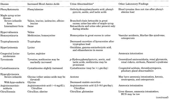
|
|
Table 12-8. Summary of Primary Overflow
Aminoacidurias (Increased Blood Concentration with Overflow into Urine)
|
P.534
|
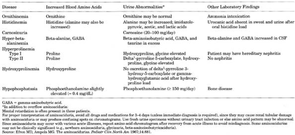
|
|
Table 12-8. (Continued)
|
P.535
|
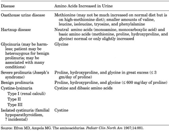
|
|
Table 12-9. Summary of Renal or Gut
Transport Aminoacidurias (Blood Amino Acids Are Normal or Low)
|
Citrullinemia
- (Rare
autosomal recessive deficiency of argininosuccinate synthetase with
metabolic block in citrulline utilization and associated mental
retardation)
- See Table
12-8.
- Genetically heterogeneous
(like other disorders of urea cycle) with various clinical pictures and
onset from neonatal to adult period
- Massive hyperammonemia (>1000 mg/dL) in neonatal form
- Markedly increased citrulline levels in blood, CSF, and urine
- Serum levels of glutamine, alanine, and aspartic acid are usually
increased; arginine is usually decreased.
- Urine orotic acid is
increased.
- Laboratory findings due
to liver disease
- Deficient enzyme activity can be demonstrated in liver cells and cultured
fibroblasts.
- Prenatal diagnosis by assay of citrulline in amniotic fluid or of enzyme in cultured
amniocytes
Cystathioninuria
- (Rare
autosomal recessive deficiency of cystathionase)
- Increased cystathionine
in urine
Cystinuria
- (Autosomal
recessive disorder characterized by failure of amino acid transport; renal
tubular reabsorption and intestinal uptake of cystine and dibasic amino
acids)
P.536
- Markedly increased cystine in urine (2030× normal). May also be increased in organic
acidemias, hyperuricemia, trisomy 21, hereditary pancreatitis, muscular
dystrophy, hemophilia, retinitis pigmentosa.
- Confirm diagnosis by identifying increased urinary arginine, lysine, and
ornithine in urine after age 6 months.
- Cystine renal and bladder stones
- Laboratory findings due
to GU tract infections. Bacteria can degrade cystine.
Hartnup
Disease
- (Autosomal
recessive disorder characterized by defect in renal or GI transport of
neutral amino acids)
- Urine contains increased (510×) amounts of alanine, threonine, valine, leucine,
isoleucine, phenylalanine, tyrosine, tryptamine, histidine.
Histidinemia
- (Rare
autosomal recessive aminoacidopathy due to deficiency of histidase in
liver and skin that causes histidine to be converted to urocanic acid.
Incidence is 1 in 14,00020,000 live births in United
States and 1 in 8000 in Japan.)
- Plasma histidine is increased to 5001000 µmol/L (normal = 85120 µmol/L).
- Urine
histidine is increased to 0.54.0 gm/day (normal <0.5 gm/day).
Histidine metabolites (imidazole acetic, imidazole lactic, and imidazole
pyruvic acids) are also increased in urine; alanine may be increased.
- Urine may show positive
Phenistix or ferric chloride test because of imidazole pyruvic acid.
- With oral histidine load,
no formiminoglutamic acid appears in urine.
- Most children show no
sequelae; therefore neonatal screening is not performed.
- Heterozygote detection is
not established yet.
Homocysteinuria/Homocysteinemia
- (Homocysteine
is reduced [sulfhydryl] form and homocystine is oxidized [disulfide] form
of homologues cysteine and cystine. Term refers to combined pool of
homocystine and homocysteine and their mixed disulfides.)
- Is
independent risk factor for premature arteriosclerosis of coronary,
cerebral, peripheral vessels
Due To
Autosomal recessive error of methionine
metabolism with deficient cystathionine synthetase in liver and brain with
inability to catalyze homocysteine to cystathionine. Incidence of mild form is
57% among general population; severe form is rare.
Increased
In
- Deranged vitamin B metabolism or block in folate metabolism or deficiency of vitamin
B , folate, or vitamin B
- Chronic renal or liver
failure, postmenopausal state, drug use (e.g., methotrexate, phenytoin,
theophylline, cigarette smoking)
- Various neoplastic
diseases (e.g., ALL, cancers of breast, ovary, pancreas).
- Urine excretion of homocysteine is increased (creased methionine and other amino acids.
- Increased serum homocysteine (up to 250 mg/day; normal = trace or not detected) and
methionine (≤2000 mg/day; normal is ≤30 mg/day); also
increased in CSF.
- Abnormal homocysteine metabolism may be shown only after methionine-loading test.
Blood samples before and at 4- to 8-hr intervals after 100 mg/kg
methionine oral load. Normal = transient increase in free and
protein-bound homocysteine peaking between 48 hrs. Abnormal = plasma
homocysteine >2 SD greater than that of normal controls.
P.537
- In homozygous form
laboratory findings due to associated clinical conditions
- Mild variable
hepatocellular dysfunction
- Mental retardation,
Marfan's syndrome, osteoporosis, etc.
- Serum methionine levels
should be kept at 20150 µmol/L by low-methionine diet and pyridoxine
therapy.
- Patients have enzyme
activity levels of 010% in fibroblasts and lymphocytes; heterozygotes
(their parents) have levels <50% of normal.
- For neonatal detection, measure methionine in filter paper specimen of blood;
confirm by measuring blood and urine amino acids.
- Can also measure specific enzyme in cultured fibroblasts.
Hydroxyprolinemia
- Increased hydroxyproline in blood
Hyperglycinemia
- (Long-chain
ketosis [without hypoglycemia] and ketonuria accentuated by leucine
ingestion)
- Same findings
(neutropenia, thrombocytopenia, hypogammaglobulinemia, increased glycine
in blood and urine, osteoporosis, hypoglycemia) may occur in
propionicacidemia, methylmalonicacidemia, isovalericacidemia,
3-ketothiolase deficiency.
Hyperprolinemia
- (Types
I and II)
- Increased proline in blood
- Increased glycine and
hydroxyproline in urine
Iminoglycinuria,
Familial
- (Inherited
autosomal defect of renal amino acid transport; may be associated with
mental retardation)
- Increased urine glycine
- Increased urine imino
acids (proline, hydroxyproline)
Joseph's
Syndrome (Imminoglycinuria)
- (Asymptomatic
malabsorption of proline, hydroxyproline, and glycine)
- Urine shows marked increase in proline, hydroxyproline, and glycine.
- Heterozygotes may show
mild prolinuria.
Lesch-Nyhan
Syndrome
- (X-linked
recessive trait of complete absence of hypoxanthine-guanine
phosphoribosyltransferase [HGPRT] that catalyzes hypoxanthine and guanine
to their nucleotides, causing accumulation of purines. The syndrome
appears in male children, with choreoathetosis, mental retardation, and
tendency to self-mutilating, biting, and scratching.)
- Increased serum uric acid levels (912 mg/dL)
- Hyperuricuria
- 34 mg of uric acid/mg
creatinine
- 4070 mg of uric acid/kg
body weight
- 6001000 mg/24 hrs in
patients weighing ≥15 kg
- Marked variation in
purine diet causes very little change
- Orange crystals or sand
in infants' diapers
- Deficient HGPRT in RBCs and fibroblasts; also allows carrier detection
and prenatal diagnosis.
- Laboratory findings due
to secondary gout (tophi after 10 yrs, crystalluria, hematuria, urinary
calculi, UTI, gouty arthritis, response to colchine); patients die of
renal failure by age 10 yrs unless treated.
- Deficiency of HGPRT in
RBCs and fibroblasts; also allows carrier detection and prenatal
diagnosis.
P.538
- Deficiency of HGPRT activity detected in cultured fibroblasts (<1.2% of normal) and
in RBC hemolysates (0%) establishes the diagnosis; in amniotic cells
allows diagnosis in utero. DNA probes allow prenatal diagnosis.
- Heterozygotes can be detected by study of individual hair follicles.
- Variants with partial deficiency of HGPRT show 050% of normal activity in RBC
hemolysates and >1.2% in fibroblasts; patients accumulate purines but
no orange sand in diapers; no abnormality of CNS or behavior.
L-Glycericaciduria
- (Genetic
variant of primary hyperoxaluria; autosomal trait that causes disease only
when homozygous)
- Renal
calculi composed of calcium oxalate
- L-Glyceric acid in urine (not found in normal urine
- Increased urinary oxalic acid (35× normal)
Maple
Syrup Urine Disease (Ketoaciduria)
- (Autosomal
recessive disorder characterized by deficiency of branched-chain keto acid
decarboxylase; incidence is 1 in 216,000 live births; characteristic maple
syrup or curry odor in urine, sweat, hair, and cerumen)
- Chromatography of urine and plasma show greatly increased urinary excretion of ketoacids
of leucine, isoleucine, and valine. Presence of alloisoleucine
(stereoisomeric metabolite of isoleucine) is characteristic.
- Metabolic acidosis and ketoacidosis occur.
- Ferric chloride test of urine produces green-gray color.
- Hypoglycemia is usual.
- The disease may be severe
or intermittent.
- Patient should be
monitored by daily urine testing with dinitrophenylhydrazine; because
urine levels correlate with plasma levels, plasma levels can be measured
once a month if urine is negative or shows only traces. (Control plasma
ranges: leucine = 180700 µmol/L, isoleucine = 70280 µmol/L, valine =
200800 µmol/L.)
- Measurement of amount of defective enzyme in leukocytes and fibroblasts shows
classic form (enzyme level 02% of normal), intermittent form (enzyme
level 28% of normal), and intermediate form (enzyme level 816% of
normal). Blood levels are normal in intermittent form except during acute
episodes caused by infection, surgery, vaccination, or sudden increased
intake of protein, which in children resemble classic form. Intermediate
form shows persistent elevation of blood amino acids, which can be kept in
normal range by maintaining dietary protein at <2 g/kg/day.
- Prenatal diagnosis by measurement of enzyme concentration in cells cultured from
amniotic fluid.
Methylmalonicaciduria
- (Very
rare autosomal recessive error of metabolism with neonatal metabolic
acidosis and mental and somatic retardation; at least four distinct forms;
screening incidence is 1 in 48,000 in infants 34 wks of age)
- Metabolic acidosis
- Increased methylmalonic acid in urine and plasma
- Long-chain ketonuria
- Intermittent
hyperglycinemia
- All findings accentuated by high-protein diet or supplemental
ingestion of valine or isoleucine.
- Hypoglycemia,
neutropenia, thrombocytopenia may occur.
- Heterozygote detection is
not reliable.
- Prenatal diagnosis by assay of methylmalonyl-CoA mutase in cultured amniocytes,
increased methylcitric or methylmalonic acids in amniotic fluid, or (late
in pregnancy) increased methylmalonic acid in maternal urine
- Methylmalonicaciduria
also occurs in vitamin B deficiency
P.539
Oasthouse
Urine Disease
- (Disorder
of methionine absorption in gut with distinctive odor of urine)
- Increase of various amino acids in blood and also in urine (e.g., phenylalanine, tyrosine,
methionine, valine, leucine, isoleucine)
Organic
Acidemias
- (E.g.,
methylmalonic, propionic, isovaleric)
- Show
- Metabolic acidosis and
ketoacidosis.
- Ketonuria.
- Hyperammonemia.
- Hypoglycemia.
- Sweat and urine have
odor of sweaty feet.
Ornithine
Transcarbamylase Deficiency
- (X-linked
recessive disorder characterized by deficiency of ornithine
transcarbamylase, an enzyme in urea cycle that converts ornithine to
citrulline)
- Increased blood ammonia,
usually 210× normal
- Decreased citrulline in blood
- Increased orotic acid in blood and urine. May also be increased in
lysinuric protein intolerance.
- Decreased ornithine transcarbamylase in biopsy of liver.
- Ornithine transcarbamylase
deficiency can occur after a bacterial or viral infection, thereby causing
confusion with Reye's syndrome.
- To detect asymptomatic carriers, measurement of urine orotic acid before and 6 hrs after
an oral protein loading test may be required for female heterozygotes. Can
also be detected by a complementary DNA probe for the ornithine
transcarbamylase gene using restriction fragment length polymorphism
analysis.
- Prenatal diagnosis using restriction fragment length polymorphism analysis for chorionic
villus DNA analysis.
Phenylketonuria
(Pku)
- (Inherited
autosomal recessive disorder [due to a variety of mutations on chromosome
12]; absence of phenylalanine hydroxylase activity in liver causes
increase in phenylalanine and its metabolites [phenylpyruvic acid,
orthohydroxyphenylacetic acid] in blood, urine, and CSF; tyrosine and the
derivative catecholamines are deficient. Results in mental retardation.
Among whites, 1 in 50 persons is a carrier and 1 in 10,000 is affected
with PKU; see Fig. 12-6
- Unrestricted protein diet
- Normal blood
phenylalanine = 2 mg/dL.
- Classic PKU: high blood phenylalanine (usually >30 mg/dL and always
>20 mg/dL in infancy) with phenylalanine and its metabolites in urine
(incidence is 1 in 14,000); normal or decreased tyrosine concentration.
- Less severe variant form of PKU: blood phenylalanine levels are 1530 mg/dL and
metabolites may appear in urine (incidence is 1 in 15,000).
- Mild persistent hyperphenylalaninemia: blood phenylalanine may be 212 mg/dL and
metabolites are not found in urine (incidence is 1 in 30,000); diet
restriction is not required for this form.
- For screening of
newborns, urine amounts of phenylpyruvic acid may be insufficient for
detection by colorimetric methods when blood level is <15 mg/dL. May
not appear in urine until 23 wks of age.
- Phenylpyruvic acid in
urine is significant (gives positive ferric chloride test) but may not be
present in some patients.
- Preliminary blood
screening tests (inhibition assay, fluorometry, paper chromatography) detect
levels >4 mg/dL. Screening should be performed after protein-containing
feedings have begun.
P.540
|
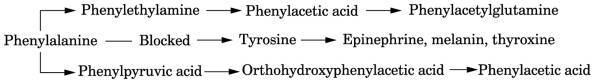
|
|
Fig. 12-6. Pathways of phenylalanine
metabolism.
|
- When repeat screening test is positive, quantitative blood phenylalanine and
tyrosine measurements are performed to confirm phenylalaninemia and
exclude transient tyrosinemia of newborn, which is most common cause of
positive screening. Diagnosis requires serum phenylalanine level ≥20
mg/dL. Urine ferric chloride is positive and chromatography confirms
orthohydroxyphenylacetic acid.
- Serial determinations should be performed in untreated borderline cases because blood
levels may change markedly with time or due to stress and infection.
- Diagnosis of PKU may be confirmed by giving 100 mg of ascorbic acid and collecting
blood and urine 24 hrs later.
- False-negative
results on Guthrie PKU test for screening newborns may occur if blood is
collected using capillary tubes and venipuncture rather than applied
directly to filter paper, especially if level is within 0.2 mg of cutoff
value.
- Adjust diet by frequent
monitoring of blood phenylalanine (e.g., 10 mg/dL) with consistently
negative ferric chloride urine test.
- In women with untreated
PKU and increased serum phenylalanine, frequency of mental retardation,
microcephaly, and congenital heart disease in offspring is greatly
increased.
- Detection of heterozygotes in 75% of families and prenatal diagnosis are now
possible using complementary DNA probe.
- Laboratory findings due
to congenital heart disease in ≤15% of PKU patients
Comparison
of PKU and Transient Tyrosinemia
|
|
PKU
|
Transient Tyrosinemia
|
|
Serum phenylalanine
|
>15 mg/dL
|
>4 mg/dL (1520 mg/dL)
|
|
Serum tyrosine
|
<5 mg/dL (is never increased)
|
>4 mg/dL (520 mg/dL)
|
|
Urine orthohydroxy-phenylacetic acid
|
Present
|
Absent
|
|
Urine
|
Phenylalanine is >100 µg/mL
|
Large amounts of tyrosine and its metabolites
|
|
Propionicacidemia
- (Rare
autosomal recessive disorder with deficiency of propionyl-CoA carboxylase,
which prevents degradation and therefore causes intolerance of isoleucine,
valine, threonine, methionine)
- Recurrent episodes (often after infections) of massive ketosis,
metabolic
acidosis, hyperammonemia, vomiting, dehydration progressing to coma.
- Same picture as
hyperglycinemia (see above).
- Increase plasma and urine glycine
- Urine is tested (daily in
infants) for ketones (e.g., Acetest reagent strips or tablets) and blood
is tested for propionic acid to monitor treatment.
- Laboratory findings of
complications (e.g., sepsis, ventricular hemorrhage)
- Prenatal diagnosis is available
- Positive assay of enzyme in cultured fibroblasts can indicate heterozygosity but
negative assay may not be reliable to indicate absence.
Tyrosinemia
Occurs as both persistent hereditary and
transient forms
P.541
Tyrosinemia
Type I (Tyrosinosis): Persistent Hereditary Form
- (Rare
autosomal recessive condition due to defect in fumarylacetoacetase;
incidence is 1 in 100,000 live births; usually fatal in first year)
- Increased blood and urine tyrosine; methionine may also be markedly increased;
increased blood phenylalanine may cause positive test when screening for
PKU.
- Urinary excretion of tyrosine metabolites p-hydroxyphenylpyruvic and
p-hydroxyphenylacetic acids (detected by
chromatography of urine) is increased; may be due to deficiency of enzyme
fumarylacetoacetate hydrolase. May also be increased in
myasthenia gravis, liver disease, ascorbic acid deficiency, malignancies.
- Detection of succinylacetone in urine is virtually diagnostic.
- Acetic and lactic acids
may be increased in urine.
- Anemia, thrombocytopenia,
and leukopenia are common.
- Urine
delta-aminolevulinic acid (delta-ALA) may be increased.
- Laboratory findings due
to Fanconi's syndrome hepatic cirrhosis and liver carcinoma are noted.
- Dietary restriction of
tyrosine, phenylalanine, and methionine can correct biochemical and renal
abnormalities but does not reverse or prevent progression of liver
disease. Liver transplant can correct biochemical abnormalities.
- Prenatal diagnosis by measurement of succinylacetone in amniotic fluid has been used.
Tyrosinemia
Type II
- (Rare
condition due to defect in tyrosine aminotransferase)
- Plasma tyrosine is markedly increased (3050 mg/dL)
- Tyrosine is found in
urine.
- No findings of liver or
kidney disease
Transient
Tyrosinemia
- (E.g.,
incomplete development of tyrosine-oxidizing system, especially in
premature or low-birth-weight infants)
- Serum phenylalanine is >4 mg/dL (520 mg/dL).
- Serum tyrosine is between 10 and 75 mg/dL
- Tyrosine metabolites in urine are ≤1 mg/mL
(parahydroxyphenyl-lactic and parahydroxyphenylacetic acids can be
distinguished from orthohydroxyphenylacetic acid by paper chromatography).
- Orthohydroxyphenylacetic acid is absent from urine
- Without administration of
ascorbic acid, 25% of premature infants may have increased serum
phenylalanine and tyrosine for several weeks (but condition is reversed in
24 hrs after ascorbic acid administration) and increased urine tyrosine
and tyrosine derivatives. Rarely seen now because of breast feeding and
low-protein formulas.
- Similar blood and urine
findings that are not reversed by administration of ascorbic acid may
occur in untreated galactosemia, tyrosinemia, congenital cirrhosis, and
giant-cell hepatitis; jaundice occurs frequently.
- Serum serotonin
(5-hydroxytryptophan) is decreased.
- Urine 5-HIAA excretion is
decreased.
- Blood levels of
phenylalanine deficiency should be monitored frequently during treatment
(e.g., twice a week during first 6 mos, once a week during next 6 mos,
twice a month up to age 18 mos, once a month thereafter).
Xanthinuria
- (Rare
autosomal recessive disorder of purine metabolism with deficiency of
xanthine oxidase in tissues, which catalyzes conversion of hypoxanthine to
xanthine and xanthine to uric acid)
- Decreased serum uric acid; <1 mg/dL strongly suggests this diagnosis.
- Decreased urine uric acid
(usually <30 mg/24 hrs; normal = ≤500 mg/24 hrs).
- Increased urine and serum
levels of xanthine and hypoxanthine
- Laboratory findings due to urinary xanthine calculi
- Enzyme activity <10% of normal in biopsy of liver and jejunal mucosa
P.542
Disorders
of Carbohydrate Metabolism
Alkaptonuria
- (Autosomal
recessive disorder in which absence of liver homogentisic acid oxidase
causes excretion of homogentisic acid in urine)
- Cardinal features are
urine changes, scleral pigmentation, lumbosacral spondylitis (Ochronosis).
May also cause deformity of aortic valve cusps.
- Presumptive diagnosis by urine that becomes brown-black on
standing and reduces Benedict's solution (urine turns brown) and Fehling's
solution, but glucose-oxidase methods are negative. Ferric chloride test is positive
(urine turns purple-black).
- Thin-layer chromatography and spectrophotometric assay identify urinary homogentisic
acid but are not generally necessary for diagnosis.
- An oral dose of homogentisic acid is largely recovered in the urine of affected
patients but not in that of healthy persons.
Fructose
Intolerance, Hereditary
- (Severe
autosomal recessive disease of infancy due to virtual absence of fructose
1-phosphate aldolase causing fructose 1-phosphate accumulation in liver;
clinically resembles galactosemia)
- Fructose in urine of 100300 mg/dL gives a positive test for
reducing substances (Benedict's reagent, Clinitest) but not with glucose
oxidase methods (Clinistix, Tes-Tape).
- Fructose is identified by paper chromatography
- Fructose tolerance test shows prolonged elevation of blood fructose and marked
decrease in serum glucose that may cause convulsions and coma. Serum
phosphorus shows rapid prolonged decrease. Aminoaciduria and proteinuria
may occur during test.
- Increased serum ALT, AST,
bilirubin; cirrhosis may occur.
- Asymptomatic carriers
have ~50% of enzyme activity.
Fructosuria,
Essential
- (Benign
asymptomatic autosomal recessive disorder due to fructokinase deficiency)
- Large amount of fructose in urine gives a positive test for reducing
substances (Benedict's reagent, Clinitest) but not with glucose oxidase
methods (Clinistix, Tes-Tape).
- Fructose is identified by paper chromatography
- Fructose tolerance test shows that blood fructose increases to 4× more than in normal
persons, blood glucose increases only slightly, and serum phosphorus does
not change.
Galactosemia
- (Inherited
defect in liver and RBCs of galactose 1-phosphate uridyltransferase, which
converts galactose to glucose, causing accumulation of galactose
1-phosphate. Rarer variant forms due to galactokinase deficiency and
uridine diphosphategalactose 4-epimerase deficiencies.)
- Increased blood galactose of ≤300 mg/dL (normal is <5 mg/dL).
- Increased urine galactose of 5002000 mg/dL (normal is <5 mg/dL). Positive
urine reaction with Clinitest but negative with Clinistix and Tes-Tape;
may be useful for pediatric screening up to 1 yr of age.
- Reduced RBC galactose 1-phosphate uridyltransferase establishes diagnosis.
- Serum glucose may appear
to be elevated in fasting state but falls as galactose increases;
hypoglycemia is usual.
- Galactose tolerance test
is positive but not necessary for diagnosis and may be hazardous because
of induced hypoglycemia and hypokalemia.
- Use an oral dose of 35
gm of galactose/sq m of body area.
- Normal: Serum galactose
increases to 3050 mg/dL; returns to normal within 3 hrs.
- Galactosemia: Serum
increase is greater, and return to baseline level is delayed.
- Heterozygous carrier:
Response is intermediate.
- The test is not specific
or sensitive enough for genetic studies.
- Albuminuria
- General ammoaciduria is
identified by chromatography.
P.543
- Laboratory findings due
to complications
- Jaundice (onset at age
410 days)
- Liver biopsydilated
canaliculus filled with bile pigment with surrounding rosette of liver
cells
- Severe hemolysis
- Coagulation
abnormalities
- Vomiting, diarrhea,
failure to thrive
- Hyperchloremic metabolic
acidosis
- Cataracts
- Mental and physical
retardation
- Decreased immunity (~25%
of infants develop Escherichia coli sepsis that
may cause death)
- Findings disappear (but
are not reversed) when galactose (e.g., milk) is eliminated from diet.
Efficacy of diet is monitored by measuring RBC level of galactose
1-phosphate (desired range <4 mg/dL or <180 µg/gm hemoglobin).
- Screening incidence is 1
in 62,000 live births. Cord blood is preferred but this prevents also
screening for PKU, because latter test is normal in neonatal cord blood.
Filter paper blood may show false-positive results for PKU, tyrosinemia,
and homocystinuria. Test is invalidated by exchange transfusion.
- Prenatal diagnosis is made by measurement of galactose 1-phosphate
uridyltransferase in cell culture from amniotic fluid. Parents show
<50% enzyme activity in RBCs.
Lactase
Deficiency; Intestinal Deficiency Of Sugar-Splitting Enzymes (Milk Allergy;
Milk Intolerance; Congenital Familial Lactose Intolerance; Disaccharidase
Deficiency)
- (Familial
disease that often begins in infancy with diarrhea, vomiting, failure to
thrive, malabsorption, etc.; patient becomes asymptomatic when lactose is
removed from diet)
- Oral lactose tolerance
test shows a rise in blood sugar of <20 mg/dL in blood drawn at 15, 30,
60, 90 mins (usual dose = 50 gm).
- In
diabetics, blood sugar may increase >20 mg/dL despite impaired lactose
absorption. Test may also be influenced by impaired gastric emptying or
small bowel transit
- If test is positive,
repeat using glucose and galactose (usually 25 gm each) instead of
lactose; subnormal rise indicates a mucosal absorptive defect; normal
increase (>25 gm/dL) indicates lactase deficiency only.
- Biopsy of small intestine mucosa shows low level of lactase in homogenized tissue. Is
used to assess results of other diagnostic tests but is seldom required
except to exclude secondary lactase deficiency with histologic studies.
- Hydrogen breath test (measured by gas chromatography) is noninvasive, rapid,
simple, sensitive, quantitative. Patient expires into a breath-collecting
apparatus; complete absorption causes no increase of H
formed in colon to be excreted in breath. Malabsorption causes H
production by fermentation in colon that is proportional to amount of test
dose not absorbed. False-negative test in ~20% of patients due to absence
of H -producing bacteria in colon or prior antibiotic
therapy.
- Lactose in urine amounts to 1002000 mg/dL. It produces a positive
test for reducing sugars (Benedict's reagent, Clinitest) but a negative
test with glucose oxidase methods (Tes-Tape, Clinistix).
- After ingestion of milk
or 50100 gm of lactose, stools have a pH of 4.56.0 (normal pH is
>7.0) and are sour and frothy.
- Fecal studies are of
limited value in adults.
Mannoheptulosuria
Mannoheptulose in urine after consumption
of avocados occurs in some persons; not clinically important.
Pentosuria
- (Deficiency
in L-xylitol dehydrogenase, which catalyzes reduction of xylulose to
xylitol in metabolism of glucuronic acid)
- Urinary excretion of L-xylulose is increased (14 gm/day), and the increase is
accentuated by administration of glucuronic acid and glucuronogenic drugs
(e.g., aminopyrine, antipyrine, menthol).
P.544
|
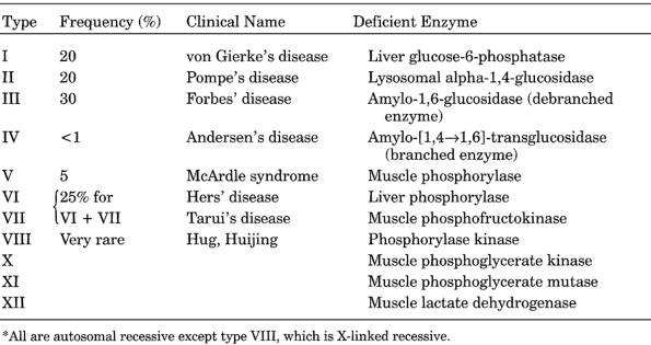
|
|
Table 12-10. Classification of Glycogen
Storage Diseases*
|
- Urine positive for reducing substances but negative for glucose
using glucose oxidase enzymatic strips.
- Heterozygotes detected by glucuronic acid loading followed by measurement of serum
xylulose or assay of reduced nicotinamide-adenine dinucleotide
phosphateL-xylulose dehydrogenase in RBCs.
Differential
Diagnosis
- Alimentary
pentosuriaarabinose or xylose excreted after ingestion of large amount of
certain fruits (e.g., plums, cherries, grapes)
- Healthy personssmall
amounts of D-ribose or trace amounts of ribulose in urine
- Muscular dystrophysmall
amounts of D-ribose in urine (some patients)
Sucrosuria
- Urine specific gravity is
very high (≤1.07).
- Urine tests for reducing
substances are negative.
- Sucrosuria may follow IV
administration of sucrose or the factitious addition of cane sugar to
urine.
Glycogen
Storage Diseases
See Table 12-10.
Type I
Glycogen Storage Disease; Glucose-6-Phosphatase Deficiency (Von Gierke's
Disease)
- (Autosomal
recessive disorder characterized by lack of glucose-6-phosphatase in liver
and kidney with an incidence of 1 in 200,000 births; may appear in first
days or weeks of life)
- Blood glucose is markedly decreased.
- After overnight fast, marked hypoglycemia and increased blood
lactate and occasionally pyruvate with severe metabolic acidosis,
ketonemia, and ketonuria. (Recurrent acidosis is
most common cause for hospital admission.)
- Blood triglycerides are
very high; cholesterol is moderately increased and serum free fatty acids
are increased. Results in xanthomas and lipid-laden cells in bone marrow.
P.545
- Mild anemia is present.
- Impaired platelet
function may cause bleeding tendency.
- Increased serum uric
acid, which may cause clinical gout, nephrocalcinosis, proteinuria.
- Serum phosphorus and ALP
are decreased.
- Urinary nonspecific amino
acids are increased, without increase in blood amino acids.
- Other renal function
tests are relatively normal despite kidney enlargement; Fanconi's syndrome
is rare.
- Liver function test
results (other than those related to carbohydrate metabolism) are
relatively normal but serum GGT, AST, and ALT may be increased.
- Glucose tolerance may be
normal or diabetic type; diabetic type is more frequent in older children
and adults.
- Functional tests
- Administer 1 mg of
glucagon IV or IM after 8-hr fast. Blood glucose increases 5060% in
1020 mins in the normal person. Little or no increase occurs in infants
or young children with von Gierke's disease; delayed response may occur
in older children and adults.
- IV administration of
glucose precursors (e.g., galactose or fructose) causes no rise in blood
glucose in von Gierke's disease (demonstrating block in gluconeogenesis),
but normal rise occurs in limit dextrinosis (type III glycogen storage
disease).
- Biopsy of liver
- Absent or markedly decreased glucose-6-phosphatase on assay of frozen liver provides
definitive diagnosis.
- Increased glycogen
content (>4% by weight) but normal biochemically and structurally.
- Other enzymes (other
glycogen storage diseases) are present in normal amounts.
- Histologic findings are
not diagnostic; vacuolization of hepatic cells and abundant glycogen
granules are seen; confirm with Best's stain.
- Biopsy of jejunum
- Intestinal
glucose-6-phosphatase is decreased or absent.
- Biopsy of muscle shows no
abnormality of enzyme activity or glycogen content.
- Can be cured by liver
transplant.
Type
Ib Glycogen Storage Disease
- (Shows
all the clinical and biochemical features of von Gierke's disease except
that liver biopsy does not show deficiency of glucose-6-phosphatase)
- Patient may have
maturation arrest neutropenia; varies from mild to agranulocytosis;
usually constant but may be cyclic. Associated increased frequency of
staphylococcal and candida infection.
- Diagnosis established by finding of impaired function of glucose-6-phosphate
activity in granulocytes.
Type
II Glycogen Storage Disease; Generalized Glycogenosis; Alpha-1,4-Glucosidase
Deficiency (Pompe's Disease)
- (Autosomal
recessive disease. Classic infantile form [Type IIA] characterized by
neurological, cardiac, and muscle involvement, frequent liver enlargement,
death within first year; juvenile form [Type IIB] shows muscle disease
resembling pseudohypertrophic dystrophy; adult form [Type IIC] characterized
by progressive myopathy)
- Fasting blood sugar, GTT,
glucagon responses, and rises in blood glucose after fructose infusion are
normal. No acetonuria is present.
- General hematologic
findings are normal.
- Staining of circulating leukocytes for glycogen shows massive deposition.
- Confirm diagnosis by absence of alpha-1,4-glucosidase in muscle and liver biopsy or
cultured fibroblasts. Assay in amniotic cell culture allows prenatal
diagnosis. Special assay of peripheral leukocytes for diagnosis of
heterozygotes.
P.546
Type
III Glycogen Deposition Disease (Forbes' Disease; Debrancher Deficiency; Limit
Dextrinosis)
- (Autosomal
recessive disease with enlarged liver, retarded growth, chemical changes,
and benign course)
- Serum CK may be
increased.
- Mild increase in
cholesterol and triglycerides are less marked than in type I disease.
- Marked fasting acetonuria
(as in starvation).
- Fasting hypoglycemia is
less severe than in type I disease.
- Normal blood lactate;
uric acid is usually normal.
- Serum AST and ALT are
increased in children but normal in adults.
- Diabetic type of glucose
tolerance curve with associated glucosuria.
- Infusions of
gluconeogenic precursors (e.g., galactose, fructose) causes a normal
hyperglycemic response unlike in type I disease.
- Low fasting blood sugar does not show expected rise after
administration of subcutaneous glucagon or epinephrine but does increase 2 hrs
after high-carbohydrate meal.
- Confirm diagnosis by liver and muscle biopsy that show biochemical findings of
increased glycogen, abnormal glycogen structure, absence of specific
enzyme activity. Normal phosphorylase and glucose-6-phosphatase activity.
Type
IV Glycogen Deposition Disease (Andersen's Disease; Brancher Deficiency;
Amylopectinosis)
- (Extremely
rare fatal condition that is due to absence of amylo-[1,4 1.6]-transglucosidase)
- Hypoglycemia is not
present.
- Liver function tests may
be altered as in other types of cirrhosis (e.g., slight increase in serum
bilirubin, reversed A/G ratio, increased AST, decreased cholesterol).
Blood glucose response to epinephrine and glucagon may be flat.
- Biopsy of liver may show a cirrhotic reaction to the presence of glycogen
of abnormal structure, which stains with Best's carmine and periodic
acid-Schiff stain, but normal glycogen concentration.
- WBC may be increased and
Hb may be decreased.
Type V
Glycogen Deposition Disease (Mcardle's Disease; Myophosphorylase Deficiency)
- (Autosomal
recessive disease due to absent myophosphorylase in skeletal muscle;
patient shows very limited ischemic muscle exercise tolerance despite normal
appearance of muscle)
- Epinephrine or glucagon
causes a normal hyperglycemic response.
- Biopsy of muscle is
microscopically normal in young; vacuolation and necrosis are seen in
later years. Increased glycogen is present.
- Definitive diagnosis is made by finding of absence of phosphorylase.
- After exercise that quickly causes muscle cramping and weakness, regional blood
lactate and pyruvate do not increase (in a normal person they increase 25
times). Similar abnormal response occurs in type III disease involving
muscle and in types VII, VIII, X.
- Myoglobulinuria may occur
after strenuous exercise.
- Increased serum muscle enzymes (e.g., LD, CK, aldolase) for
several hours after strenuous exercise.
Type
VI Glycogen Storage Disease (Hepatic Phosphorylase Deficiency)
- Enlarged liver present
from birth is associated with hypoglycemia.
- Serum cholesterol and
triglycerides are mildly increased.
- Serum uric acid and
lactic acid are normal.
- Liver function tests are
normal.
P.547
- Fructose tolerance is
normal.
- Response to glucagon and
epinephrine is variable but tends to be poor.
- Diagnosis is based on decreased phosphorylase activity in liver, leukocytes, and RBC
hemolysate, but muscle phosphorylase is normal.
Type
VII Glycogen Storage Disease (Muscle Phosphofructokinase Deficiency; Tarui's
Disease)
- (Autosomal
recessive disease with deficiency of muscle phosphofructokinase)
- Fasting hypoglycemia is
marked.
- Other members of family
may have reduced tolerance to glucose.
- RBCs show 50% decrease in phosphofructokinase activity
- Biopsy of muscle shows marked decrease (13% of normal) in
phosphofructokinase activity.
- Clinically identical to
type V disease.
Type
VIII Glycogen Storage Disease
- (Very
rare X-linked recessive disease with deficiency of phosphorylase b
kinase)
- Blood glucose is markedly
decreased, causing hypoglycemic seizures and mental retardation.
- Glucagon administration
causes no increase in blood glucose (see von Gierke's
disease), but ingestion of food causes a rise in 23 hrs.
- Biopsy of liver shows marked decrease in glycogen synthetase.
Porphyrias
See Fig. 12-7 and and Table 12-11.
Porphyrin
Tests of Urine (Fluorometric Methods)
- May Be
Positive Due To
- Drugs that produce
fluorescence, e.g.,
- Acriflavine
- Ethoxazene
- Phenazopyridine
- Sulfamethoxazole
- Tetracycline
- Drugs that may
precipitate porphyria, e.g.,
- Antipyretics
- Barbiturates
- Phenylhydrazine
- Sulfonamides
(1)
Congenital Erythropoietic Porphyria
- (Extremely
rare disorder due to decreased activity of uroporphyrinogen III synthase
in RBCs; usual onset in infancy, extreme cutaneous photosensitivity with
mutilation, red urine and teeth)
- Ultraviolet fluorescence of urine, teeth, and bones
- Variable number of RBCs
and marrow normoblasts
- Normocytic, normochromic,
anicteric hemolytic anemia that tends to be mild; may be associated with
hypersplenism, increased reticulocytes and normoblasts.
- Urinemarked increase of uroporphyrin I is characteristic; coproporphyrin shows
lesser increase. Excretion of porphobilinogen and delta-ALA is normal.
Watson-Schwartz test is negative.
- RBCs and plasmamarked increase of uroporphyrins; increased coproporphyrin
- Stoolmarked increase of porphyrins, especially coproporphyrins
P.548
|
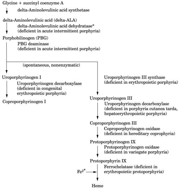
|
|
Fig. 12-7. Heme biosynthesis pathway
showing site of enzyme action and disease caused by enzyme deficiency.
Accumulation of porphyrins and their precursors preceding the enzyme block
are responsible for the clinical and laboratory findings in each syndrome.
Porphobilinogen (PBG) and aminolevulinic acid (ALA) cause abdominal pain and
neuropsychiatric symptoms. Increased porphyrins (with or without increased
PBG or ALA) cause photosensitivity. Thus, deficiencies near the end of the
metabolic path cause more photosensitivity and fewer neuropsychiatric
findings.
|
(2)
Erythropoietic Protoporphyria
- (Relatively
common type of porphyria due to deficiency of ferrochelatase activity in
bone marrow, reticulocytes, liver, and other cells)
- Mild microcytic
hypochromic anemia in 2030% of patients
- Laboratory findings due
to liver disease (severe in 10% of cases) with increased serum direct
bilirubin, AST, ALP (due to intrahepatic cholestasis), and gallstones
containing porphyrins may be found.
- Urineporphyrins within
normal limits
- RBCsmarked increase of free protoporphyrin in symptomatic patients
(zinc-chelated form may also be increased in iron-deficiency anemia and
lead poisoning
P.549
|
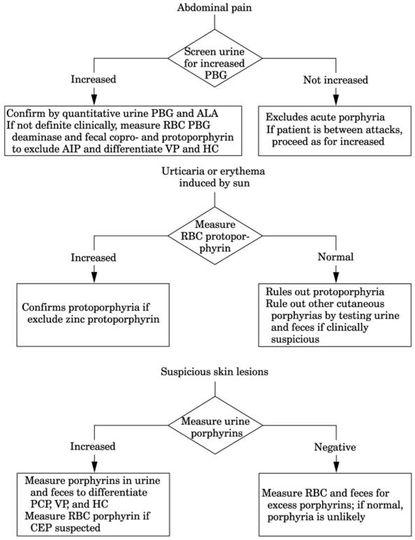
|
|
Fig. 12-8. Diagnostic strategy for
suspected porphyria according to symptoms. Excess production of porphyrins is
associated with cutaneous photosensitivity. Excess production of only
porphyrin precursors is associated with neurologic symptoms. Excess
production of both is associated with both types of clinical symptoms. (AIP =
acute intermittent porphyria; ALA = aminolevulinic acid; CEP = congenital
erythropoietic porphyria; HC = hereditary coproporphyria; PBG =
porphobilinogen; PCT = porphyria cutanea tarda; VP = variegate porphyria.)
|
P.550
|
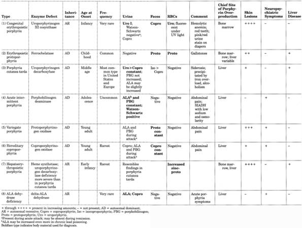
|
|
Table 12-11. Comparison of Porphyrias
|
P.551
P.552
but nonchelated form is present in protoporphyria). May
be normal or slightly increased in asymptomatic carriers. Examination of dilute
blood by fluorescent microscopy may show rapidly fading fluorescence in
variable part of RBCs.
- Stoolprotoporphyrin is usually increased in symptomatic patients and in some carriers
even when carrier RBC porphyrins are normal.
- Three chemical patterns consist of increased free RBCs alone, stool
protoporphyrin alone, and both together.
(3)
Porphyria Cutanea Tarda
- (Most
common porphyrin disorder. Inherited form [autosomal dominant] is
expressed in ~20% of patients with this gene and is due to deficiency of
uroporphyrinogen decarboxylase in liver in toxic/sporadic forms and in all
tissues in familial form. Associated with alcoholic liver disease and
hepatic siderosis. Acquired form [inhibitor of uroporphyrinogen
decarboxylase may be generated in liver] may be due to hepatoma,
cirrhosis, chemicals [an epidemic in Turkey was caused by contamination of
wheat by hexachlorobenzene]. May be activated by increased ingestion of
iron, alcohol, estrogens.)
- Urinemarked increase of uroporphyrin (frequently up to 10003000 µg/24 hrs; normal is
<300 µg) with only slight increase of coproporphyrin and
uroporphyrin/coproporphyrin ratio of >7.5 (ratio is <1 in variegate
porphyria). In biochemical remission, 24-hr uroporphyrin is <400 µg.
- Stoolisocoproporphyrins
are present.
- Plasmaincreased
protoporphyrin
- Distinguished from variegate porphyria in which fecal protoporphyrins are
increased and urine coproporphyrins exceed uroporphyrins during cutaneous
symptoms
- Serum iron and
transferrin saturation are increased in ~50% of cases.
- Laboratory findings of
underlying liver disease
- Liver biopsy shows
morphologic changes of underlying disease and fluorescence under
ultraviolet light; usually shows iron overload.
- Diabetes mellitus in
≤33% of patients
- Phlebotomy therapy to
remove iron is monitored by decreased urine uroporphyrins excretion.
(4)
Acute Intermittent Porphyria
- Most
frequent and severe form of porphyria in United States. Deficiency of
porphobilinogen deaminase. Adult onset with acute attacks of various
neuropsychiatric and abdominal symptoms. No photosensitivity
- Can be diagnosed in acute or latent states by finding of decreased delta-ALA
dehydratase activity and porphobilinogen deaminase activity (~50% of
normal) in RBCs (test performed in special laboratories); normal in other
porphyrias.
- UrineDiagnostic finding is marked increase of porphobilinogen and, to a lesser extent,
of delta-ALA; these decrease during remission but are rarely normal; not
increased in silent carriers; also increased in plasma. Watson-Schwartz
screening test for porphobilinogen should be confirmed by quantitative
test. Coproporphyrin and uroporphyrin may be increased.
- RBCsdecreased porphobilinogen activity is used to confirm diagnosis because urine
findings may occur during acute attacks of variegate porphyria and
hereditary coproporphyria.
- Stoolprotoporphyrin and
coproporphyrin are usually normal.
- Urine may be of normal color when fresh and become brown, red, or
black on standing.
- During acute attack,
slight leukocytosis, decreased serum sodium (may be marked), chloride, and
magnesium, and increased BUN may be seen.
- Liver function tests are
normal.
- Other frequent laboratory
abnormalities are increased serum cholesterol, hyperbetalipoproteinemia
(type II-a), increased serum iron, abnormal glucose tolerance, increased
T4, and thyroxine-binding globulin (TBG) without hyperthyroidism.
(5)
Variegate Porphyria
- Deficiency of protoporphyrinogen oxidase, which also occurs in cultured fibroblasts,
liver tissue, peripheral blood lymphocytes. Skin or neurologic
manifestations may occur. Precipitated by same factors as acute
intermittent porphyria.
P.553
- Stoolcharacteristic change is marked increase of protoporphyrin, which is found during
attack, remission, or only with skin manifestations. When stool is normal
or borderline, or in asymptomatic patients, increased porphyrins can be
demonstrated in bile.
- Urinemarked increase of
delta-ALA and porphobilinogen during an acute attack; levels are usually
normal after acute episode in contrast to acute intermittent porphyria and
hereditary coproporphyria.
- Bloodporphyrin levels
are not increased.
(6)
Hereditary Coproporphyria
- Deficiency
of coproporphyrinogen oxidase. Disease is latent in two-thirds of
patients. Precipitated by same factors as acute intermittent porphyria
- Stoolcoproporphyrin is always increased, very markedly during an acute attack; also
increased in plasma. Protoporphyrin is normal or only slightly increased.
- Urinecoproporphyrin may
be increased or not; is usually normal during remission. Isolated increase
may be secondary to liver, hematologic, neoplastic, and toxic conditions.
Increased ALA and porphobilinogen during acute attacks.
- RBCsdiminished coproporphyrinogen oxidase is strongly indicative.
- Liverdiminished coproporphyrinogen oxidase is diagnostic.
(7)
Hepatoerythropoietic Porphyria
- Severe
deficiency of uroporphyrinogen decarboxylase (510% of normal); 50% of
normal in parents.
- Porphyrin abnormalities resemble those in porphyria cutanea tarda but in addition zinc
protoporphyrin is increased in RBCs.
- Adults usually have mild
normochromic anemia; fluorescent normoblasts appear in bone marrow.
- Serum GGT and
transaminase may be increased. Liver disease may progress to cirrhosis.
- Severe skin involvement
(8)
ALA Dehydrase Deficiency
- 98%
deficiency of enzyme; parents had 50% of normal activity.
- Acute porphyria-type
symptoms
- Urineincreased ALA and coproporphyrin (resembles lead intoxication)
- RBC,
but not plasma, protoporphyrins are also increased in iron-deficiency
anemia and lead intoxication. Screening tests using fluorescence
microscopy of RBCs or Wood's lamp viewing of treated whole blood may also
be positive in iron-deficiency anemia, lead intoxication, and other
dyserythropoietic states. In congenital erythropoietic porphyria, 520% of
RBCs show fluorescence that lasts up to a minute or more in contrast to
erythropoietic protoporphyria in which fluorescence is half that and lasts
~30 secs and in lead poisoning in which almost all RBCs fluoresce for only
a few seconds. Fluorescence of hepatocytes occurs in erythropoietic
protoporphyria, porphyria cutanea tarda, porphyria variegata, and
hereditary coproporphyria
- Laboratory evaluation for
porphyrias may include: 24-hr urine for quantitative ALA, porphobilinogen,
uroporphyrin, and coproporphyrin (urine should be kept refrigerated as
porphyrins deteriorate quickly, especially at room temperature); plasma
porphyrin; free RBC protoporphyrin; spot stool quantitative coproporphyrin
and protoporphyrin; Watson-Schwartz test to demonstrate porphyrin
precursors in urine (Ehrlich's reagent and sodium acetate added to urine;
when positive, urine turns cherry red with addition of chloroform); search
for evidence of hemolytic anemia, liver disease; fluorescence of
appropriate tissues; enzyme activity assay of RBCs, liver tissue, or
cultured fibroblasts. Urine delta-ALA and porphobilinogen should be
measured during episodes.
- Acute episodes (which may
include abdominal pain and psychiatric symptoms; hypertension,
paresthesias, fever, neuromuscular weakness; seizures are less frequent)
are characteristic of acute intermittent porphyria, coproporphyria, and
variegate porphyria; may be precipitated by certain drugs (especially
barbiturates, alcohol, and sulfonamides; also diphenylhydantoin,
chlordiazepoxide, ergots, certain steroids), infection, starvation.
P.554
Lysosomal
Storage Disorders11
|
Disorder
|
Deficient Enzyme
|
Major System, Organ, or Tissue
Involved
|
|
Glycoprotein degradation
|
|
Fucosidosis
|
Alpha-fucosidase
|
CNS, high sweat electrolytes
|
|
Mannosidosis
|
Alpha-mannosidase
|
CNS, mild bone changes,
hepatosplenomegaly
|
|
Sialidosis (mucolipidosis
I)
|
Alpha-N-acetylneuraminidase
|
CNS, bone, liver, spleen
|
|
Glycogen storage disease
|
Alpha-glucosidase
|
Muscle, heart
|
|
Aspartylglycosaminuria
|
Aspartylglucosaminidase
|
CNS, bone marrow, connective tissue;
prominent inclusions in leukocytes
|
|
Enzyme localization
|
|
Mucolipidosis II (I-cell
disease) (formerly muco-polysaccharidosis VII)
|
N-Acetylglucosaminyl-phosphor
transferase
|
CNS, bone, connective tissue
|
|
Mucolipidosis III
(pseudoHurler's polydystrophy)
|
N-Acetylglucosamine-1-transferase
|
Predominantly joint and connective
tissue
|
|
Lysosomal efflux
|
|
Cystinosis
|
|
Kidney
|
|
Salla disease
|
|
CNS
|
|
- Mucopolysaccharidoses
(see Table 12-12)
- Sphingolipidoses (see Table 12-13)
- Lipidoses
- Chédiak-Higashi syndrome
Cystinosis
- (Autosomal
recessive lysosomal storage disease due to impaired transport of cystine
out of lysosomes; only this one amino acid is accumulated)
- Infants (acute
nephropathic form)
- Fanconi-like syndrome
(aminoaciduria, glycosuria, proteinuria, phosphaturia, polyuria)
- Metabolic acidosis
- Polyuria
- Vitamin Dresistant
rickets
- Diagnosis by finding of high cystine content in leukocytes or cultured fibroblasts
- Crystalline inclusions in conjunctiva and cornea, and leukocytes,
bone marrow, rectal mucosa
- Adults (benign disease)
- Urinary tract calculi
- Cystinuria (cystine
crystals in urine; >200 mg of cystine in 24-hr urine specimen)
- Asymptomatic cystine
crystals also present in eye
Fabry's
Disease (Alpha-Galactosidase A Deficiency)
- (X-linked
recessive disease with deficiency of alpha-galactosidase A that causes
skin lesions and accumulation of ceramide in various organs, affecting
function [e.g., kidney, heart, lung, brain]. Symptoms due to involvement
of these organ systems in infancy and cherry-red spots in macula.)
- Prenatal
diagnosis by demonstration of enzyme deficiency in cultured amniotic fluid
cells
- Heterozygote detection by enzyme assay of cultured fibroblasts or individual hair roots or
by assay of glycolipid content of urine sediment
P.555
Gaucher's
Disease
- (Rare
autosomal recessive deficiency of beta-glucosidase; most frequent storage
disease; may be present in 10,00020,000 Americans, with highest
prevalence in Ashkenazi Jews; gene on chromosome band 1q21)
- Measurement of decreased beta-glucosidase activity in leukocytes or fibroblasts is
reliable diagnostic method; substantial overlap between heterozygotes and
healthy persons.
- Diagnostic Gaucher's cells are seen in bone marrow aspiration, needle biopsy, or
aspiration of spleen, liver, or lymph nodes examined for thrombocytopenia
or unrelated disorder and cause the nonneurologic manifestations.
- Serum acid phosphatase is increased in most patients (if substrate
for test is different from that for prostatic acid phosphatase; i.e., use phenyl phosphate
or p-nitrophenyl phosphate instead of glycerophosphate). It may return to
normal after splenectomy.
- Serum ACE is increased in
most patients.
- Serum cholesterol and
total fats are normal.
- Laboratory findings due
to involvement of specific organs
- Spleenhypersplenism
occurs with anemia (normocytic normochromic), leukopenia (with relative
lymphocytosis; monocytes may be increased), thrombocytopenia without
bleeding.
- Boneserum ALP may be
increased.
- Liverserum AST may be
increased.
- CSFAST may be increased.
- Laboratory findings due
to increased incidence of lymphoproliferative disorders (e.g., multiple
myeloma, CLL).
- Prenatal diagnosis by enzymatic determination of cultured amniotic fluid cells. If
both parental mutations have been identified at the DNA level, chorionic
villus sampling for fetal DNA can be done.
- Enzymatic methods do not
detect carriers reliably. Molecular methods accurately detect carriers.
- Phenotype cannot be predicted from genotype. Common mutations can be detected
using PCR and aid in genetic counseling for general risk of transmitting
the gene but not specific prognosis for future affected children.
- Type 1 (99% of patients):
no neurologic involvement
- Type 2: fulminating
disorder with severe neurologic involvement and death within first 18 mos
- Type 3: juvenile form
with later onset of neurologic symptoms and milder course with death in
early childhood
- Bone marrow
transplantation is effective therapy but has associated morbidity and
mortality. Enzyme replacement therapy usually obviates need for
splenectomy.
Gm
Gangliosidosis (Landing's Disease, Systemic Late Infantile Lipidosis)
- (Rare
autosomal recessive deficiency of acid beta-galactosidase with no racial
predilection, characterized by psychomotor deterioration, enlargement of
liver and/or spleen, cherry-red macular spots, dysostosis multiplex;
infantile, juvenile, and adult forms)
- Diagnosis by absence of lysosomal acid beta-galactosidase enzyme in leukocytes, cultured
fibroblasts, or brain. Tissue biopsy or culture of marrow or skin
fibroblasts shows accumulation of ganglioside GM ;
also can demonstrate GM in brain and viscera and
mucopolysaccharides in viscera.
- Heterozygote carriers can be detected by enzyme assay in leukocytes.
- Vacuolated lymphocytes
may be found.
- Abnormal leukocytic granulations (Alder-Reilly bodies) may be present.
- Serum LD, AST, and
fructose 1-phosphate aldolase are normal.
- Foam cell histiocytes (resembling Niemann-Pick cells) may be seen
in biopsy from bone marrow, liver, or rectum.
P.556
P.557
|
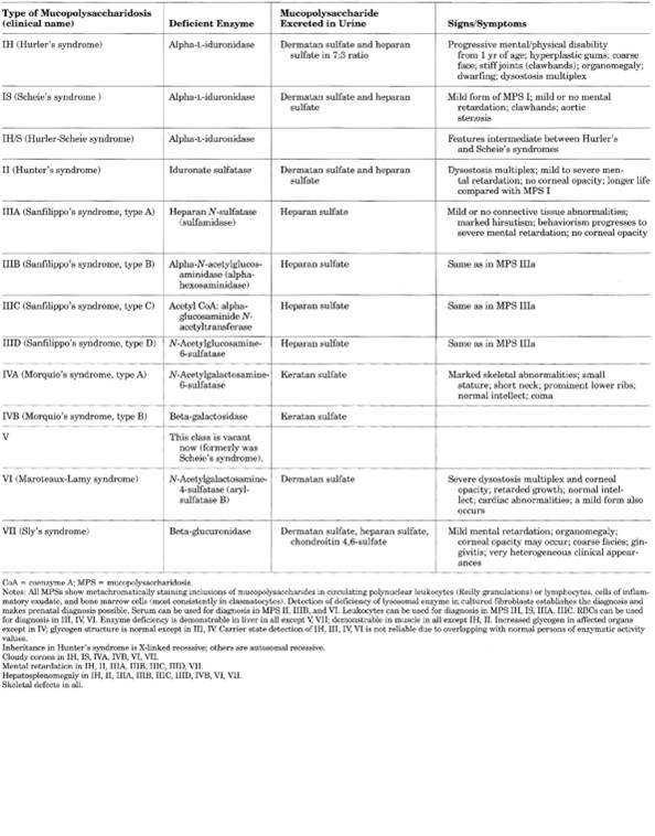
|
|
Table 12-12. Classification of
Mucopolysaccharidoses
|
P.558
P.559
|
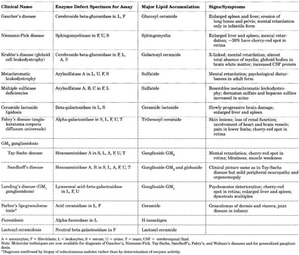
|
|
Table 12-13. Classification of
Sphingolipidosis
|
P.560
- Prenatal diagnosis by enzyme assay in cultured amniotic fluid cells or by HPLC
analysis of galactosyl oligosaccharides in amniotic fluid.
I-Cell
Disease (Mucolipidosis II)
- (Autosomal
recessive disease with defect in recognition and uptake of certain lysosomal
enzymes due to deficient activity of
N-acetylglucosaminylphosphotransferase. Clinical features resemble those
of Hurler's syndrome but without corneal changes or increased
mucopolysaccharides in urine.)
- Deficiency of N-acetylglucosaminylphosphotransferase in cultured
fibroblasts establishes the diagnosis.
- Vacuolation (cytoplasmic inclusions) in lymphocytes, fibroblasts, and liver and kidney
cells show positive reaction to Sudan black and acid phosphatase.
Lysosomal enzyme activity (hexosaminidase A and B and alpha-galactosidase)
is low in these cells but high in serum or culture medium.
- Urine mucopolysaccharides
are not increased.
- Prenatal diagnosis by finding of high levels of multiple acid hydrolases in amniotic
fluid or deficiency of them in cultured amniocytes.
- Some heterozygotes have
abnormal inclusions in fibroblasts. Some heterozygotes have intermediate
enzyme levels in leukocytes and cultured fibroblasts.
Krabbe's
Disease (Globoid Cell Leukodystrophy; Galactosylceramide Lipidosis)
- (Autosomal
recessive disorder characterized by deficiency of galactosylceramidase,
causing progressive CNS disease from ~3 mos of age and death by ~2 yrs)
- Diagnosis by finding of deficiency of this enzyme (510% of normal) in leukocytes or
cultured fibroblasts
- Conjunctival biopsy shows characteristic ballooned Schwann cells.
- Brain biopsy (massive infiltration of unique multinucleated inclusion-containing
globoid cells in white matter due to accumulation of galactosylceramide;
also diffuse loss of myelin, severe astrocytic gliosis)
- CSF protein
electrophoresis shows increased albumin and alpha globulin and decreased
beta and gamma globulin (as in metachromatic leukodystrophy).
- Prenatal diagnosis by measurement of enzyme activity in cultured
amniotic fluid cells.
Mucolipidosis
III (N-Acetylglucosaminylphosphotransferase
Deficiency; PseudoHurler's Polydystrophy)
- (Clinical
features resemble those in Hurler's syndrome but without increased
mucopolysaccharides in urine
- Autosomal
recessive transmission of fundamental defect in recognition or catalysis
and uptake of certain lysosomal enzymes due to deficient activity of
N-acetylglucosamine-1-transferase.
- Heterozygotes may have intermediate enzyme levels in leukocytes and cultured
fibroblasts.
Mucopolysaccharidoses,
Genetic
- All mucopolysaccharidoses show metachromatically staining inclusions of
mucopolysaccharides in circulating PMNs (Reilly granulations) or
lymphocytes, cells of inflammatory exudate, and bone marrow cells (most
consistently in clasmatocytes). Mucopolysaccharide is also deposited in
various parenchymal cells. Detection of deficiency of lysosomal enzyme in
cultured fibroblasts establishes the diagnosis and makes prenatal
diagnosis possible. Serum can be used for diagnosis in
mucopolysaccharidoses II, IIIB, VI. Leukocytes can be used for diagnosis
in mucopolysaccharidoses IH, IS, IIIA, IIIC. RBCs can be used for
diagnosis in mucopolysaccharidoses III, IV, VI. Enzyme deficiency is
demonstrable in liver in all types except V, VII; demonstrable in muscle
in all types except IH, II. Increased glycogen in affected organs except
in type IV; glycogen structure is normal except in types III, IV. Carrier
state detection of types IH, III, IV, VI is not reliable due to overlap
with normal persons in enzymatic activity values.
- Inheritance is X-linked
recessive in Hunter's syndrome; autosomal recessive in others.
P.561
- Cloudy cornea in types
IH, IS, IVA, IVB, VI, VII.
- Mental retardation in
types IH, II, IIIA, IIIB, IIIC, IIID, VII.
- Hepatosplenomegaly in
types IH, II, IIIA, IIIB, IIIC, IIID, IVB, VI, VII.
- Skeletal defects in all.
Hurler's
Syndrome (Mucopolysaccharidosis IH)
- (Most
patients die by age 10 yrs.)
- Initial diagnosis by quantitative increase of mucopolysaccharide in urine;
confirmed by assay of alpha-L-iduronidase in cultured fibroblasts or
leukocytes.
- Similar enzyme assay
detects carriers who have ~50% activity, but the wide range and overlap
between normal persons and carriers may make the diagnosis difficult in
individual cases.
- Prenatal diagnosis by assay of enzyme or mucopolysaccharide in amniocytes.
Hunter's
Syndrome (Mucopolysaccharidosis II)
- (Clinically
similar to Hurler's syndrome but milder and no corneal opacity)
- Initial diagnosis by quantitation of total glucosaminoglycans in urine and accumulation
of keratan sulfate in tissues is confirmed by enzyme assay in fibroblasts.
- Heterozygous female carriers recognized by presence of mucopolysaccharide in
fibroblasts or enzyme assay of individual hair roots.
- Prenatal diagnosis by enzyme assay of amniotic fluid should be confirmed by assay of
cultured cells.
- Maternal serum shows increased activity of iduronate sulphate sulfatase with a normal
or heterozygous fetus but no increase if fetus has Hunter's syndrome.
- Mild and severe subtypes
Sanfilippo's
Syndrome Type A (Mucopolysaccharidosis III)
- (The
four types of Sanfilippo's syndrome cannot be distinguished clinically)
- Only mucopolysaccharidosis in which finding only heparan sulfate in urine
confirms diagnosis.
- Assay of fibroblasts shows deficiency of enzyme in patients and decrease of normal
activity in carriers, who also show mucopolysaccharide accumulation.
- Metachromatic inclusion bodies in lymphocytes are coarser and
sparser than in Hurler's syndrome and may be seen in bone marrow cells.
Severe cerebral changes with relatively mild changes in other body
tissues.
Morquio's
Syndrome (Mucopolysaccharidosis IV)
- Keratan sulfate is increased in urine (often 23× normal)
- Metachromatic granules may be seen in PMNs.
- Diagnosis by enzyme assay in fibroblasts and leukocytes
- Prenatal diagnosis by assay of enzymes in cultured amniocytes
Maroteaux-Lamy
Syndrome (Mucopolysaccharidosis VI)
- Metachromatic cytoplasmic inclusions (Alder granules) may be seen
in 50% of lymphocytes and 100% of granulocytes, and are more marked than
in other mucopolysaccharidoses.
- Large amount of dermatan sulfate occurs in urine.
- Diagnosis is established by a finding of deficiency of specific enzyme in cultured
fibroblasts.
- Enzyme assay also allows diagnosis of heterozygotes and prenatal diagnosis.
- Other rare diseases due
to enzyme deficiencies that resemble these conditions include I-cell
disease (mucolipidosis I) and mucolipidosis III and related disorders.
Niemann-Pick
Disease
- (Sphingomyelin
lipidosis)
- Diagnosis by demonstration of sphingomyelinase deficiency in cultured fibroblasts or
circulating leukocytes
P.562
- Foamy histiocytes may be found in bone marrow aspiration, liver,
spleen, skin, skeletal muscle, and eye and may appear in peripheral blood
terminally.
- Peripheral blood
lymphocytes and monocytes may be vacuolated (220% of cells).
- WBC is variable.
- Rectal biopsy may show
changes in ganglion cells of myenteric plexus.
- Laboratory findings due
to involvement of specific organs
- Anemia is due to hypersplenism
or microcytic anemia associated with anisocytosis, poikilocytosis, and
elliptocytosis.
- AST may be increased in
serum and CSF.
- Enzyme changes in CSF
are same as in Tay-Sachs disease, except that LD is normal.
- Acid phosphatase is
increased (as in Gaucher's disease).
- LD is normal in serum and
CSF.
- Different isoenzyme
activities result in different clinical forms.
- Acute infantile form
(type A): acute progressive neuropathic loss of motor and intellectual
function early in life with death common in infancy. Cherry-red macula is
often present.
- Subacute/juvenile forms
(types C and D): not neuropathic; later onset.
- Chronic forms (types B
and E): similar to acute type but later in onset and not neuropathic.
- Types A and B show
primary sphingomyelinase deficiency; type C shows defect in cholesterol
esterification (autosomal recessive inheritance).
Oligosaccharidoses
With Increased Urinary Oligosaccharides
- Sialidosis
- I-cell disease
(mucolipidosis II)
- Fucosidosis
- Mannosidosis
- Galactosialidosis
- PseudoHurler's
polydystrophy (mucolipidosis III)
- GM
gangliosidosis
- Aspartylglucosaminuria
Tay-Sachs
Disease (Gm Gangliosidosis)
- (Autosomal
recessive trait [chromosome 15] found predominantly in Ashkenazi Jews,
French Canadians, and Cajuns characterized in infantile form by appearance
of psychomotor deterioration, blindness, cherry-red spot in the macula,
and an exaggerated extension response to sound, with death by age 4 yrs;
patients with juvenile form die by age 15 yrs; chronic form in adults;
macula spot occurs only in infantile form.)
- Diagnosis is established by absence of hexosaminidase A activity in
serum (also absent in all tissues of body and tears). Accumulation of GM
ganglioside in brain is due to deficiency or absence of hexosaminidase A.
Electron microscopy shows characteristic cytoplasmic bodies in brain. (In
Sandhoff's disease, a variant of Tay-Sachs disease, both hexosaminidase A
and B are defective and globoside is accumulated in other tissues as well
as brain.)
- Heterozygotes can be identified by plasma assay showing 50% decrease in activity of
hexosaminidase A; screening should be done before pregnancy, which may
cause false-positive results; use of oral contraceptives, diabetes
mellitus, and liver disease may also cause false-positive results; in
these cases WBCs are used for hexosaminidase A assay.
- Prenatal diagnosis using cultured amniotic cells is superior to testing of
amniotic fluid or uncultured amniotic cells; false-negative results can
occur due to contamination with maternal blood or tissue or bacteria.
- PCR for specific DNA mutations in WBCs or fibroblasts is more specific than
enzyme assay, and can detect various mutations and predict severity of
disease in affected child.
- Early marked increase of
serum LD and AST is seen; levels return to normal if patient survives 34
yrs.
- Decrease in serum
fructose 1-phosphate aldolase; also decreased in heterozygotes
P.563
- CSF AST parallels serum
AST.
- Occasional vacuolated
lymphocytes are seen.
- Liver function tests are
normal.
- Serum acid phosphatase is
normal.
Other
Genetic Disorders
Batten
Disease (Batten-Spielmeyer-Vogt Disease)
- (Autosomal
recessive type of juvenile amaurotic idiocy)
- Azurophilic hypergranulation of leukocytes occurs in patients and in heterozygous and
homozygous members of their families. In Giemsa- and Wright's-stained
smears, it resembles toxic granulation but differs by the absence of
supravital staining in Batten disease and by normal leukocyte ALP activity
(markedly increased in toxic granulation). This granulation occurs in
≥15% of neutrophils.
D
Trisomy (Trisomy 13; Patau's Syndrome)
- See Table
12-14.
- In peripheral blood smears, ≤80% of PMNs (neutrophils and
eosinophils) show an increased number of anomalous nuclear projections
(tags, threads, drumsticks, clubs); the nuclear lobulation may appear
abnormal (nucleus may look twisted without clear separation of individual lobes,
coarse lumpy chromatin, etc.). Present in almost all complete trisomic
cases. Nuclear coils of chromatin by electron microscopy.
- Fetal hemoglobins may
persist longer than normal (i.e., be increased); these include HbF,
Bart's, Gower II.
- Decreased AFP in maternal serum and amniotic fluid
- Laboratory findings due
to multiple congenital abnormalities (including almost pathognomonic
tetrad of narrow palpebral fissures and microphthalmos, cleft palate,
parieto-occipital scalp defect, polydactyly).
- Karyotyping shows numerical abnormality in 80% of cases: 47 XX,+13, or 47 XY,+13.
Due to translocations in 20% of cases.
Down
Syndrome (Trisomy 21; Mongolism)
- See Table
12-14.
- Karyotyping
shows 47 chromosomes with trisomy 21 in most patients; due to
translocation, usually to chromosome 14, to other D group chromosome in
<5% of cases. 2% have mosaicism with one cell population trisomic.
- Increased leukocyte ALP
score.
- Leukocytes show decreased
incidence of drumsticks and mean lobe counts.
- Serum acid phosphatase
may be decreased.
- Risk of developing acute
lymphocytic or nonlymphocytic leukemia is increased (~1%).
- Incidence 1020× greater
than in general population.
- Congenital AML occurs
within several months of birth; always fatal.
- Transient leukemoid reaction
(WBC ≤400,000/cu mm) occurs only with trisomy 21; differentiated
from congenital leukemia by bone marrow biopsy including cytogenetic and
immunohistochemical studies. ≤25% of these Down syndrome infants
develop acute megakaryocytic leukemia within 3 yrs.
- Increased susceptibility
to infection (e.g., hepatitis is common in institutionalized patients, in
whom HBsAg was first noted).
- Laboratory findings due
to associated congenital abnormalities (e.g., GI, GU, cardiovascular
systems).
Prenatal
Screening and Diagnosis
- See Table
12-6 and Fig. 12-2.
- Optimal
screening combines measurement of hCG, AFP, and unconjugated estriol
levels in maternal serum in pregnant patients aged >35 yrs
P.564
|
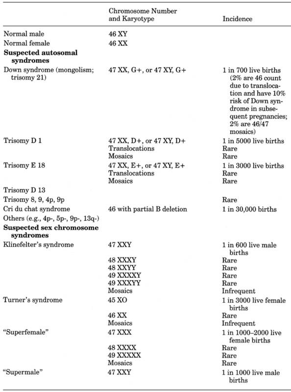
|
|
Table 12-14. Chromosome Number and
Karyotype in Various Clinical Conditions
|
Maternal
Serum AFP
- Interpretation
- Use of maternal serum
AFP alone is not recommended; should be
combined with measurements of hCG and unconjugated estriol when maternal
age is >35 yrs; this combination can identify ~60% of cases of Down
syndrome with false-positive rate of 6.6%; ultrasonography to verify
gestational age (which has profound effect on calculated risk of Down
syndrome) reduces false-positive rate to 3.8%.
P.565
- Decreased
maternal blood level of AFP in pregnancy is a valuable screening test,
but diagnosis should be confirmed by finding of increased levels in
amniotic fluid and by ultrasonography (to rule out missed abortion, molar
pregnancy, absent pregnancy), as well as by chromosomal studies to
confirm or refute the diagnosis. Lower AFP value makes Down syndrome more
likely.
- In midtrimester,
usual range is 10150 ng/mL; is usually reported as multiple of median
(MoM) (normal 0.42.5 MoM) to minimize interlaboratory variability and
correct for patient's race, diabetes mellitus, and gestational age.
- Decreased
In
- Down syndrome (trisomy
21) and trisomy 18
- Long-standing death of
fetus
- Overestimation of
gestational age (underestimation of age in amniotic fluid sample)
- Choriocarcinoma,
hydatidiform mole
- Increased maternal
weight (does not affect amniotic fluid concentration)
- Pseudopregnancy,
nonpregnancy
- Various drugs (therefore
no medications should be taken for at least 12 hrs before test)
- Other unknown factors
- Women with diabetes
mellitus have values 2040% less than those of nondiabetic women.
- Increased
In
- (Should confirm by
increase in amniotic fluid)
- Twin pregnancy (>4.5
MoM)
- Gestational age (for
which values must be adjusted)
- Race (1015% higher in
blacks) (for which values must be adjusted)
- Open neural tube defects
(e.g., open spina bifida, anencephaly, encephalocele, myelocele); 80% of
severe cases are detected by AFP. Occurs in 2 in 1000 births in the United
States. Women with one affected child have 5% chance of giving birth to
another; affected families make up 10% of these cases. Optimal screening
is in 16th20th week of gestation. Hydrocephaly. Microcephaly.
- Ventral wall defects
associated with exposed fetal-membrane and blood-vessel surfaces, e.g.,
- Omphalocele (incidence
13 in 10,000)
- Gastroschisis (incidence
110 in 10,000)
- Hydrops fetalis
- Intrauterine death
- Fetal-maternal hemorrhage
- Esophageal or duodenal
atresia
- Cystic hygroma
- Renal disorders, e.g,
- Congenital proteinuric
nephropathies
- Polycystic kidneys
- Renal agenesis
- Aplasia cutis
- Sacrococcygeal teratoma
- Tetralogy of Fallot
- Turner's syndrome
- Oligohydramnios
- Maternal causes (e.g.,
neoplasm that produces AFP, hepatitis)
- Very rare benign
hereditary familial elevation of serum AFP
Maternal
hCG
- (Appears
in maternal serum soon after pregnancy and reaches peak by 811 wks of
gestation, decreases to nadir at 18 wks, and then remains constant to end
of pregnancy)
- Use
- Best single
marker for Down syndrome screening
- Increase of >2.5× MoM
at 1825 wks of gestation detects ~56% of cases. One study that detected
73% of cases had a 4% false-positive rate at that serum level.
P.566
- Diagnosis of early
pregnancy
- Diagnosis of germ cell
tumors and monitoring of treatment effectiveness (see Chapter
14)
Maternal
Serum Unconjugated Estriol
- (Is of
fetal origin from fetal adrenal, liver, and placental function. Begins to
appear by seventh to ninth week of gestation.)
- Decreased
In
- Fetal Down syndrome
- Low values at 3536 wks
of gestation identify up to one-third of light for dates infants.
- Interpretation
- Value >12 ng/mL rules
out postmaturity in cases of prolonged gestation if no other diseases are
present (e.g., diabetes mellitus, isoimmunization).
- Decreased value detects
45% of cases with a 5.2% false-positive rate.
- ≤0.6 MoM in 5% of
unaffected pregnancies and 26% of Down syndrome cases.
- Safe levels indicate
fetal well-being.
- Increasing serial values
rule out prolonged pregnancy and postmaturity.
- Constant normal values
are consistent with 4041 wks of gestation.
- Declining values are
consistent with prolonged gestation.
- Low or significantly
falling values are seen in fetal distress and postmaturity.
Amniotic
Fluid Estriol
- Interpretation
- Values are not meaningful
before 20 wks' gestation (<1.0 µg/dL); gradual increase to 35th week
and then rapid increase to 40th week. Each laboratory must establish its
own reference ranges.
- Decreasing levels are
associated with fetal distress, and failure to increase with fetal death.
Human
Placental Lactogen
- Appears by fifth week of
gestation and increases progressively thereafter.
- Values correlate better
with placental than with fetal weight. Therefore useful to evaluate
placental function; sudden decrease in concentration before fetal death. Use only as adjunct to other tests.
- Useful
In
- Diabetes mellitus, severe
- Hypertension
- Postmaturity syndrome
- Idiopathic placental
failure
- Not
Useful In
- Diabetes mellitus, mild
or moderately severe
- Rh sensitization disease
Maternal
Serum PSA
Recent report of ultrasensitive assay of
PSA suggests that second trimester amniotic fluid concentrations are low
(normally increases between gestational weeks 11 and 21) and are increased in
maternal serum in Down syndrome cases.16
Chromosomal
Analysis of Amniotic Fluid
Detects ~20% of cases because 80% of Down
syndrome infants are born to women <35 yrs old.
Dysautonomia,
Familial (Riley-Day Syndrome)
- (Autosomal
recessive disorder occurring in Ashkenazi Jews; patients show difficulty
in swallowing, corneal ulcerations, insensitivity to pain, motor
incoordination, excessive sweating, diminished gag reflex, lack of tongue
papillae, progressive kyphoscoliosis, pulmonary infections, etc.)
P.567
- Urine VMA
(3-methoxy-4-hydroxymandelic acid) may be low, and HVA increased.
- Urine VMA may be lower in
asymptomatic carriers than in healthy adults.
Fragile
X Syndrome Of Mental Retardation
- (Most
common form of inherited mental retardation; due to mutations that
increase the size of a specific DNA fragment of the X chromosome [in
Xq27.3])
- Direct diagnosis by DNA analysis using Southern blot test but PCR is often done
simultaneously. Can also be used to establish prenatal diagnosis and to
detect asymptomatic carriers. Can distinguish between full mutation, in
which 100% of males and ~50% of females are mentally impaired, and
premutation, in which only ~3% are impaired.
Mediterranean
Fever, Familial (Familial Paroxysmal Peritonitis; Periodic Disease)
- (Autosomal
recessive disorder characterized by recurrent polyserositis occurring
predominantly in Sephardic Jews, Arabs, and Armenians)
- WBC is increased
(10,00020,000/cu mm); eosinophils may be increased during an attack but a
return to normal between attacks. ESR is increased during an attack but
normal between attacks.
- Mild normocytic
normochromic anemia is occasionally seen.
- Serum glycoprotein is
increased in patients and their relatives.
- Increased alpha2 globulin
and fibrinogen are common.
- Amyloid nephropathy that
is usually fatal develops in 1040% of patients; it is not related to
frequency or severity of clinical attacks.
- PCR amplification of DNA identifies one of three common mutations.
Trisomy
18
- (Usually
sporadic; due to nondisjunction; associated with increased maternal age)
- Decreased AFP, hCG, and unconjugated estriol in maternal serum.
- Laboratory findings due
to congenital abnormalities (e.g., cardiovascular, GU, GI systems).
REFERENCES
1. McAuliffe JJ, et al.
Hypoproteinemic alkalosis. Am J Med
2. Wax JR, Blakemore KJ.
What can be learned from cordocentesis? Clin Lab Med
3. Winchester B, Young E.
Prenatal diagnosis of enzyme defectsan update. Arch
Dis Child
4. D'Alton ME, DeCherney
AH. Prenatal diagnosis. N Engl J Med
5. Cole HM, ed. Chorionic
villus sampling: a reassessment. Diagnostic and therapeutic technology
assessment. JAMA
6. Cleary MA, Wraith JE.
Antenatal diagnosis of inborn errors of metabolism. Arch
Dis Child
7. Naito HK. The clinical
significance of apolipoprotein measurements. J Clin
Immunoassay
8. Summary of the second
report of the National Cholesterol Education Program (NCEP) Expert Panal on
Detetion, Evaluation, and Treatment of High Blood Cholesterol in Adults. JAMA
9. Guba SC, Fink LM,
Fonseca V. Hyperhomocysteinemia. An emerging and important risk factor for
thromboembolic and cardiovascular disease. Am J Clin
Pathol
10. Welch GN, Loscalzo J.
Homocysteine and atherothrombosis. N Engl J Med
11. Classification adapted
from Kornfeld S, Sly WS. Lysosomal storage defects. Hosp
Pract Aug 15, 1985:7182.
12. Data from Beutler E.
Gaucher's disease. N Engl J Med
13. National Institutes of
Health. Gaucher disease: current issues in diagnosis and treatment. Statement
presented at: Technology Assessment Conference, Feb 27Mar 1, 1995.
14. Sundaram SG, Goldstein
PJ, Manimekalai S, Wenk RE. Alpha-fetoprotein and screening markers of
congenital disease. Clin Lab Med
15. D'Alton ME, DeCherney
AH. Prenatal diagnosis. N Engl J Med
16. Lambert-Messerlian GM,
et al. Increased concentrations of prostate-specific antigen in maternal serum
from pregnancies affected by fetal Down syndrome. Clin
Chem
17. Rousseau F, et al.
Direct diagnosis by DNA analysis of the fragile X syndrome of mental
retardation. N Engl J Med
Footnotes
*Values are for serum unless otherwise
indicated.
**Measure serum total cholesterol, HDL
cholesterol, and triglycerides after 12- to 13-hr fast. Average results of two
or three tests; if difference of ≥30 mg/dL, repeat tests 18 wks apart
and average results of three tests. Use total cholesterol for initial case
finding and classification and monitoring of diet therapy. Do not use age- or
sex-specific cholesterol values as decision levels.
***Predominantly hypertriglyceridemia.
Predominantly hypercholesterolemia.
****Also characterized by lactic acidosis.
Cystathionine.
Hydroxyproline.
