|
ABC of clinical electrocardiography Exercise tolerance testing Jonathan Hill and Adam Timmis
|
|
||||||||||||||
|
Exercise tolerance testing is an important diagnostic and prognostic tool for assessing patients with suspected or known ischaemic heart disease. During exercise, coronary blood flow must increase to meet the higher metabolic demands of the myocardium. Limiting the coronary blood flow may result in electrocardiographic changes. This article reviews the electrocardiographic responses that occur with exercise, both in normal subjects and in those with ischaemic heart disease.
|
|||||||||||||||
|
|
Clinical relevance Exercise tolerance testing (also known as exercise testing or exercise stress testing) is used routinely in evaluating patients who present with chest pain, in patients who have chest pain on exertion, and in patients with known ischaemic heart disease.
Exercise testing has a sensitivity of 78% and a specificity of 70% for detecting coronary artery disease. It cannot therefore be used to rule in or rule out ischaemic heart disease unless the probability of coronary artery disease is taken into account. For example, in a low risk population, such as men aged under 30 years and women aged under 40, a positive test result is more likely to be a false positive than true, and negative results add little new information. In a high risk population, such as those aged over 50 with typical angina symptoms, a negative result cannot rule out ischaemic heart disease, though the results may be of some prognostic value. Exercise testing is therefore of greatest diagnostic value in patients with an intermediate risk of coronary artery disease. |
||||||||||||||
|
|
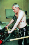
The test Protocol The Bruce protocol is the most widely adopted protocol and has been extensively validated. The protocol has seven stages, each lasting three minutes, resulting in 21 minutes' exercise for a complete test. In stage 1 the patient walks at 1.7 mph (2.7 km) up a 10% incline. Energy expenditure is estimated to be 4.8 METs (metabolic equivalents) during this stage. The speed and incline increase with each stage. A modified Bruce protocol is used for exercise testing within one week of myocardial infarction.
Preparing the patient Blockers should be discontinued the day before the test, and dixogin (which may cause false positive results, with ST segment abnormalities) should be stopped one week before testing.
The patient is first connected to the exercise electrocardiogram machine. Resting electrocardiograms, both sitting and standing, are recorded as electrocardiographic changes, particularly T wave inversion, may occur as the patient stands up to start walking on the treadmill. A short period of electrocardiographic recording during hyperventilation is also valuable for identifying changes resulting from hyperventilation rather than from coronary ischaemia.
During the test the electrocardiogram machine provides a continuous record of
the heart rate, and the 12 lead electrocardiogram
is recorded intermittently. Blood pressure must be measured before
the exercise begins and at the end of each exercise stage. Blood
pressure may fall or remain static during the initial stage of
exercise. This is the result of an anxious patient relaxing. As the test
progresses, however, systolic blood pressure should rise as
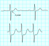
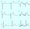
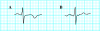
exercise increases. A level of up to 225 Safety If patients are carefully selected for exercise testing, the rate of serious complications (death or acute myocardial infarction) is about 1 in 10 000 tests (0.01%). The incidence of ventricular tachycardia or fibrillation is about 1 in 5000. Full cardiopulmonary resuscitation facilities must be available, and test supervisors must be trained in cardiopulmonary resuscitation. Limitations The specificity of ST segment depression as the main indicator of myocardial ischaemia is limited. ST segment depression has been estimated to occur in up to 20% of normal individuals on ambulatory electrocardiographic monitoring. There are many causes of ST segment changes apart from coronary artery disease, which confound the result of exercise testing. If the resting electrocardiogram is abnormal, the usefulness of an exercise test is reduced or may even be precluded. Repolarisation and conduction abnormalities-for example, left ventricular hypertrophy, left bundle branch block, pre-excitation, and effects of digoxin-preclude accurate interpretation of the electrocardiogram during exercise, and as a result, other forms of exercise test (for example, adenosine or dobutamine scintigraphy) or angiography are required to evaluate this group of patients. Normal trace during exercise The J point (the point of inflection at the junction of the S wave and ST segment) becomes depressed during exercise, with maximum depression at peak exercise. The normal ST segment during exercise therefore slopes sharply upwards. By convention, ST segment
depression is measured relative to the isoelectric baseline (between the T
and P waves) at a point 60-80
Abnormal changes during exercise The standard
criterion for an abnormal ST segment response is horizontal
(planar) or downsloping depression of >1 mm. If 0.5 mm of
depression is taken as the standard, the sensitivity of the test increases
and the specificity decreases (vice versa if 2
Other recognised abnormal responses to exercise include ST elevation of >1 mm, particularly in the absence of Q waves. This suggests severe coronary artery disease and is a sign of poor prognosis. T wave changes such as inversion and pseudo-normalisation (an inverted T wave that becomes upright) are non-specific changes. A highly specific sign for ischaemia is inversion of the U wave. As U waves are often difficult to identify, especially at high heart rates, this finding is not sensitive. The presence of extrasystoles that have been induced by exercise is neither sensitive nor specific for coronary artery disease. Stopping the test In clinical practice, patients rarely exercise for the full duration (21 minutes) of the Bruce protocol. However, completion of 9-12 minutes of exercise or reaching 85% of the maximum predicted changes in heart rate is usually satisfactory. An exercise test should end when diagnostic criteria have been reached or when the patient's symptoms and signs dictate.
After the exercise has stopped, recording continues for up to 15 minutes. ST segment changes (or arrhythmias) may occur during the recovery period that were not apparent during exercise. Such changes generally carry the same significance as those occurring during exercise. |
||||||||||||||
|
|
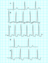
Interpreting the results Diagnostic testing Any abnormal electrocardiographic changes must be interpreted in the light of the probability of coronary artery disease and physiological response to exercise. A normal test result or a result that indicates a low probability of coronary artery disease is one in which 85% of the maximum predicted heart rate is achieved with a physiological response in blood pressure and no associated ST segment depression.
A test that indicates a high probability of coronary artery disease is one in which there is substantial ST depression at low work rate associated with typical angina-like pain and a drop in blood pressure. Deeper and more widespread ST depression generally indicates more severe or extensive disease. False positive results are common in women, reflecting the lower incidence of coronary artery disease in this group. Prognostic testing Exercise
testing in patients who have just had a myocardial infarction is
indicated only in those in whom a revascularisation procedure is
contemplated; a less strenuous protocol is used. Testing provides prognostic
information. Patients with low exercise capacity and hypotension
induced by exercise have a poor prognosis.
Screening Exercise testing of asymptomatic patients is controversial because of the high false positive rate in such individuals. Angina remains the most reliable indicator of the need for further investigation. In certain asymptomatic
groups with particular occupations (for example, pilots) there is a role
for regular exercise testing, though more stringent criteria for an abnormal
test result (such as ST segment depression of >2 mm) should be applied. In
the |
||||||||||||||
|
|||||||||||||||
|
Footnotes Jonathan Hill is specialist registrar in cardiology
at Barts and The London NHS Trust; Adam Timmis is a consultant cardiologist
at the The ABC of clinical electrocardiography is edited by Francis Morris, consultant in emergency medicine at the Northern General Hospital, Sheffield; June Edhouse, consultant in emergency medicine, Stepping Hill Hospital, Stockport; William J Brady, associate professor, programme director, and vice chair, department of emergency medicine, University of Virginia, Charlottesville, VA, USA; and John Camm, professor of clinical cardiology, St George's Hospital Medical School, London. The series will be published as a book in the summer |
Performance of exercise ECG testing
Outline of Topic
INTRODUCTION TEST LIMITATIONS . Patient selection . Test accuracy INDICATIONS CONTRAINDICATIONS CHOICE OF EXERCISE DEVICE ECG LEAD SYSTEMS EXERCISE TEST PROTOCOLS . Treadmill testing . Bruce protocol . Cornell protocol . Naughton protocol . Bicycle testing EXERCISE TEST PROCEDURE . Patient interview and examination . ECG data collection . ECG interpretation . BP measurements . Exertional hypotension . Exertional hypertension . Chest discomfort . Test end points . Submaximal testing . Procedures after exercise . Life-threatening complications . The exercise test summary report REFERENCES
Graphics
. ACC AHA ETT diagnosis CHD . Choice stress test modality . Treadmill score survival . Treadmill score gender . Pretest prob of CHD in CASS . Pretest probability of CHD . ACC AHA gas exchange exercise . ACC AHA exercise arrhythmia . Contraindications to ETT . Exercise testing protocols . Exercise test endpoints . ACC AHA indication stopping ETT . Minimizing exercise test risk
Related Topics
. Stress testing for the diagnosis of coronary heart disease . Electrocardiographic changes during exercise ECG testing . Pharmacologic stress myocardial perfusion imaging in the diagnosis and prognosis of coronary heart disease . Exercise myocardial perfusion imaging in the diagnosis and prognosis of coronary heart disease . Stress echocardiography in the diagnosis and prognosis of coronary heart disease . Exercise ECG testing to determine prognosis of coronary heart disease . Stress testing to determine prognosis and management of patients with known or suspected coronary heart disease . Screening for coronary heart disease . Functional exercise testing: Ventilatory gas analysis . Exercise capacity and VO2 in heart failure . Overview of the management of acute ST elevation (Q wave) myocardial infarction . Overview of the management of unstable angina and acute non-ST elevation (non-Q wave) myocardial infarction . Exercise in the treatment of hypertension . Ventricular arrhythmias in heart failure and cardiomyopathy . Risk stratification for cardiac events after acute ST elevation (Q wave) myocardial infarction . Risk stratification after unstable angina or non-ST elevation (non-Q wave) myocardial infarction
|