ALTE DOCUMENTE
|
||||||||||
Anatomy of the Masticatory System
A rational clinical examination of the masticatory system requires a sound basic knowledge of the anatomy. As will become clear later in the discussion of clinical examination procedures, the foundation of manual functional analysis is a good knowledge of the functional anatomy. In this chapter the individual anatomical structures will be described in a sequence corresponding to the later examination steps and separated according to their physiology and stages of adaptation and compensation. Knowledge of the different progressive and regressive tissue reactions is not only relevant to the diagnostic interpretation of the findings, but it also decisively influences the treatment strategy. The division into physiological, compensated, and adapted masticatory systems is necessary not only for diagnostic purposes but, more importantly, for the determination of what treatment goals are attainable for the individual.
The human jaw articulation is a so-called secondary joint (Gaupp 1911)
because it developed separately and not as a modification of a primary joint (Dabelow 1928). The
essential morphogenetic events
in the formation of the joints of the jaw occur
between the seventh and twentieth embryonic weeks
(Baume 1962, Furstman 1963, Moffet 1957, Baume and Holz 1970, Blackwood et al. 1976, Keith 1982,
Perry et al. 1985, Burdi 1992,
Klesper and Koebke 1993, Valenza et al. 1993, Bach-Petersen et al. 1994, Ogutcen-Toller and Juniper 1994, Bontschev 1996, Rodriguez-Vazquez et al. 1997).
The critical period for the
appearance of malformations in the joints of the jaw is reported differently in different studies. According to Van
der Linden et al. (1987) it is between the seventh and eleventh weeks, according to Furstman (1963)
between the eighth and twelfth weeks,
and according to
Formation of the bony mandible begins in weeks 6-7 lateral to Meckel's cartilage in both halves of the face. A double anlage of Meckel's cartilage is extremely rare (Rodriguez-Vazquez et al. 1997). Its effect on embryonic development is unknown. By about the twelfth week the two palatal processes have united at the midline to complete the separation of the oral and nasal cavities. At the same time, bony anlagen of the maxilla form in the region of the future infraorbital foramina. These spread rapidly in a horizontal direction and progressively fill the space between the oral
cavity and the eyes. When the crown-rump length (CRL) is approximately 76 mm (weeks 10-12), the anlagen of the maxillary bone, the zygomatic bone, and the temporal bone come into contact with one another. Ossification of the base of the cranium and of the facial portion of the skull follows in a strict, genetically determined sequence (Bach-Petersen et al. 1994). First to ossify is the mandible, followed by the maxilla, medial alar process of the sphenoid bone, frontal bone, zygomatic bone, zygomatic arch, squamous part of the occipital bone, greater wing of the sphenoid bone, tympanic bone, condyles of the occipital bone, lesser wing of the sphenoid bone, and finally the dorsolateral portion of the sphenoid bone.
In an embryo with a CRL of approximately 53 mm the coro-noid process and the condylar process can already be clearly distinguished from one another. The biconcave form of the articular disk becomes apparent at a CRL of 83 mm. In histological preparations, fibers of the pterygoid muscle can also be seen streaming in quite early (Radlanski et al. 1994). At this stage the superior belly of the lateral pterygoid muscle inserts at the middle and central third of the disk and the lower belly inserts at the condyle (Merida-Velasco et al. 1993). At a CRL of 95 mm all structures of the temporomandibular joint can be clearly identified and thereafter undergo no essential change other than an increase in size (Bontschev 1996).
Anatomy of the Masticatory System
![]() Embryology of the Temporomandibular Joint
and the Muscles of Mastication
Embryology of the Temporomandibular Joint
and the Muscles of Mastication
During the development of the temporomandibular joint the articular fossa is the first structure to become recognizable. This occurs during weeks 7-8 (Burdi 1992). It first appears as a concentration of mesenchymal cells over an area of tissue that later differentiates into disk and capsule. Between the tenth and eleventh weeks the fossa begins to ossify. Development of the cortical layer and the bony tra-beculae is more rapid in the fossa than in the condyle. The fossa develops first as a protrusion on the original site of the zygomatic arch and grows in a medial-anterior direction (Lieck 1997). At the same time the articular eminence
begins to develop. The condyle, at first cartilaginous, develops between the tenth and eleventh weeks from an accumulation of mesenchymal cells lateral to Meckel's cartilage (Burdi 1992). Enchondral ossification progresses apically, creating a bony fusion with the body of the mandible. After the fifteenth week the chondrocytes have differentiated enough so that the cartilage already exhibits the typical postnatal organization of structure (Perry et al. 1985), and from the twentieth prenatal week onward only the superficial portion of the process consists of cartilage.
|
|
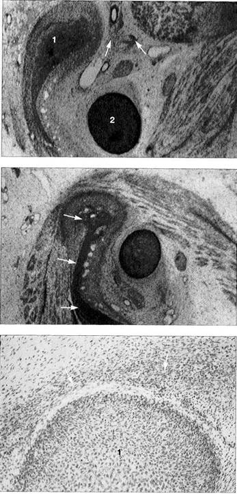
Joint development
Tenth week
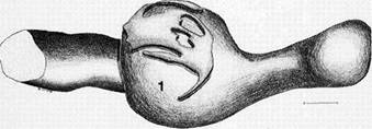
A histological section in the frontal plane showing the condylar process (1) and Meckel's cartilage (2) at the tenth week of embryonic development.
The condylar process is rounded over and surrounded by a layer of especially dense mesenchyme (arrows). It lies lateral to Meckel's cartilage. The fast-growing dorsocra-nial portion of the accumulation of cartilage cells creates the distinctive shape of the condyle.
Eleventh week
Above: A human temporomandibular joint in the frontal plane at the eleventh week of development. This represents the same area shown in Figure 19 only 10 days further along. The condylar process is beginning to ossify (arrows). At this time the swallowing reflex is also developing and is accompanied by the formation of secondary cartilage in the temporomandibular joint (Lakars 1995).
Contributed by R. Wurgaft Dreiman
Below: Sagittal section of a temporomandibular joint at the same stage of development. Above the condyle (1) is a distinct concentration of mesenchymal cells (arrows). At its inferior region the mesenchymal thickening is already beginning to detach from the condyle as the lower joint space forms. During this time the first collagen fibers of the disk become visible and increase greatly in number until the twelfth week.
Contributed by R.J. Radlanski
Embryology of the Temporomandibular Joint and the Muscles of Mastication
![]() The articular disk can first be
identified after 7.5 weeks in utero
as a horizontal concentration of mesenchymal cells
(Burdi 1992). Between weeks 19
and 20 its typical fibrocartilaginous structure is
already evident.
The articular disk can first be
identified after 7.5 weeks in utero
as a horizontal concentration of mesenchymal cells
(Burdi 1992). Between weeks 19
and 20 its typical fibrocartilaginous structure is
already evident.
The joint capsule first appears between weeks 9 and 11 as thin striations around the future joint region (Burdi 1992). After 17 weeks the capsule is clearly demarcated, and after 26 weeks all of its cellular and synovial parts are completely differentiated.
In weeks 9-10 the lateral pterygoid muscle is recognizable with its superior head inserting on the disk and capsule and
its inferior head inserting on the condyle. Fibers of the mas-seter and temporal muscles also insert on the disk (Merida Velascoetal. 1993).
During the tenth week the first blood vessels become organized around the joint. The disk has small blood vessels only at its periphery and is itself avascular (Valenza et al. 1993). Branches of the trigeminal and auriculotemporal nerves are clearly visible in the twelfth week (Furstman 1963). The numerous nerve endings that can still be seen in the disk in the twentieth week diminish rapidly so that after birth the disk is no longer innervated (Ramieri et al. 1996).
|
|
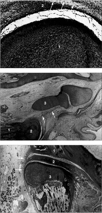
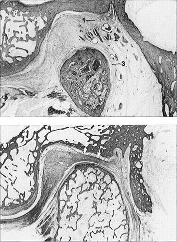
Fourteenth week
Sagittal section of a human disk-condyle complex. A distinct joint space has now formed between the condyle (1) and the disk (2). Above the disk the temporal blastema begins to split away to form the upper joint space (arrows). The cartilage of the condyle is increasingly replaced by bone from below. However, remnants of the original cartilage remain in the neck of the condyle until past puberty.
Sixteenth week
Horizontal section of a human temporomandibular joint during the sixteenth week of embryonic development. Insertion of the lateral pterygoid muscle (1) onto the condyle (2) can be clearly identified. In agreement with reports in the literature (Ogutcen-Toller and Juniper 1994, Ogutcen-Toller 1995), the discomaleolar ligament (arrows) runs from the joint capsule through the tympanosquamosal fissure to the malleus (3) as an extension of the muscle.
Eighteenth week
Frontal section through a human temporomandibular joint in the eighteenth week of embryonic development. The fossa (1), disk (2) and condyle (3) are completely developed and from now on will experience only an increase in size. The joint capsule (arrows) can also be clearly identified. The cartilaginous condyle will ossify further. Distribution of cartilage at this stage indicates that future growth will be primarily in the laterosuperior direction.
Contributed by R. Wurgaft Dreiman
Anatomy of the Masticatory System
![]() Development of the Upper and Lower Joint Spaces
Development of the Upper and Lower Joint Spaces
The upper and lower joint spaces arise through the formation of multiple small splits in the dense mesenchyme from which the condyle, disk, and fossa arose previously.
The lower joint space appears first at about the tenth week (50-65 mm CRL), but later the upper joint space overtakes it in its development (Burdi 1992). At first the space is extensively compartmentalized, and it is only later that the individual cavities merge (Bontschev 1996). The lower joint space lies close to the embryonic condyle.
The upper joint space appears after about the twelfth week (60-70 mm CRL) and spreads posteriorly and medially over Meckel's cartilage with its contour corresponding to that of the future fossa. After week 13 the lower joint space is already well formed as the upper joint space continues to take shape. From its beginning, the upper joint space has fewer individual islands of space and grows more rapidly than the lower joint space. After week 14 both joint spaces are completely formed. During weeks 16-22 the lumens of the chambers become adapted to the contours of the sur-
|
|
Joint development
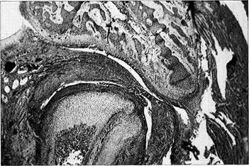
Twenty-sixth week
Completely formed human 22422v213w temporomandibular joint with physiol-gical lower and upper joint spaces. Trabecula-like structures can be identified in both joint spaces where the disk has not yet separated completely from the temporal and condylar portions. At present it has not been conclusively determined whether or not this type of incomplete separation could be one cause of disk adhesions.
|
|
Development of the joint spaces
Above: Three-dimensional reconstruction from a series of histological sections of the developing joint space (yellow) of a right temporomandibular joint. In the center of the picture is the condyle (1); to the right of it lies the coronoid process (2). To the left behind the condyle is Meckel's cartilage (3). The upper joint space arises approximately 2 weeks after the lower.
|
|
Below: Three-dimensional reconstruction of the lower joint space (green) of the same joint. Initially the mesenchyme in the condylar region (1) is still uniformly structured, but in weeks 10-12 it begins to tear in several places mesial and distal to the condyle. The resulting clefts run together to form the lower joint space. A region of concentrated mesenchyme remains between the two joint spaces, from which the fibrocartilaginous articular disk is later formed.
Contributed byR.]. Radianski (Figs. 23-25)
Development of the Upper and Lower Joint Spaces
![]() rounding bone. The fibrocartilaginous articular disk develops from the concentrated mesenchyme between the two joint spaces. The articular disk is not visible until the CRL is 70 mm. Even before formation of the joint spaces
the disk is already thinner at its
center than at the periphery and this leads to its final biconcave form (Bontschew 1996). The peripheral portions are not sharply demarcated
from the surrounding loose mesenchyme. In
fetuses with a CRL of 240 mm, the mesenchymal tissue changes into dense fibrous connective tissue. At this stage the
peripheral region has a greater blood
supply than the central region. According to Moffet (1957), compression of the disk between the
rounding bone. The fibrocartilaginous articular disk develops from the concentrated mesenchyme between the two joint spaces. The articular disk is not visible until the CRL is 70 mm. Even before formation of the joint spaces
the disk is already thinner at its
center than at the periphery and this leads to its final biconcave form (Bontschew 1996). The peripheral portions are not sharply demarcated
from the surrounding loose mesenchyme. In
fetuses with a CRL of 240 mm, the mesenchymal tissue changes into dense fibrous connective tissue. At this stage the
peripheral region has a greater blood
supply than the central region. According to Moffet (1957), compression of the disk between the
temporal bone and the condyle results in an avascular central zone. At the beginning of its development the disk lies closer to the condylar process than to the future fossa. At this stage there is still a layer of loose mesenchyme between the temporal bone and the upper joint space. It is only after a CRL of 95 mm has been reached that the condylar process and the fossa become closer and the mesenchymal layer disappears.
|
|
Development of the lateral pterygoid muscle
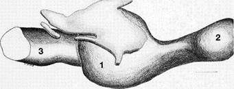
Three-dimensional representation of the insertion of the lateral pterygoid muscle (1) onto a left temporomandibular joint. As the muscle develops from the eleventh week, its upper belly attaches to the condyle, capsule, and disk while its lower belly attaches only to the condyle (2). At no time during development do the fibers of the lateral pterygoid muscle make direct contact with Meckel's cartilage (Ogutcen-Toller and Juniper 1994).
|
|
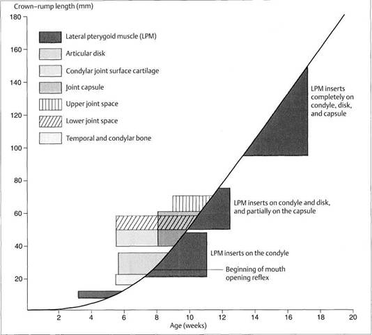
Development of the human temporomandibular joint
Graphic representation (modified from van der Linden et al. 1987) of prenatal development of the human temporomandibular joint showing its relationship to the CRL and age. First to form are the bony structures and the disk. Development of the joint capsule is accompanied by development of the upper and lower joint spaces. It is most interesting that prenatal mandibular movements can be observed as early as weeks 7-8 (Hooker 1954, Humphrey 1968), even though most of the joint structures and even the muscle insertions do not develop until a few weeks later. It is assumed that the movements are made possible by the primary jaw joint between Meckel's cartilage and malleus-incus (Burdi
Anatomy of the Masticatory System
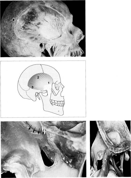
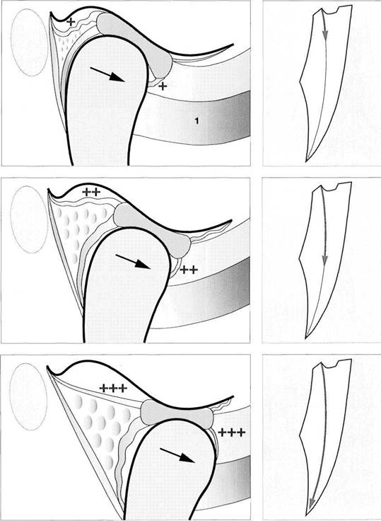
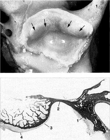
Glenoid Fossa and Articular Protuberance
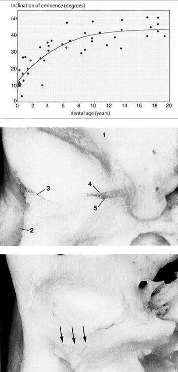
The temporal portion of the joint can be divided into four functional parts from posterior to anterior: postglenoidal process, glenoid fossa, articular protuberance, and apex of the eminence. The inclination of the protuberance to the occlusal plane varies with age and function (Kazanjian 1940), but is 90% determined at the age of 10 years (Nickel et al. 1988). Three fissures can be found at the transition to the tympanic plate of the temporal bone: the squamotym-panic, petrotympanic, and petrosquamous fissures (Fig. 28). In patients with disk displacement, these fissures are fre-
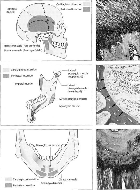
quently ossified (Bumann et al. 1991). Under physiological conditions the only parts of the temporal portion of the joint that are covered with secondary cartilage are the protuberance and the eminence (Fig. 31). Secondary cartilage is formed only when there is functional loading. Before the fourth postnatal year stimulation of the cells of the perio-seum leads to the formation of secondary cartilage (Hall 1979, Thorogood 1979, Nickel et al. 1997). With no persisting functional load the chondrocytes of the condyle would differentiate into osteoblasts (Kantomaa and Hall 1991).
![]()
|
Inclination
of the articular
protuberance to the occlusal
plane
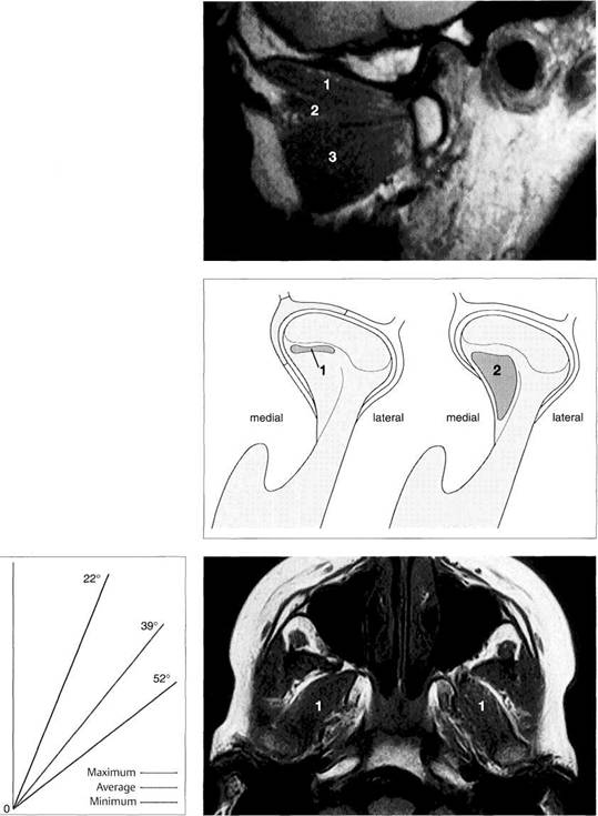
This graph (adapted from that of Nickel et al. 1988) indicates the inclination of the posterior slope of the eminence (articular protuberance) in relation to the occlusal plane. Accordingly, at the age of 3 years the eminence has reached 50% of its final shape (Nickel et al. 1997). Between the tenth and twentieth year there is a difference of only 5°. The study material originates from the osteological collection of Hamman-Todd and Johns Hopkins, Cleveland Museum of Natural History.
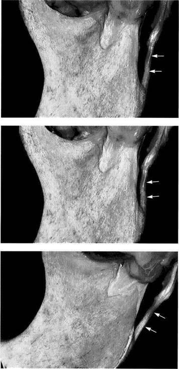
Joint
region of the temporal
bone
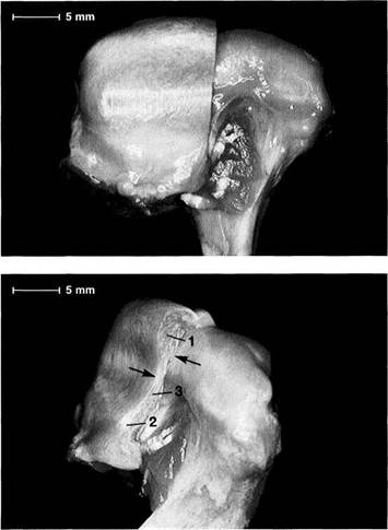
Inferior view of the temporal portion of a defleshed temporomandibular joint. Near the upper border of the picture is the articular eminence (1) and at the far left is the external auditory meatus (2). In the posterior portion of the fossa the squamotympanic fissure (3) is found laterally, and the petrosquamous (4) and petrotympanic (5) fissures are found medially. Both the superior stratum of the bilaminar zone and the posterior portion of the joint capsule, and sometimes also the fascia of the parotid gland can insert into these fissures.
Ossification
of the fissures
and disk displacement
Inferior view of a temporal bone with partially ossified fissures. The lateral half of the squamotympanic fissure is completely ossified (arrows). The superior stratum of the bilaminar zone can now insert only into the periosteum in this region. It has been shown that these fissures are ossified in more than 95% of patients with disk displacement, whereas in joints without disk displacement normal fissure formation prevails (Bumann et al. 1991).
Glenoid Fossa and Articular Protuberance
![]() However, the maturation process of these cells
is delayed by functional demands (Kantomaa and Hall
1988). Loading reduces the intracellular concentration of cyclic adenosine monophosphate (cAMP). This increases the rate of mitosis and suppresses the ossification process relative
to the proliferation of cartilage
(Copray et al. 1985). Furthermore, the proteoglycane content of
cartilage correlates with its ability to withstand compressive loads (Mow et al. 1992).
However, the maturation process of these cells
is delayed by functional demands (Kantomaa and Hall
1988). Loading reduces the intracellular concentration of cyclic adenosine monophosphate (cAMP). This increases the rate of mitosis and suppresses the ossification process relative
to the proliferation of cartilage
(Copray et al. 1985). Furthermore, the proteoglycane content of
cartilage correlates with its ability to withstand compressive loads (Mow et al. 1992).
The hypothesis that structures of the temporomandibular joint are subjected to compressive loads during function has been around for many decades and is supported by a num-
ber of experimental studies (Hylander 1975, Hinton 1981, Taylor 1986, Faulkner et al. 1987, Boyd et al. 1990, Mills et al. 1994a). Studies using finite element analysis (FEA) also verify that during function, temporomandibular joint structures are subject to variable loads depending upon the individual static and dynamic occlusion (Korioth et al. 1994a, b). Different types of loads also bring about different responses in bone. When erosive changes are found in the condyle, the trabecular bone volume (TBV) of the temporal portion of the joint is significantly higher (25%) than when the condyle is unchanged (16%; Flygare et al. 1997).
![]()
![]()
|
Function Movement with little friction Progressive adaptation Cartilage hypertrophy Bone apposition |
|
Regressive adaptation Cartilage degeneration Bone deformation Inflammation Ankylosis |
Inferior view of the temporal cartilaginous joint surface and capsule attachment
Caudal view of the left temporomandibular joint of a newborn. The bony portions have been separated from the periosteum up to the circular insertion of the capsule and bilaminar zone. Part of the zygomatic arch (1) can be seen near the right border of the photograph. The fibrocartilaginous articular surfaces over the articular protuberance are thickened medially and laterally (arrows). When covered with synovial fluid they allow movements with virtually no friction (Smith 1982).
Sagittal
histological section
showing buildup of the temporal
joint components
The temporal portion of the joint can be divided into four functional components: 1 postglenoidal process, 2 glenoid fossa, 3 articular protuberance, and 4 apex of the eminence. As a rule, no cartilage can be identified within the fossa. The average thickness of the fibrous cartilage over the protuberance and the eminence is between 0.07 and 0.5 mm (Hansson et al. 1977). As this photograph shows, there can be considerable variation in thickness within the same individual.
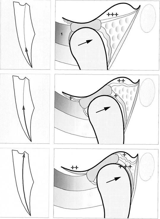
Function
and structural
adaptation of the articular
eminence
A summary of the basic anatomical changes in the temporal joint tissues. Increased functional loading will cause hypertrophy through secondary cartilage formation and bone deposition (progressive adaptation). Persistent nonphysiological loading (massive influences) leads to deforming or degenerative changes. This regressive adaptation is accompanied by more or less noticeable rubbing sounds, sometimes in combination with pain.
![]() Mandibular Condyle
Mandibular Condyle
Human condyles differ greatly in their shapes and dimensions (Solberg et al. 1985, Scapino 1997). From the time of birth to adulthood the medial-lateral dimension of the condyle increases by a factor of 2 to 2.5, while the dimension in the sagittal plane increases only slightly (Nickel et al. 1997). The condyle is markedly more convex in the sagittal plane than in the frontal plane.
The articulating surfaces of the joint are covered by a dense connective tissue that contains varying amounts of chondrocytes, proteoglycans, elastic fibers and oxytalan fibers
(Hansson et al. 1977, Helmy et al. 1984, Dijkgraaf et al. 1995). The composition and geometric arrangement of the extracellular matrix proteins within the fibrous cartilage determine its properties (Mills et al. 1994 a,b). Cartilage that can absorb and distribute compressive loads is characterized by a matrix with high water content and high molecular weight chondroitin sulfate in a network of type II collagen (Maroudas 1972, Mow et al. 1992). A low level of functional demand upon the joint leads to an increase of type I collagen and a reduction of type II (Pirttiniemi et al. 1996). Inter-leukin la inhibits the matrix synthesis of chondrocytes,
|
|
|
|
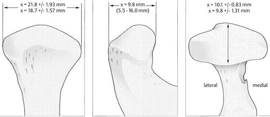
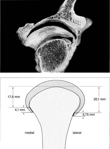
Condyle dimensions
Left: Width of condyle in the frontal plane (Solberg et al. 1985). The average condylar width is significantly greater in men (21.8 mm) than in women (18.7 mm).
Center: Anteroposterior dimension of the central portion shown in the sagittal plane (Oberg et al. 1971; minimum and maximum in parentheses).
Right: Anteroposterior dimension of the condyle in the horizontal plane. There is no significant difference between men (10.1 mm) and women (9.8 mm).
Functional joint surface
Histological preparation showing a physiological fibrocartilaginous joint surface (thin arrows) of the condyle of a 58-year-old individual. In spite of the intact joint surface on the condyle, the pars posterior (1) of the disk is flattened and the functional fibrocartilaginous temporal surface of the joint on the articular protuberance shows degenerative changes (outlined arrows). The subchondral cartilage has not yet been affected and would appear intact on a radiograph.
Buildup of the condylar cartilage
Histologically, the secondary cartilage of the condyle is made up of four layers:
![]() Fibrous connective-tissue zone
Fibrous connective-tissue zone
Proliferation
zone with undiffer
entiated connective-tissue
cells
Fibrous cartilage zone
Enchondral ossification zone
Other structures shown are:
Eminence
Disk
Condyle
![]() Contributed
by R. Ewers
Contributed
by R. Ewers
Mandibular Condyle
![]() while the transforming growth factor TGF-b promotes
it (Blumenfeld et al. 1997). The
collagen fibers of the fibrocartilaginous joint surfaces are oriented mainly
in a sagittal plane (Steinhardt 1934).
while the transforming growth factor TGF-b promotes
it (Blumenfeld et al. 1997). The
collagen fibers of the fibrocartilaginous joint surfaces are oriented mainly
in a sagittal plane (Steinhardt 1934).
Joint surface cartilage must permit frictionless sliding of the articulating structures while at the same time it must be able to transmit compressive forces uniformly to the subchondral bone (Radin and Paul 1971). Hypomobility of the mandible results in a more concentrated loading of the joint surfaces. Even if the forces in the masticatory system remain the same, the load per unit of area on the cartilage will be increased when there is hypomobility. The amount
of structural change depends upon the amplitude, frequency, duration, and direction of the loads (Karaharju-Suvanto et al. 1996).
In joints that have undergone erosive changes, the percentage of trabecular bone volume (21%) and the total bone volume (54%) are significantly higher than the corresponding 15% and 40% found in condyles without these changes (Fly-gare et al. 1997). Degenerative changes therefore are closely associated with nonphysiological loading of the joint surfaces.

|
|
Function Movement with little friction Progressive adaptation Cartilage hypertrophy Bone apposition |
|
Regressive adaptation Cartilage degeneration Bone deformation (osteophytes) Inflammation Ankylosis |
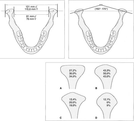
Intercondylar distance
Left: Sex-specific data on the distances between pairs of medial poles and lateral poles of the condyle (after Christiansen et al. 1987). The numbers given are average values. A difference of 5-10 mm in the intercondylar distance will have a corresponding effect on the tracings of condylar movements and the accuracy of simulated movements in the articulator (see pp. 216 and 243).
Right: Schematic drawing illustrating the intercondylar angle.
Condylar shapes in
the
frontal plane
According to Yale et al. (1963) 97.1 % of all condyles fall into one of four groups based upon their frontal profile. These are described as either flat (A), convex (B), angled (C), or round (D). The relative frequencies of occurrence are taken from the works of Yale et al. (1963), Solberg et al. (1985), and Christiansen et al. (1987). The condyle form affects the radiographic image of this partofthejointinthe Schuller projection (Bumann et al. 1999) and the loading of the joint surfaces (Nickel and McLachlan
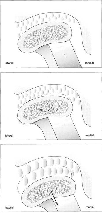
Function
and structural
adaptation of the condyle
Summary of the basic anatomical and functional changes in the condylar portion of the joint. Increased functional loading will stimulate cartilaginous hypertrophy (= progressive adaptation) that is not noticeable clinically. Continuous nonphysiological loading of the condyle can lead to degeneration, deformation, and even ankylosis (Dibbets 1977, Stegenga 1991). These changes may be accompanied by pain or, with sufficient adaptation, they may progress painlessly.
Anatomy of the Masticatory System
Positional Relationships of the Bony Structures
The position of the condyle relative to the articular protuberance has been a subject of controversy in dentistry for many years (Lindblom 1936, Pullinger et al. 1985). A well-defined condylar position oriented to the maximal occlusion is especially relevant to extensive dental treatment (Spear 1997). In the past, to transfer the jaw relations to an articulator the condyles were always placed in their most posterosuperior position because this relationship could be most easily reproduced (Celenza and Nasedkin 1979). Under purely static conditions the condylar position is dependent
upon the shape of the fossa, the inclination of the protuberance, and the shape of the condyle. In the 1970s this led to the assignment of a geometric centric position of the condyle in the fossa (Gerber 1971). However, the dimensions of the joint space are quite variable in both the sagittal plane (anterior, posterior, and superior) and the transverse plane (medial, central, and lateral) (Pullinger et al. 1985, Hatcher et al. 1986, Christiansen et al. 1987, Bumann et al. 1997). For this reason the concept of an anatomical orientation is untenable, and the radiographic techniques
|
|
Sagittal relationships
Macroscopic anatomical preparation showing the relation of the fossa, disk, and condyle to one another in the sagittal plane. Because the shapes of fossae and condyles vary so greatly, it is not possible to determine a universally applicable measurement of the condylar position. Although the physiological (i.e. centric) condylar position is defined as the most anterosuperior position with no lateral displacement (arrows), this position depends upon the basic neuromuscular tonus.
Frontal relationships
Macroscopic anatomical preparation showing the relation of the fossa, disk, and condyle to one another in the frontal plane. In this plane, too, there is no standard geometric arrangement of condyle and fossa because of the variability of the hard and soft tissues (Yung et al. 1990). In this preparation the disk (arrows) is displaced laterally. Structures of the bilaminar zone (1) can be identified in the medial portion of the joint. The close proximity of the joint to the middle (2) and inner ear (3) can also be observed.
Horizontal relationships
A right temporomandibular joint viewed from above showing the relation of the fossa, disk, and condyle to one another in the horizontal plane. The lateral portion of the joint is near the left border of the picture. Near the upper border a section through the external auditory meatus can be seen (1). The roof of the fossa has been removed. Near the center of the picture lies the transition from the pars posterior (2) to the bilaminar zone (3). The central perforation was created during sectioning, and through it can be seen the upper surface of the condyle (arrow).
Positional Relationships of the Bony Structures
![]() (p. 148) are unsuitable for determining a
therapeutic condylar position (Pullinger and Hollender 1985).
Therefore the current definitions
of centric relation are geared more toward the functional conditions (van Blarcom 1994, Daw-son 1995,
Lotzmann 1999). It has been demonstrated experimentally that the surfaces of the temporomandibular joint are subjected to loads of 5-20 N (Hylanderl979, Brehnan et al. 1981, Christensen et al. 1986). In a patient's
habitual occlusion this force is
partially intercepted by the occluding premolars
and molars. Tooth loss can lead to higher joint loading and regressive adaptation (van den Hemel 1983, Christensen et al. 1986, Seligman and Pullinger
1991). How-
(p. 148) are unsuitable for determining a
therapeutic condylar position (Pullinger and Hollender 1985).
Therefore the current definitions
of centric relation are geared more toward the functional conditions (van Blarcom 1994, Daw-son 1995,
Lotzmann 1999). It has been demonstrated experimentally that the surfaces of the temporomandibular joint are subjected to loads of 5-20 N (Hylanderl979, Brehnan et al. 1981, Christensen et al. 1986). In a patient's
habitual occlusion this force is
partially intercepted by the occluding premolars
and molars. Tooth loss can lead to higher joint loading and regressive adaptation (van den Hemel 1983, Christensen et al. 1986, Seligman and Pullinger
1991). How-
ever, if the joint's capacity for adaptation is sufficiently great, degenerative changes may be avoided (Helkimo 1976, Kirveskari and Alanen 1985, Roberts et al. 1987). The direction of functional loading is anterosuperior against the articular protuberance (Dauber 1987). Clear evidence for this is the presence of the load-induced secondary cartilage on the joint surfaces in this region.
Positioning of the condyles on the protuberances is accomplished exclusively through the antagonistic activity of the neuromuscular system and from a functional standpoint requires no border position.
|
|
Relationships in the frontal
plane
Schematic depiction of the joint space relationships in the frontal plane. A number of studies have reported that the dimensions found in the lateral, central, and medial parts may vary greatly (Christiansen et al. 1987, Vargas 1997). Although the lateral portion is affected more frequently by degenerative changes, the width of the joint space is usually least at its center (blue line).
Contours
on the temporal
surface of the joint
Schematic drawing (modified from Hassoetal. 1989) of the contours in the lateral (green), central (blue), and medial (red) regions of the joint. The entire protrusive functional path is represented as a convex bulge that can vary markedly as the result of regressive or progressive adaptation. Therefore, the loads borne by the lateral and medial portions of the joint during function are also influenced by the morphology of the articular protuberance (Oberg et al. 1971, Hylan-der 1979, Hinton 1981).
Relationships
in the medial
part of the joint
Schematic drawing (modified from Christiansen et al. 1987) of the positional relationships in the medial portion of a left temporomandibular joint. This finding also emphasizes the fundamental principles of physiological joint movements. As with all other joints, the temporomandibular joint has a passive "play" space in all directions and is thus not confined to any border position.
Average values: 1 = 3.4 mm; 2 = 4.4 mm
Anatomy of the Masticatory System
![]() Articular Disk
Articular Disk
The articular disk can be divided into three regions based upon their function: the partes anterior, intermedia, and posterior. The primary functions of the disk are to reduce sliding friction and to dampen load spikes (McDonald 1989, Scapino et al. 1996). The extracellular matrix of the fibrocartilaginous disk consists primarily of type I and type II collagen (Mills et al. 1994b). The orientation of the collagen fibers in the disk displays a typical pattern (Knox 1967, Scapino 1983). In the pars intermedia dense bundles of collagen fibers run approximately in a sagittal direction. These intertwine with the exclusively transverse fibers of the pars
anterior and pars posterior (Takisawa et al. 1982). Elastic fibers are found in all parts of the disk (Nagy and Daniel 1991) but are more numerous in the pars anterior and in the medial portion of the joint (Luder and Babst 1991). A reduction in the thickness of the disk results in an exponential increase in the load it experiences (Nickel and McLachlan 1994). The more rapidly a load is applied, the "stiffer" the disk reacts (Chin et al. 1996). The inferior stratum and the convexity of the pars posterior help stabilize the disk on the condyle.
|
|
Alignment
of fibers within
the disk and their attachment to
the condyle
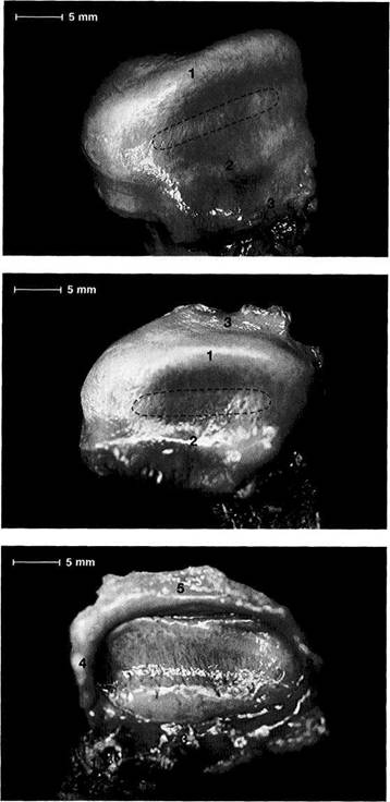
Macroscopic anatomical preparation of the disk-condyle complex of a right temporomandibular joint. The collagen fibers of the pars posterior (1) and the pars anterior (2) run from the medial to the lateral pole of the condyle (Moffet 1984), making possible a wide range of movement of the disk relative to the condyle in the sagittal plane. The fibers of the pars intermedia (outlined area), on the other hand, run in a more sagittal direction. The medial pterygoid muscle (3) makes its insertion at the anteromedial region.
Cranial view
A view from above of the disk in Figure 45 after removal of the condyle. In this view the transverse course of the fibers in the pars posterior (1) and pars anterior (2) can be seen more clearly. Histologically the disk is composed of dense collagenous connective tissue with a few embedded chondrocytes (Rees 1954). In the pars anterior and pars posterior the chondrocytes are found in clusters, but in the pars intermedia (outlined) they are arranged uniformly. Part of the bilaminar zone
can be seen
attached at the dis
tal border of the pars posterior.
Inferior
view of the same
disk
In this view the insertion of a portion of the superior head of the lateral pterygoid muscle (1) can be clearly seen. The remaining fibers of the superior head insert on the condyle. This preparation also demonstrates the insertion of the lateral (2), anterior (3), and medial
borders of
the joint capsule. In
the posterior part of the joint the
capsule is connected to the posteri
or surface of the condyle by the
stratum inferium (5) of the bilami
nar zone (see p. 47).
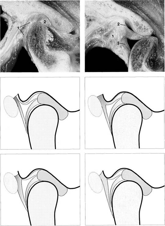
Anatomical Disk Position
![]() Anatomical Disk
Position
Anatomical Disk
Position
In a physiological temporomandibular joint, the pars posterior of the disk lies on the superior portion of the condyle. In the "centric condylar position" the thinnest part of the disk, the pars intermedia, is located between the anterosu-perior convexity of the condyle and the articular protuberance (van Blarcom 1994). This finding is also supported by studies using measurements and mathematical models (Bumann et al. 1997, Kubein-Meesenburg 1985). The pars anterior lies in front of the condyle (Steinhardt 1934, Wright and Moffet 1974, Scapino 1983). The disk is attached to the
medial and lateral poles of the condyle by means of the transversely aligned collagen fibers of the pars anterior and pars posterior. Viewed by itself, this anatomical arrangement with the condyle allows a great degree of movement during active mandibular movements (see p. 46). The disk exhibits viscoelastic properties under compressive loads. Its resistance is strengthened by the arrangement of the collagen fibers (Shengyi and Xu 1991). The elastic fibers within the disk serve primarily to restore the shape of the disk after a load has been removed (Christensen 1975).
![]()
![]()
|
Function Moveable joint surface Load distribution Progressive adaptation Reversible deformation |
|
Regressive adaptation Permanent deformation Disk perforation Ossification |
Anterosuperior aspect of
the disk-condyle complex
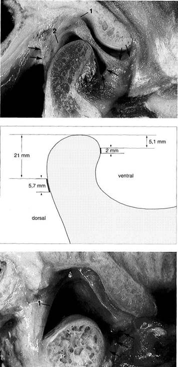
Macroscopic anatomical preparation of a left temporomandibular joint showing the relationship between disk and condyle. The lateral half of the disk has been removed fora clearer view. The dorsal border of the pars posterior is near the region of the apex of the condyle. From a functional point of view, this broad description is not very helpful for diagnostic purposes because the physiological position of the pars posterior depends to a large extent upon the inclination of the protuberance.
Anterolateral aspect
of the
disk-condyle complex
The same preparation in half profile. Here the pars posterior (1), pars intermedia (3), and pars anterior (2) can be clearly distinguished. Although the posterior border of the pars posterior lies over the apex of the condyle, the pars intermedia is in front of the anterosuperior convexity (arrows) of the condyle. The pars anterior is 2.0 mm thick, the pars intermedia 1.0 mm thick, and the pars posterior 2.7 mm thick (Gaa 1988).
Function
and structural
adaptation of the disk
Functionally, the disk serves as a "moveable fossa" for the condyle. Because of its unique tissue structure it can cushion and dampen peaks of force. Progressive adaptation differs from regressive in that the former is reversible. Strictly speaking, there is no "positive" tissue reaction in the disk because functional loads as well as continuous nonphysiological loads result in
rlpfnrmatinn
Anatomy of the Masticatory System
![]() BilaminarZone
BilaminarZone
The posterior portion of the temporomandibular joint has been variously referred to as the bilaminar zone (Rees 1954), retroarticular plastic pad (Zenker 1956), retroarticu-lar pad (DuBrul 1988), retrodiskal fat pad (Murikami and Hoshino 1982), or trilaminar zone (Smeele 1988). It consists of an upper layer (superior stratum) and a lower layer (inferior stratum) (Rees 1954, Griffin and Sharpe 1962). Between these two layers lies the genu vasculosum with its numerous vessels, nerves, and fat cells (Griffin and Sharpe 1962). The superior stratum is composed of a loose network of elastic and collagen fibers, fat, and blood vessels (Zenker 1956).
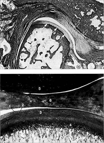
By contrast, the inferior stratum is made up of tight collagen fibers (Rees 1954, Wilkes 1978, Luder and Bobst 1991). In the bilaminar zone the collagen fibers are more loosely organized and run more or less in the sagittal plane (Mills et al. 1994b). The fibers of both strata stream into the pars posterior of the disk and there intertwine with the transverse fibers of the pars posterior and the sagittal fibers of the pars intermedia (Scapino 1983). The elastic fibers in the bilaminar zone have larger diameters than those of the disk and are concentrated predominantly in the superior stratum (Rees 1954, Scapino 1983, Mills et al. 1994a).
|
|
Macroscopic
anatomical
preparation
Left: With the jaws closed the bil-aminar zone (1) fills the space posterior to both the pars posterior (2) and the condyle (3). The inferior stratum stabilizes the disk on the condyle in the sagittal plane. An overextension of the bilaminar zone through posterosuperior displacement of the condyle is an essential precondition for an anterior disk displacement to occur. Right: With the mouth open the genu vasculosum (1) fills with blood. The superior stratum (2) and inferior stratum (3) can be easily identified.
Variants
of the postero
superior attachment
Left: Type A insertion. The superior stratum and the posterior joint capsule run separately to their insertions in the fissures. This type of insertion occurs most often in the medial portion of the joint.
Right: Type B insertion. Here the superior stratum and the posterior joint capsule merge before reaching the fissures and continue posterosuperiorly as one uniform, undifferentiated structure. This variant is the second most common in the medial portion of the joint.
Variants
of the postero
superior attachment
Left: Type C insertion. The superior stratum inserts on the glenoid process because the fissures are completely filled by the posterior portion of the joint capsule. This type of insertion is found most frequently in the lateral part of the joint.
Right: Type D insertion. In this rare variant no posterior capsule structure can be demonstrated histologically. The posterior boundary is formed by the parotid fascia.
BilaminarZone
![]() The superior stratum is attached posteriorly to
the bony auditory meatus,
the cartilaginous part of the auditory meatus, and the fascia of the parotid gland (Scapino 1983). Four insertion variations can be distinguished (Bumann
et al.
The superior stratum is attached posteriorly to
the bony auditory meatus,
the cartilaginous part of the auditory meatus, and the fascia of the parotid gland (Scapino 1983). Four insertion variations can be distinguished (Bumann
et al.
The inferior stratum inserts on the posterior side of the condyle below the fibrocartilaginous articulating surface and is responsible for stabilizing the disk on the condyle. Anterior disk displacement is possible only when the predominantly collagenous inferior stratum becomes overstretched. The superior stratum, on the other hand, is responsible for retracting the articular disk, especially dur-
ing the initial phase of closure, but is of lesser importance in the occurrence of anterior disk displacement (Eriksson et al. 1992). These facts are very important to consider in the diagnosis and treatment of disk displacements. Continuous posterior or posterosuperior loading of the bilaminar zone eventually leads to fibrosis and sometimes to the formation of a pseudodisk (Hall et al. 1984, Isberg et al. 1986, Kurita et al. 1989, Westesson and Paesani 1993, Bjornland and Refsum 1994).
![]()
![]()
|
Function Stabilization of the disk in the sagittal plane Proprioception Cell nutrition Progressive adaptation Fibrosis |
|
Regressive adaptation Inflammation Overextension with inflammation Perforation |
Histology of the trilaminar
zone
The superior stratum (1), genu vas-culosum (2), and inferior stratum (3) can be clearly distinguished from one another. Sensory and sympathetic nerve fibers provide pain perception and regulation of blood-vessel tonus. Here the neuropeptides A and Y effect vasoconstriction (Lundberg et al. 1990, Grundemar and Hakanson 1993) while vasodilation is brought about by the vasoactive intestinal peptide, the peptide histidine-isoleucine amide and acetylcholine (Widdicombe1991).
Progressive
adaptation
(fibrosis)
Chronic overloading brings about fibrosis (arrows) and reduction of the number of blood vessels. Such fibrosis can be seen in 64-90% of patients, depending on the position of the disk. Posterior and pos-terosuperior condylar displacement without adaptation of the bilaminar zone is a common cause of joint pains. Therefore, previous adaptation of the bilaminar zone can be considered a favorable factor for treatment.
Function
and structural
adaptation of the bilaminar zone
In addition to supplying nutrients and proprioception, the inferior stratum is of special importance in stabilizing the disk in the sagittal plane. Increased functional loading can lead to its fibrosing. Our own studies indicate that in spite of mechanical loading, fibrosis does not occur in 10-36% of joints. Chronic nonphysiological overloading usually results in perforation, overexte-nion. or inflammation.
Anatomy of the Masticatory System
![]() Joint Capsule
Joint Capsule
The bony parts of the temporomandibular joint are enclosed in a thin fibrous capsule. In addition to lateral, medial, and posterior capsule walls, there is an anterior wall that can be divided into upper and lower portions. The medial and lateral walls are reinforced by the similarly named medial and lateral ligaments (Schmolke 1994, Loughner et al. 1997). Attachment of the disk to the lateral and medial poles of the condyle is independent of the capsular structure (Fig. 60). The boundaries of the superior attachment of the capsule to the temporal bone are shown in Figure 30.
Because of its loose connective-tissue structure the anterior capsule wall cannot withstand as much loading as the other parts of the capsule (Koritzer et al. 1992, Johannson and Isberg 1991). The insertion of the capsule on the condyle is superficial and it lies at different levels on different sides of the condyle (Figs. 58, 61). Anterior disk displacements are accompanied not only by overextension of the inferior stratum, but also by stretching of the lower anterior capsule wall (Scapino 1983). The amount of extension is directly related to the amount of anterior disk displacement (Katzberg et al. 1980).
|
|
Joint
capsule in the sagittal
plane
By applying artificial traction on the specimen, the anterior portions of the upper and lower joint capsules (arrows) have been made more clearly visible. Posteriorly the joint spaces are bounded by the superior stratum (1) and inferior stratum (2). The posterior capsule wall lies behind the genu vasculosum. The type-Ill receptors of the capsule are only activated by heavy tensile loads on the lateral ligament and serve then to stimulate the elevator muscles (Kraus 1994).
Attachment
of the capsule
to the condyle
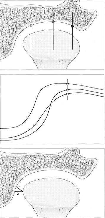
Schematic representation of the attachment of the joint capsule in the sagittal plane. The band-like insertion is significantly broader posteriorly than anteriorly. Because of the insertion of the lateral pterygoid muscle on the anterior surface of the condyle, the anterior part of the joint capsule attaches much higher anteriorly than posteriorly. The values given are based on measurements made on 39 human temporomandibular joints (Brauck-mann 1995).
Overdistended capsule
Anterior disk displacement requires not only a stretching of the inferior stratum (1), but also a distention of the lower anterior wall of the joint capsule (arrows). However, because the connective tissue of the anterior capsule wall is much looser, disk displacement depends almost exclusively on posterior loading vectors and the adaptability of the inferior stratum. A downward movement of the condyle as shown here without downward movement of the disk is possible only with extensive stretching of the inferior stratum.
Joint Capsule
The interior surface of the capsule is covered by synovial membrane (Dijkgraaf et al. 1996a, b). The synovial cells form synovial fluid which serves to bring nutrients to the avascular cartilage of the joint surfaces and to reduce friction. Lubrication of the joint surfaces is accomplished through two mechanisms (Okeson 1998). One is the displacement of synovial fluid from one area to another by jaw movements. The other is the ability of the cartilage to store a limited amount of synovial fluid. Under functional pressure the fluid is again released to ensure minimal friction within the joint, in spite of static and dynamic loads (Shengyi and Xu
A second important function of the joint capsule is proprioception. Receptors are divided into four types (Wyke 1972, Clark and Wyke 1974, Zimny 1988). Type I have a low threshold, adapt slowly, provide postural information, and have a reflexive inhibiting effect on the antagonistic muscles. Likewise, type II have a low threshold but adapt quickly and provide information about movements. Type III have a high threshold and are slow to adapt. Type IV receptors stand ready for sensory pain perception and do not "fire" under normal conditions.
|
|
Disk and capule attachments in the frontal plane
Macroscopic anatomical preparation of a temporomandibular joint in the frontal plane. Although the insertion of the disk on the condyle at the condylar poles has been described by some as an attachment through the joint capsule in the form of a "diskocapsular system" (Dauber 1987), other studies (Sol-berg et al, 1985, Bermejo et al. 1992) identify two separate connective-tissue structures for attachment to the condyle, one for the disk (1) and the other for the capsule (2).
Attachment of the joint capsule to the condyle
Schematic representation of the capsule attachment in the frontal plane. The collagen fibers of the disk and capsule insert somewhat lower on the lateral than on the medial surface of the condyle. It is not known to what extent the band of insertion is shifted superiorly when there is contracture of the capsule. However, shortening of the capsule walls does change the activity of the mechanoreceptors and thereby the activity of the muscles of mastication (Kraus 1994).
![]()
|
Function Proprioception Cell nutrition Progressive adaptation Overextension Constriction |
|
Regressive adaptation Inflammation Rupture Ankylosis |
Function and structural adaptation of the joint capsule
The primary functions of the capsule are proprioception and nourishment of the fibrocartilaginous joint surfaces. Increased functional loading of the joint can result in either stretching or contraction of the capsule. A chronic loading that exceeds the physiological limits activates the type-IV receptors through inflammation or rupture, resultina in Dain.
Anatomy of the Masticatory System
![]() Ligaments of the Masticatory System
Ligaments of the Masticatory System
The ligaments of the masticatory system, as in all other freely movable joints, have three main functions: stabilization, guidance of movement, and limitation of movement. From a functional view, limitation of movement is the most important function (Mankin and Radin 1979, Osborn 1995). There are different interpretations concerning the number and nomenclature of the ligaments found within the masticatory system (Sato et al. 1995). Five or six ligaments have been described: lateral ligament, stylomandibular ligament, sphenomandibular ligament, discomalleolar (Pinto's) liga-
ment and Tanaka's ligament. Sometimes the collateral attachment fibers between disk and condyle are included in the list as the lateral and medial collateral ligaments of the disk (Yung et al. 1990, Kaplan and Assael 1991, Okeson 1998), although from a functional viewpoint, this is not accurate.
The lateral ligament or temporomandibular ligament is made up of two parts: a deep, more horizontal part and a superficial, more vertically oriented part (Arstad 1954, Sicher and
Lateral ligament
Situation with jaws closed
Unlike in formalin-fixed preparations, the lateral ligament (arrows) is usually clearly distinguishable in fresh preparations. The initial rotation during an opening movement is limited by the superficial part of the lateral ligament (von Hayek 1937, Burch and Lundeen 1971). Further opening of the jaws can occur only after protrusion has relieved tension on the ligament, following which the ligament is again stressed by renewed rotation (Osborn 1989).
Situation with jaws open
Jaw opening is restricted by the length of the lateral ligament from its origin to its insertion. However, if the condyle can slip past the apex of the tubercle (eminence), the ligament (arrows) will no longer have this limiting effect. In addition, the lateral ligament will now impede retrusive and laterotrusive movements of the condyle (Posselt 1958, Brown 1975, Osborn 1989).
Function
and structural
adaptation of the ligaments
The chief function of the ligaments is to limit movement and thereby protect sensitive structures. In addition, they stabilize the joint and take over guidance functions (Rocabado and Iglarsh 1991). Depending upon the proportions of the types of collagen within the ligament and the direction of the functional overload on the joint, ligaments may become either stretched or shortened.

|
Function Limitation of movement (proprioceptive and mechanical) Progressiva adaptation Stretching Constriction |
|
Regressive adaptation Inflammation Rupture Ossification |
Ligaments of the Masticatory System
![]() DuBrul 1975, Kurokawa 1986). The horizontal
part limits retrusion (Hylander 1992) as well as
laterotrusion (DuBrul 1980) and thereby
protects the sensitive bilaminar zone from injury. The vertical part of
the lateral ligament, on the other hand,
limits jaw opening (Osborn 1989, Hesse and Hansson 1988). The
superficial portions of the lateral ligament contain Golgi tendon organs
(Thilander 1961). These nerve endings are
very important for the neuromuscular monitoring
of mandibular movements (Hannam and
Sessle 1994, Sato et al. 1995). For this
reason, anesthetizing the lateral portion of the joint permits a 10-15%
increase in jaw opening (Posselt and Thilander 1961).
DuBrul 1975, Kurokawa 1986). The horizontal
part limits retrusion (Hylander 1992) as well as
laterotrusion (DuBrul 1980) and thereby
protects the sensitive bilaminar zone from injury. The vertical part of
the lateral ligament, on the other hand,
limits jaw opening (Osborn 1989, Hesse and Hansson 1988). The
superficial portions of the lateral ligament contain Golgi tendon organs
(Thilander 1961). These nerve endings are
very important for the neuromuscular monitoring
of mandibular movements (Hannam and
Sessle 1994, Sato et al. 1995). For this
reason, anesthetizing the lateral portion of the joint permits a 10-15%
increase in jaw opening (Posselt and Thilander 1961).
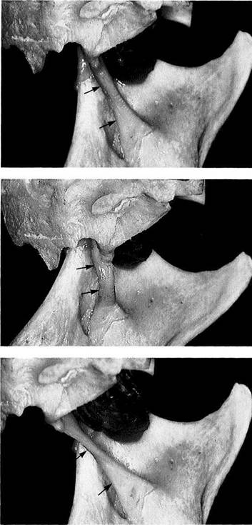
The stylomandibular ligament is a part of the deep fascia of the neck and runs from the styloid process to the posterior edge of the angle of the mandible. While part of the ligament inserts onto the mandible, its largest part radiates into the fascia of the medial pterygoid muscle (Sicher and DuBrul 1975). Although the stylomandibular ligament is relaxed during jaw opening, it restricts protrusive and mediotrusive movements (Burch 1970, Hesse and Hansson 1988). Even so, it should prevent excessive upward rotation of the mandible (Burch 1970), which sometimes causes problems in patients with a significantly reduced vertical dimension.
|
|
Stylomandibular ligament
Situation with jaws closed
Lateral view of a macroscopic anatomical preparation approximating the habitual condylar position. The ligament runs from the styloid process (1) to the posterior border of the angle of the jaw. In this mandibular position the ligament (arrows) is essentially free of tension.
Chronic nonphysiological loading (Fig. 68) can lead to insertion tendi-nosis (Ernest syndrome; Brown
Situation during rotational
jaw opening
Preparation shown in Figure 66 after the initial opening rotation. Rotational movement of the condyle against the articular protuberance causes a relaxation of the ligament (arrows). With further rotational opening, the angle of the jaw would swing farther posteriorly and allow even more slack in the ligament.
Situation during translation
Same preparation following anterior translation (= protrusion). Anterior translational movements in the temporomandibular joint always increase tension in the ligament (arrows). This helps to protect more sensitive structures (such as the superior stratum) from overextension during protrusion. Excessive closing rotation of edentulous jaws can likewise produce tension in the ligament.
Anatomy of the Masticatory System
![]() The sphenomandibular ligament has its sole origin on the sphenoidal spine in only about one-third of patients (Burch
1966). In the majority of individuals it also inserts into the medial wall of
the joint capsule, in the petrotympanic fissure
or on the anterior ligament of malleus (Cameron 1915, Loughner et al. 1989, Schmolke 1994). By means of its insertion on the lingula of the mandible, the
sphenomandibular ligament limits
protrusive and mediotrusive movement (Langton
and Eggleton 1992) as well as passive jaw opening (Hesse and Hansson 1988,
Osborn 1989). The importance of the sphenomandibular ligament to the
physiology of movement is negligible in
comparison with the previously
The sphenomandibular ligament has its sole origin on the sphenoidal spine in only about one-third of patients (Burch
1966). In the majority of individuals it also inserts into the medial wall of
the joint capsule, in the petrotympanic fissure
or on the anterior ligament of malleus (Cameron 1915, Loughner et al. 1989, Schmolke 1994). By means of its insertion on the lingula of the mandible, the
sphenomandibular ligament limits
protrusive and mediotrusive movement (Langton
and Eggleton 1992) as well as passive jaw opening (Hesse and Hansson 1988,
Osborn 1989). The importance of the sphenomandibular ligament to the
physiology of movement is negligible in
comparison with the previously
described ligaments (Williams et al. 1989), as is confirmed by the lack of related clinical symptoms.
The discomalleolar ligament (= Pinto's ligament) was described by Pinto (1962) as a connection between the malleus and the medial wall of the joint capsule. However, a separate ligament can be demonstrated here in only 29% of temporomandibular joints (Loughner et al. 1989).
Tanaka's ligament represents a cord-like reinforcement of the medial capsule wall, similar to the lateral ligament (Tanaka 1986,1988).
|
|
Sphenomandibular ligament
Situation at the habitual
condylar position
Macroscopic anatomical preparation displaying the left sphenomandibular ligament from the medial. The ligament runs from the spine on the underside of the sphenoid bone (spina ossis sphenoidalis) to the lingula of the mandible (Rodriguez-Vazquez et al. 1992). With the jaws in this position the ligament (arrows) is essentially relaxed. Besides the ligament, a section of the lateral pterygoid muscle can be seen.
Situation during opening
rotation
Same preparation after opening rotation. As long as the condlye is rotating against the articular protuberance without leaving the fossa, the ligament (arrows) becomes progressively more relaxed. Only after translation begins does the ligament re-acquire the same degree of tension it had when the jaws were closed.
Situation during translation
Macroscopic anatomical preparation after anterior translation (= protrusion). In synergy with the stylomandibular ligament, tension in this ligament is increased as translation progresses. The stylomandibular and sphenomandibular ligaments together restrain protrusive and mediotrusive movements. If a pain-producing lesion in one of these ligaments is suspected, passive movements must be used to
fp^ffhp lirmmpnt*;
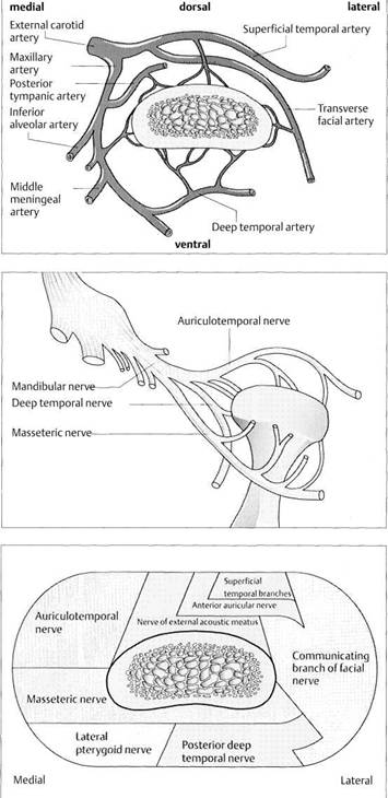
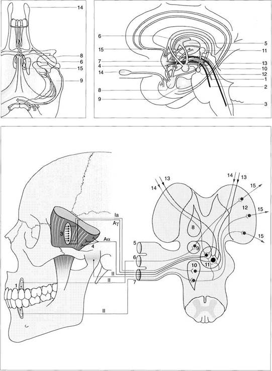
Arterial Supply and Sensory Innervation of the Temporomandibular Joint
Arterial Supply and Sensory Innervation of the Temporomandibular Joint
The arterial blood supply of the temporomandibular joint is provided primarily by the maxillary artery and the superficial temporal artery (Boyer et al. 1964). Both of these arteries are also the principle supply for the muscles of mastication. Apart from the network of arteries surrounding it, the condyle is also supplied from the inferior alveolar artery through the bone marrow (Okeson 1998). The venous drainage is through the superficial temporal vein, the maxillary plexus, and the pterygoid plexus. The sensory innervation of the joint capsule and its receptors has already been addressed briefly on page 27. The tem-
poromandibular joint is innervated predominantly by the auriculotemporal, masseter, and temporal nerves (Klineberg et al. 1970, Harris and Griffin 1975). Proprioception occurs through four types of receptors (Thilander 1961, Clark and Wyke 1974, Zimny 1988): Ruffini mechanoreceptors (type I), pacinian corpuscles (type II), Golgi tendon organs (type III), and free nerve endings (type IV). These receptors are located in the joint capsule, the lateral ligament, and in the bilami-nar zone and its genu vasculosum. The anteromedial portion of the capsule contains relatively few pain receptors, of type IV (Thilander 1961).
|
|
Arterial supply
Diagram of the arterial blood supply of a left temporomandibular joint (modified after Voy and Fuchs 1980). The condyle is supplied with blood from all four sides. In addition, there are anastomoses with the inferior alveolar artery within the marrow spaces. Because of the abundant blood supply, avascular necrosis is rarely found in the condyle (Hatcher etal. 1997). Compression of the anterior vessels by anterior disk displacement (Schell-has et al. 1992) will not interfere with the condyle's blood supply.
Sensory innervation of a left
temporomandibular joint
The afferent nerve fibers arise from the mandibular branch of the trigeminal nerve and exhibit four types of nerve endings. In rats, free nerve endings (type IV), which are potential pain receptors, have been found in the capsule, lateral ligament, bilaminar zone, and in the pars anterior and pars posterior of the disk (Ichikawa et al. 1990, Kido et al. 1991). This has not been verified for human disk structures, however.
Innervation in the
capsule
region
Schematic diagram of the different areas of innervation (modified from Ishibashi 1974, Schwarzer 1993). Activation of the type-IV receptors in the capsule increases the activity of sympathetic efferent fibers (Roberts and Elardo 1985). Because of the sympathetic innervation of the intrafusal muscle fibers (Grassi et al. 1993), a secondary rise in muscle tone is brought about by activation of the afferent fibers of the muscle spindles and the efferent α-motoneurons (Schwarzer
Anatomy of the Masticatory System
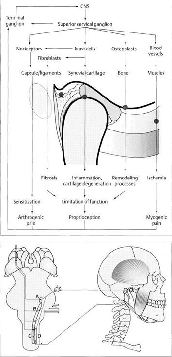
Sympathetic Innervation of the Temporomandibular Joint
The sympathetic innervation of the temporomandibular joint comes from the superior cervical ganglion (Biaggi 1982, Widenfalk and Wiberg 1990). Neurons with the neuropeptides CGRP (calcitonin gene-related peptide) and SP (substance P), that are associated with the sensory nervous system, are found chiefly in the anterior part of the joint capsule (Kido et al. 1993). Sympathetic fibers containing neuropeptide A (NA), Y (NPY), or VIP (vasoactive intestinal peptide) are more numerous in the posterior part of the joint. The ratio of sympathetic to sensory nerve fibers is approximately 3:1 in the temporomandibular joint
(Schwarzer 1993). Sympathetic neurons serve primarily for monitoring the vasomotor status. This monitoring allows optimal adjustment of the blood volume in the genu vascu-losum during excursive and incursive condylar movements. There is evidence that, in addition to the vasomotor effect, the sympathetic nervous system also plays a role in pain perception (Roberts 1986, Jahnig 1990, McLachlan et al. 1993). Both NA and SP effect the release of prostaglandins, which heighten the sensitivity of pain receptors (Levine et al. 1986, Lotz et al. 1987).
|
|
Effects
of the sympathetic
nervous system on the temporo
mandibular joint
Certain neuropeptides can increase the sensitivity of nociceptors and thereby directly influence pain perception. Special importance has been attributed to the neuropeptides SP and CGRP in the production of synovial cells (Shimizu et al. 1996). Bone remodeling processes are likewise guided by neuropeptides. A nonphysiological increase of pressure within the genu vascu-losum caused by a sympathetic or hormonal malfunction during in-cursive condylar movement results in an anteriorly directed force on the articular disk (Ward et al. 1990, Graber 1991) and this can contribute to an anterior disk displacement. (Revised drawing from Schwarzer 1983.)
Afferent paths of the
trigeminal nerve and conections
of neurons in the brain stem
Schematic representation of the interrelationships between afferent fibers of the trigeminal nerve and the so-called nociceptive specific (NS) neurons and/or the wide dynamic range (WDR) neurons (Dub-ner and Bennett 1983, Sessle 1987a, b) in the region of the cervical spinal column. The specific connections (A-D) in the sensory trigeminal nucleus in the brain stem can result in identical perceptions in the cortex regardless of where the pain was first perceived.
Muscles of Mastication
![]() Muscles of Mastication
Muscles of Mastication
Anatomically the muscles of mastication can be divided into simple and complex muscles (Hannam 1994,1997). The lateral pterygoid and the digastric muscles are counted among the simple muscles. These muscles work through a favorable lever arm relative to the joint and so do not have to produce a great deal of force to bring about functional mandibular movements. The parallel muscle fibers in these muscles have their sarcomeres arranged in series, and these are responsible for the adequate muscle contraction. During contraction, the diameter of each muscle increases and is at its greatest near the midpoint of the muscle.
In contrast, the complex muscles include the temporal, masseter, and medial pterygoid muscles with their many aponeuroses and varying sizes. During function the aponeuroses can shift and become deformed (Langenbach et al. 1994). The muscle fibers in this group run obliquely and increase their angle to one another during contractions. A complex muscle can produce a force of approximately 30-40 N per cm2 of cross-section (Korioth et al. 1992, Weijs and van Spronsen 1992). The orientation of the muscle fibers and their facultative activation during various mandibular movements is one of the reasons that muscle symptoms can be reproducibly provoked by loading in one certain direction but not in others. Although there are recur-
rent principles in muscle architecture (Hannam and McMillan 1994), variations in the areas of muscle attachment and differences in intramuscular structure have an effect on craniofacial development (Eschler 1969, Lam et al. 1991, Tonndorf 1993, Holtgrave and Muller 1993, Goto et al. 1995).
The motor units in the muscles of mastication are small and seldom extend beyond the septal boundaries (Tonndorf and Hannam 1997). The "red" muscle fibers (with higher myoglobin content) contract slowly. They maintain postural positions and are slow to become fatigued. The "white" fibers (with lower myoglobin content) have fewer mitochondria and can contract more rapidly, but they fatigue sooner because of their predominantly anaerobic metabolism. The muscles of mastication are composed of varying mixtures of fibers of types I, IIA, IIB, IIC, and IM (Mao et al. 1992, Stal 1994).
Next, the four chewing muscles proper (temporal, masseter, medial pterygoid, and lateral pterygoid) and the suprahyoid and perioral musculature will be described in preparation for the clinical examination that will be addressed later.
![]()
![]()
![]()
|
Function Movement Stabilization Progress! ve adaptation HypD-/hypertonicity»hypo-/hypertrophy Muscle shortening |
|
Regressive adaptation Inflammation Rupture Ossification |
Muscles of mastication
Drawing of the muscles of mastication. In the narrowest sense these include only the temporal, masseter, medial pterygoid, and lateral pterygoid muscles. The suprahy-oidal musculature is also shown here because it is of interest in the diagnosis and treatment of functional disturbances. The sternocleidomastoid muscle is not included here because it belongs to the musculature of the neck.
Function
and structural
adaptation of the muscles of
mastication
Antagonistic muscle activity serves not only to execute mandibular movements, but also helps stabilize the joints. Functional demands can bring about changes in tonus, response to stimuli, and muscle length. Adaptation depends upon the combination of fibers present. Chronic overloading may lead to inflammation, ruptures, or ossification.
Anatomy of the Masticatory System
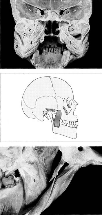
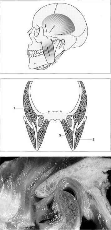
![]() Temporal Muscle
Temporal Muscle
The temporal muscle is a compartmentalized muscle that arises from the superior and inferior temporal lines of the temporal bone. It inserts on the coronoid process and on the anterior edge of the ascending ramus of the mandible. Three functional parts can be distinguished (Zwijnenburg et al. 1996). The anterior part has muscle fibers that pull upward and serve as elevators (Moller 1966). The middle part effects closure of the jaws and, with a posterior vector, retrusion (Blanksma and van Eijden 1990). According to DuBrul (1980) the posterior part is involved primarily with jaw closure and only to a minimal extent with retrusion. Neverthe-
less, experimental studies have revealed a distinct retrusion when the posterior part is activated (Zwijnenburg et al. 1996). During normal opening and closing movements, as well as during tooth clenching, the activity in all three parts is at a nearly equal high level. During chewing, however, there are great differences between the anterior and posterior parts. The activity is greater on the working side than on balancing side (Blanksma and van Eijden 1995). During lateral jaw movement, EMG activity is markedly lower where there is canine guidance than where there is group function (Manns et al. 1987).
|
|
Macroscopic anatomical
preparation
The pars anterior and pars media of the temporal muscle consist of approximately 47% fatigue-resistant type-l muscle fibers with a low threshold of stimulation (Eriksson and Thornell 1983). The content of thinner type-IIB fibers is about 45%, leading to a higher concentration of fibers in the muscle (Stalberg et al. 1986). Type-IIA muscle fibers are not present at all and those of type IIC and/or IM account for only about4%(Ringqvist1974).
From the collection ofB. Tillmann
Schematic drawing of the
right temporal muscle
The muscle comprises a pars anterior (1), pars media (2), and pars posterior (3).
Although the sarcomere lengths are the same in the various parts, there are significant differences in the lengths of the muscle-fiber bundles (21.7-28.9 mm) which indicates different functional demands (van Eijden et al. 1996).
Insertion of the temporal
muscle on the disk-capsule
complex
Left: Medial view. Some of the horizontal fibers (arrows) insert onto the middle and lateral third of the disk (Merida Velasco et al. 1993, Bade etal. 1994).
Right: Insertion of the temporal muscle viewed from above. Easily identified is the tendon (*) of the pars posterior, which inserts on the lateral portion of the disk-condyle complex.
Musculus temporalis
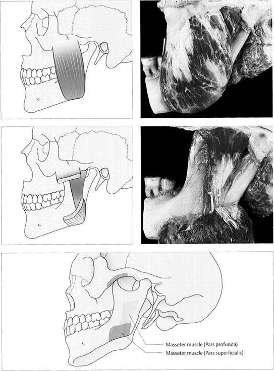
Masseter Muscle
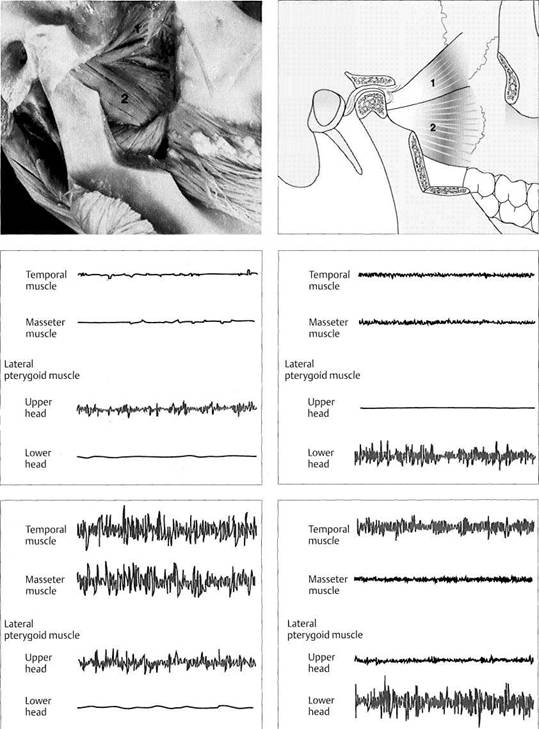
The masseter muscle consists of a superficial and a deep part. The origin of the superficial part is on the zygomatic arch and its insertion is on the lateral masseteric tuberosity at the angle of the mandible. The deep part also arises on the zygomatic arch but inserts on the lateral surface of the ascending ramus. Portions of the deep part also insert on the joint capsule and the disk (Frommer and Monroe 1966, Meyenberg et al. 1986, Dauber 1987). In this way the masseter can influence the capsule receptors by changing the capsule tension. The lowest EMG activity and the greatest chewing force in this muscle can be demonstrated at a jaw
opening of 15-20 mm (Manns et al. 1979, Lindauer et al. 1993, Morimoto et al. 1996). Seventy-four percent of the masseter's muscle spindles are to be found in the deep part (Eriksson and Thornell 1987). These muscle spindles have large diameters and a four-fold higher concentration of intrafusal fibers. From this it can be deduced that there are special functions for different areas of the muscle. The masseter muscle shows no significant difference in EMG activity between subjects with canine guidance and those with group function (Borromeo et al. 1995).
|
|
Pars
superficialis of the
masseter muscle
Left: Schematic drawing of the pars superficialis. The masseter has a higher concentration of capillaries relative to the diameter of the fibers than all the other skeletal muscles (Staletal. 1996).
Right: Macroscopic anatomical preparation of the masseter muscle. The resultant force of the pars superficialis is in an anterosuperior direction. The posterior part of the pars superficialis is composed of up to 45% type-IIB fibers (Eriksson and Thornell 1983), which have a high threshold and are fatigue-resistant.
Pars profunda
Left: Schematic drawing.
Right: The pars profunda revealed in an anatomical dissection. The muscle has a relatively broad insertion on the zygomatic arch from which it pulls on the lateral surface of the ascending ramus of the mandible. The posterior part of the pars profunda also inserts into the lateral third of the disk-capsule complex (Merida Velasco et al. 1993, Bade et al. 1994). Only approximately 25% of the muscle fibers of the pars profunda are of type IIB.
Origin
and insertion of the
masseter muscle
Schematic drawing showing the areas of origin and insertion of the masseter muscle. The origin of the pars superficialis is on the inferior surface of the zygomatic arch anterior to that of the pars profunda. The insertion of the pars superficialis lies on the lateral surface of the angle of the mandible. At the inferior border of the mandible it is continuous with the medial pterygoid muscle on the inner surface of the mandible. The pars profunda inserts above the masseteric tuberosity on the ascending ramus of the mandible.
Anatomy of the Masticatory System
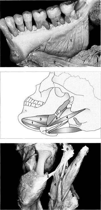
![]() Medial Pterygoid Muscle
Medial Pterygoid Muscle
The medial pterygoid muscle, together with the temporal and masseter muscles, are the jaw-closing muscles. This muscle has its origin in the pterygoid fossa of the pterygoid process of the sphenoid bone. From there it extends inferi-orly, posteriorly, and laterally to the inner side of the angle of the mandible, where it connects with the masseter to form a muscle sling. The course of the medial pterygoid muscle closely parallels that of the pars superficialis of the masseter. The medial pterygoid muscle functions primarily during jaw closure, but also takes part in protrusive movements. Unilateral contraction results in mediotrusion.
Because of its oblique course in the frontal plane, this muscle also influences the transverse position of the condyle. Unlike the temporal and masseter muscles, the medial pterygoid cannot be adequately palpated except for its insertion. Its activity in protrusive position increases with the number and size of tooth contacts (MacDonald and Han-nam 1984, Wood 1986). Tooth gnashing in a posterior direction is accompanied by a greater increase in EMG activity than in anteriorly directed gnashing (Vitti and Basmajian 1977).
|
|
Posterior
view of the medial
pterygoid muscles in the frontal
plane
This preparation clearly reveals the relationship of the medial pterygoid muscles (1) to the lateral pterygoids (2) and the masseters (3) and the muscular slings they form with the latter. The strength of a muscle depends on its cross-sectional area (Sasaki et al. 1989). In patients with dentitions damaged by periodontal disease, the chewing force was found to be reduced by 25% in the masseters and 10% in the medial pterygoids (Gilbert and Newton 1997).
Schematic drawing of the
medial pterygoid muscle
Like the pars superficialis of the masseter, the composition of the anterior part of the medial pterygoid is 64% type-l muscle fibers (low threshold, fatigue-resistant) and 27.5% type-IIB (high threshold, fatigue-resistant) (Eriksson and Thornell 1983). In the posterior part, on the other hand, 44% of the fibers are of type I and 45% are of type IIB.
Medial view of an
anatomical dissection of a right
medial pterygoid muscle (1)
The muscle runs inferiorly and posteriorly from the fossa of the pterygoid process to the inner side of the angle of the mandible. As in the masseter, the muscle fibers are extensively interlaced with no uniform direction of alignment. They are comparatively short, but have a relatively large cross-section (van Eiidenetal. 1995).
Medial Pterygoid Muscle
![]() Suprahyoid Musculature
Suprahyoid Musculature
The suprahyoid muscles, together with the lateral pterygoids, make up the jaw-opening muscles. The suprahyoid musculature includes the digastric, mylohyoid, geniohyoid, and stylohyoid muscles. The digastric muscle has two bellies. The posterior belly has its origin medial to the mastoid process and extends anteriorly, inferiorly, and medially to the hyoid bone; here it attaches to the bone and connects with the anterior belly by means of an intermediate tendon. The anterior belly inserts in the digastric fossa on the inner side of the mandible. When the hyoid bone is braced by the infrahyoid musculature, contraction of the digastric muscle
tends to open and retract the jaw. The mylohyoid muscle extends from the step-like myohyoid line on the inner aspect of the body of the mandible to the body of the hyoid bone. The geniohyoid muscle arises at the inner side of the mandibular symphysis, and from there its parallel fibers run directly to the body of the hyoid bone. The pair of geniohyoid muscles can function together as jaw openers, but they can also lift the floor of the mouth and pull the hyoid bone forward. The stylohyoid muscle runs from the styloid process of the temporal bone to the greater horn of the hyoid and serves to stabilize the hyoid.
|
|
88 Mylohyoid muscle
Anatomical preparation of a right mylohyoid muscle (1). The anterior and middle fibers of the mylohyoid muscle insert in the center at the mylohyoid raphe. The fibers of the posterior portion insert on the hyoid bone under the geniohyoid muscle (2). These muscles are active primarily during jaw opening, swallowing, and protraction of the tongue, but they also show some activity during lateral jaw movements (Vitti and Basmajian 1977).
89 Suprahyoid musculature from a left posteroinferior view
The digastric muscle with its anterior belly (1) and posterior belly (2) can be seen from this perspective. The mylohyoid (3) and stylomandibular (4) muscles are also visible. When the mandible is stabilized by the closing muscles (elevators), contraction of the suprahyoid musculature lifts the hyoid bone. This is an important part of the act of swallowing.
90 Suprahyoid musculature
Posterior view of an anatomical preparation of the left half of the jaw showing the posterior belly of the digastric (1), stylohyoid (2), styloglossus (3), and stylopharyngeus (4) muscles as well as the stylomandibular ligament (5). Except for the digastric muscle, all these structures have their origin on the stylohyoid process (6). Muscle fibers of type I, type 11 A, and type MB each make up one-third of the digastric muscle (Eriksson etal. 1982).
From the collection of B. Tillmann (Figures 85, 87, 88, 90)
Anatomy of the Masticatory System
Lateral Pterygoid Muscle
The lateral pterygoid muscle is made up of two functionally different parts (McNamara 1973): an upper (superior) head and a lower (inferior) head. In 12% of the specimens studied, Abe et al. (1993) could also identify a third, intermediate muscle belly.
The upper head arises on the greater wing of the sphenoid bone. It always has an insertion in the upper portion of the pterygoid fovea, but it may also insert to varying degrees into the disk-capsule complex (Chissin 1906, Troiano 1967, Porter 1970, Mahan et al. 1983, White 1985, Meyenburg et al.
1986, Dauber 1987, Carpentier et al. 1988,
The lower head arises on the lateral face of the lateral lamina of the pterygoid process and inserts in the pterygoid fovea. It is possible that fibers from the upper and lower head are intermingled, but in any event, fibers of the lower head can radiate into the disk-capsule complex (Dauber 1987).
|
|
Lateral pterygoid muscle
Left: Lateral view of an anatomical preparation. This muscle is characterized by relatively long fiber bundles with small cross-sections (van Eijden et al. 1995) in its upper head (1) and lower head (2). The upper head always inserts on the condyle, and in 60% of specimens it also inserts on the disk-capsule complex.
From the collection ofB. Till'mann Right: Schematic drawing.
![]() Upper
head
Upper
head
Lower head
![]() EMG activity of the muscles
EMG activity of the muscles
of mastication at rest and during
jaw opening
After Juniper 1984
Left: In the relaxed mandibular position only minimal EMG activity can be detected from the upper head of the lateral pterygoid muscle.
Right: During jaw opening the upper head is active and serves to protrude the condyles. Activity of the suprahyoid musculature was not measured in this study.
EMG
activity during
grinding of the teeth
After Juniper 1984.
Left: During tooth grinding in maximum occlusion, the two large jaw-closing muscles and the upper head of the lateral pterygoid show a high level of EMG activity.
Right: During grinding of the anterior teeth, the lower head holds the mandible forward and the activity of the elevators decreases because of the reflex inhibition from the periodontium (Widmalm etal. 1987).
Lateral Pterygoid Muscle
Fibers of the lateral pterygoid muscle were found to insert into the periphery of the disk in 22% of the joints studied. In these cases 88% of the muscle fibers were from the upper head and 12% from the lower head (Abe et al. 1993). The smaller the area of attachment of the lateral pterygoid muscle to the condyle, the greater the tendency for disk displacement (Dreger 1994). The upper and lower heads have an antagonistic action. Numerous EMG studies (Molin 1973; Gibbs et al. 1984; Juniper 1983,1984; Widmalm et al. 1987; Yoshida 1995) have revealed that the lower head is always active during excursive mandibular movements (jaw opening, protrusion, mediotrusion), whereas the upper head is active during incursive movements (jaw closing, retrusion,
laterotrusion), serving to hold the disk-condyle complex continuously against the slope of the eminence and to restrain it during incursive movements (Wood et al. 1986). The high proportion (80%) of type-I muscle fibers (low stimulation threshold, fatigue-resistant) is also indicative of a continuous holding action with a low level of force (Eriksson et al. 1981, Mao et al. 1992). Because the upper head becomes longer during its holding action, this can be referred to as eccentric muscle activity (Wilkinson 1988).
|
|
MRI of a left lateral
pterygoid muscle in the sagittal
plane
According to Abe et al. (1993), in 12% of individuals the lateral pterygoid muscle divides into three parts as seen here. According to Ogut-cen-Toller and Juniper (1994) the muscle becomes segmented into three parts in week 12 of embryonic development to form an upper (1), middle (2), and lower (3) head.
Attachment areas of the
lateral pterygoid
muscle on the
condyle
From Moritz and Ewers 1987.
In agreement with numerous other studies, the upper head always inserts on the condyle (1), and in 60% of joints it also inserts into the an-teromedial portion of the disk-capsule complex (gray). The lower head always inserts into the pterygoid fovea (2). An overly strong upper head cannot by itself displace the disk anteriorly.
Lateral pterygoid muscles in
the horizontal plane
Left: Diagram showing the angulation of the lateral pterygoid muscle to the midsagittal plane (modified from Christiansen et al. 1988). With the jaws closed, the angle averages 39Q and ranges from 22s to 529. In protrusion the origin and insertion come closer together and the angle becomes greater (Okeson 1998).
Right: MRI showing the lateral pterygoid muscles (1) in the horizontal plane.
Anatomy of the Masticatory System
![]() Gross Anatomical and Histological Studies
of the Masticatory Muscle Insertions
Gross Anatomical and Histological Studies
of the Masticatory Muscle Insertions
The tendons of the muscles of mastication attach to bone by means of special insertion structures (Goss 1940, Long 1947, Symons 1954, Cooper and Misol 1970, Chong and Evans 1982). A fundamental distinction must be made between periosteal-diaphyseal insertions (Biermann 1957) and chondral-apophyseal insertions (Knese and Biermann 1958). A periosteal-diaphyseal insertion may be flat or circumscribed. The structural makeup of an insertion should be such that it can equalize the different moduli of elasticity of the tissues. Pathological changes in these areas bring about the clinical picture of insertion tenopathy (Becker and
Krahl 1978, Tillmann and Thomas 1982). In the extremities there is a direct relationship between the mode of osteogenesis in the region and the histological makeup of the tendon attachment structures (Evans et al. 1991). This type of relationship has not been demonstrated in the masticatory structures. Insertion tenopathies occur primarily in chondral-apophyseal insertion structures. It has been suggested that the cause lies in a disturbed collagen synthesis combined with a low content of glycosaminoglycans in the fibrocartilaginous tissue (Hems 1995).
|
|
|
-^SS*j |
Muscle
origins and
insertions on the side of the skull
Left: Schematic drawing showing the origins and insertions of the masseter and temporal muscles. Both muscles have periosteal and cartilaginous areas of insertion.
Right: Histological preparation of the insertion of the tendon of the temporal muscle. The tendon inserts on the coronoid process by means of cartilaginous structural elements (arrows).
Muscle
origins and
insertions on the medial surface
of the mandible
Left: Schematic drawing of the origins and insertions of lateral and medial pterygoid muscles and the mylohyoid muscle. Except for the medial pterygoid, the insertions of all these muscles are entirely periosteal.
Right: Histological preparation of the insertion of the lateral pterygoid muscle in the pterygoid fovea. The insertion is entirely periosteal (arrows).
Muscle origins and
insertions on the posterior
mental protuberance
Schematic presentation of the origins and insertions of the geniohyoid, genioglossus, and digastric muscles. In this region there are both cartilaginous and periosteal areas of insertion.
Right: Histological preparation of the insertion of the digastric muscle. The insertion of this muscle is primarily periosteal, but it does have some regions of cartilaginous insertion.
Histology by B. Tillmann (Fiqs. 97-99, right)
Force Vectors of the Muscles of Mastication
![]() Force Vectors of the Muscles of Mastication
Force Vectors of the Muscles of Mastication
No chewing muscle contracts in isolation. Every muscle contraction contributes to a resultant force vector that acts upon the mandible, the teeth, and the temporomandibular joint (Hannam 1994). From this it follows that only limited parts of a muscle are active during certain functions. In addition, the mandible is an elastic structure and this property can cause the loading vectors to be completely different depending on the muscle activation and the function at the moment (Korioth et al. 1992). The maximum force that a muscle can develop can be calculated from the dimension-less product of its cross-sectional area with a value of
approximately 35 (Weijs and Hillen 1984, Korioth et al. 1992). As a general rule, decompensated muscles exhibit a loading vector in the direction of their contraction. Causes for this can be a muscle hypertonus related to central stimuli or the occlusion, or an inflammation of the bilaminar zone. The pattern for complex activation of the muscles of mastication depends much less upon the available muscle force than upon the direction of the resultant force. A complex pattern of activation, therefore, allows for unconscious improvement of the muscle forces for optimum chewing efficiency (Mao and Osborn 1994).
|
|
Muscle
vectors in the
sagittal plane
Directions of individual force vectors (arrows). The resultant force on the temporomandibular joint is directed anterosuperiorly. The mas-seter muscle pulls at an angle of approximately 70° to the occlusal plane and the medial pterygoid muscle at an angle of approximately 80° (Weijs and van Spronsen 1992). Individual variations in these orientations amount to only about 10° (Hagiwara et al. 1994). The physiological cross-sectional area of the temporal muscle ranges from 1.8-2.9 cm2 (van Eijden et al.
Muscle
vectors in the
frontal plane
Diagram of the force vectors in the frontal plane (redrawn from Hylan-der 1992). Through the specific arrangement and activation of the temporal (1), masseter (2), and medial pterygoid (3) muscles, the condyles are directed transversely against the articular protuberance. The direction of the force vectors in the frontal plane is not correlated with the transverse dimensions of the facial skeleton (van Spronsen
Example
of a loading
vector of the lateral pterygoid
muscle
Clinical evidence of a muscle-specific pain in the lateral pterygoid muscle (1) would point to a chronic overloading in the direction of contraction (arrow). The cause could be a centrally stimulated muscle hypertonus or inflammation of the bilaminar zone. In the latter case, the muscle reflexly seeks to relieve pressure on the painful bilaminar zone. Painful decompensation of the lateral pterygoid muscle without lesions in the bilaminar zone has not been observed clinically for a longtime.
Anatomy of the Masticatory System
![]() Tongue Musculature
Tongue Musculature
The extrinsic tongue musculature includes the styloglossus, genioglossus, and hyoglossus muscles. They connect the body of the tongue to the nearby bone structures to give it its ample mobility within the oral cavity (Thiele et al. 1992).
The intrinsic tongue musculature is made up of the longitudinal, transverse, and vertical muscles of the tongue. They help to change the shape of the tongue.
While the balanced antagonistic activity of the muscles of mastication contributes to positioning the condyles on the
articular protuberance, the activity of the tongue musculature on the lingual tooth surfaces and the muscles of the cheeks and lips on the facial surfaces influence the position of the teeth in the dental arch. Even though the tongue has little effect on the joints, afferent information from the joint capsules does indirectly alter the position of the tongue (Lowe 1978).
The occurence of dysphagia and limited motor functions of the tongue have not been proven to be age-dependent. (Caruso and Max 1997, Koshino et al. 1997).
![]()
![]()
![]()
![]()
![]() Extrinsic tongue
Extrinsic tongue
musculature
The styloglossus muscles pull the tongue back and up. If one muscle acts unilaterally, the tongue becomes concave. With hypofunction the third phase of the swallowing reflex will be absent (Thiele et al. 1992). The genioglossus can move the dorsum of the tongue downward, pull the border of the tongue forward, and help extend the tongue. Hyperfunction of the genioglossus results in tongue thrusting. The hyoglossus lowers the body of the tongue and pulls the tongue back. Its hyperfunction results in a sunken tongue (Thiele et al. 1992).
Intrinsic
tongue
musculature
Diagram of the internal tongue muscles and their positions in relation to the muscles of the floor of the mouth. The longitudinal muscles of the tongue shorten the tongue. The transverse muscles bring the borders of the tongue closer together. Hypotonus of these fibers leads to a widened tongue. The vertical muscles make the tongue flatter and wider, and their hyperfunction frequently accompanies lateral tongue thrusting, which can cause widening of the dental arch.
Function
of the tongue
muscles
A list of the specific functions of the extrinsic and intrinsic muscles of the tongue and their effects on the body of the tongue (from Thiele et al. 1992). Without the stable buttress formed by the musculature of the floor of the mouth, many dysfunctions of the tongue would not be possible. The floor of the mouth is stabilized differently in different individuals. The act of swallowing does not necessarily involve all the muscles of the floor of the mouth in lifting the hyoid bone (Spiro et al.
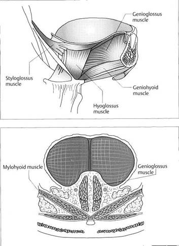
|
Acting muscle |
Effect on the body of the tongue |
|
Styloglossus muscle |
High position of the dorsum with tendency for widening; interocclusal position; lateral and palatal thrusting (unilateral and bilateral) |
|
Genioglossus muscle |
Forward thrust of the medial portion; low position of the tip; frontal thrust |
|
Hyoglossus muscle |
Low position of the body of the tongue; low, lateral tongue thrust |
|
longitudinalis muscle |
Short, thick, wide ball shape |
|
Transverse muscle of tongue |
Anterior thrust and interocclusal position |
|
Vertical muscle of tongue |
Lateral thrust and interocclusal position |
Muscles of Expression
![]() Muscles of Expression
Muscles of Expression
The orofacial musculature is made up of the following muscles: nasalis, depressor septi, levator labii superioris alaeque nasi, orbicularis oris, zygomaticus major, zygomaticus minor, risorius, levator labii superioris, levator anguli oris, depressor anguli oris, depressor labii inferioris, mentalis and platysma myoides. Disturbances in the tonus of these muscles can affect the patient's facial appearance and the position of the teeth within the dental arch (Hoyer and Lim-brock 1990, Padovan 1995). In combination with the activation of one or more of the muscles of mastication, they can also influence the condylar position and thereby the loading
of the different structures of the joint (Pahkala et al. 1995). The previous statement about the muscles of mastication also holds true for the muscles of expression: no muscle contracts in isolation (Brown 1996). Systematic observation of individual muscles of expression is not appropriate for grasping the complex interactions that occur during function (Smith 1992, Thiele et al. 1992).
A noninvasive, objective study of muscle function is possible, however, with the aid of electromagnetic articulogra-phy (Ackermann et al. 1993, Engelke et al. 1990).
|
|
Anatomical preparation of
the left buccinator muscle
This muscle (1) determines the transverse position of the posterior teeth in the dental arch by countering the pressure of the tongue. During chewing, the buccinators and the tongue work together to keep the food between the occlusal surfaces. In playing a wind instrument, each buccinator (Latin for trumpeter) supports the stream of air laterally. Their hyperfunction causes a narrowing of the upper and lower jaws with the formation of an exaggerated linea alba inside the cheeks.
From the collection ofB. Till ma nn
Orbicularis oris muscle
The orbicularis oris is the muscular foundation of the lips (Thiele et al. 1992). It is made up of a pars marginalis, pars labialis, and a system of straight radiating muscles. Strictly speaking, it consists of two arches that are connected by fascia at the corners of the mouth. Hyper-tonus results in a small, sharp mouth. The orbicularis oris in combination with interwoven surrounding muscles can position the lower lip between the incisors and cause an anterior horizontal overlap ("overjet").
Mentalis muscle
Right: Frontal view of a patient with a hyperactive mentalis muscle. The muscle is the cause of the forceful lip closure. With hyperactivity the lower lip presses in on the lower incisors and the upper lip can be forced upward. This can result in a reduction of lip pressure against the upper incisors.
Left: Same patient in profile view. Because of the hypertonic mental-is, the apex of the chin (pogonion) lies well in front of the alveolar process.
Anatomy of the Masticatory System
![]() Temporomandibular Joint and the
Musculoskeletal System
Temporomandibular Joint and the
Musculoskeletal System
The relationships between functional disturbances of the temporomandibular joint and the rest of the musculoskeletal system are well known from clinical investigations (Smith 1993, de Wijer et al. 1996b,c, Dvorak and Walchli 1997). The structural and neurophysiological connections, however, are subjects of debate (Graf-Baumann and Lohse-Busch 1997). Head and neck pain, referred pain, pain in the area innervated by the trigeminal nerve, and disturbances in the functions of speech and swallowing are often associated with the upper cervical spine (Wolff 1996). Disturbances in the cervical vertebrae C2 and C3 can bring on dysphonia and
the sensation of a lump in the throat. Up to 25% of patients with temporomandibular-joint disturbances report tinnitus (Hiilse 1998), whereas 16% of all adults in our industrial society have a long-standing history of ear noises. Lotzmann and Steinberg (1993) demonstrated changes in body posture after removal of premature tooth contacts. On the other hand, artificial alterations of the occlusion in healthy test subjects had no effect on body posture (Ferrario et al. 1996a).
|
|
Influence
of head and body
posture on the mandible
Schematic drawing
showing the muscular suspension of the mandible
and its relation to the muscles of the neck. As an example, the sternocleidomastoid
muscle is always active
during bruxism; however, an activity level of 50% is necessary in the
masseter muscle before a 5% level is
reached in the sternocleidomastoid
muscle (
Influence
of afunctional
disturbance in the cervical
spinal
column upon centric position
Changes in condylar position in the
horizontal plane following isolated
orthopedic treatment of the upper
cervical spinal column.
Red: Initial findings
Blue: Control findings after
6 weeks with no treatment Lilac: Findings after 3 weeks of
treatment Green: Control findings after
3 months
In each group the difference from habitual occlusion (HO) is given in millimeters.
Influence
of the occlusion
upon body posture
Left: Recording of changes in the upright body posture (orthostasis). In this case voluntary clenching on an unstable occlusion resulted in a repeatable posterior shift in the body's center of gravity.
Right: Clinical arrangement with the patient on the measuring platform that detects even minor changes in the patient's center of gravity projected horizontally.
Peripheral and Central Control of Muscle Tonus
![]() Peripheral and Central Control of Muscle Tonus
Peripheral and Central Control of Muscle Tonus
Muscle spindles and Golgi tendon organs are the two types of receptors in muscles that are involved in controlling muscle activity. Peripheral monitoring occurs primarily through the monosynaptic proprioceptive reflex and the polysynaptic stretch reflex (Guyton and Hall 1996). Increases in length are reported from the muscle spindles over la fibers directly to the corresponding α-motoneuron and can then be corrected (phasic proprioceptive reflex). Golgi receptors read the muscle force and transmit this information over Ib fibers to an inhibitory intermediate neuron. This again inhibits the α-motoneurons (Johnson 1998). The resting
tonus is a continuous background level of contraction that is maintained primarily through a segmental spinal reflex (Graber 1989). Supraspinal centers can both inhibit and promote muscle tone. Here the limbic system plays an essential role. It is the complex circuitry of numerous pathways and nuclei that make it possible for individuals to control their emotions and behavior. Muscle tone is influenced predominantly by the γ-motoneurons (Johnson 1998).
|
|
Limbic system
After Nieuwenhuys et al. 1980.
![]() Mamillotegmental
tract
Mamillotegmental
tract
Fasciculus telencephalicus med.
Inferior longitudinal fasciculus
Hypothalamus
Nucleus medialisdorsalisthalami
Anterior thalamic nucleus
Septal area
Amygdaloid body
Hippocampus formation
Mamillary body
Habenula
Ncl. Interpendicularis
Dorsal tegmental nucleus
Olfactory bulb
Mamillothalamic tract
![]() Central and peripheral
Central and peripheral
neuromuscular control
Muscle activity can be influenced by both peripheral and central nerves. Emotional factors increase muscle tonus through γ-neurons.
![]() Periodontal
receptors
Periodontal
receptors
Joint capsule receptors
Muscle spindles (stretching)
Motor end plates
Trigeminal
nerve (semilunar
ganglion)
Trigeminal
nerve (semilunar
ganglion)
Trigeminal
nerve (semilunar
ganglion)
Mesencephalic tract nucleus
Superior sensory nucleus (n.V)
Inferior sensory nucleus (n.V)
Motor nucleus (n.V)
Thalamus
Descending paths from cortex
(α-motor = "voluntary motor")
Descending paths from limbic
system
(γ-motor = "psychoveg-
etative impulses"
Ascending paths to cortex
("consciousness",}
Anatomy of the Masticatory System
![]() Physiology of the Jaw-Opening Movement
Physiology of the Jaw-Opening Movement
Jaw opening is accomplished through the action of the suprahyoid muscles (rotation) and the lateral pterygoid muscles (translation). In centric condylar position the elastic fibers in the temporomandibular joint are in equilibrium. The initial phase of an opening movement is primarily a rotation that always progresses with a translational component (Merlini and Palla 1988, Maeda et al. 1992, Ferrario et al. 1996a). The opening rotation of the condyle always causes the disk to lie against a more posterior region of the condyle where it is more stable. During translation the disk is passively carried along in an anterior direction (Sicher
1964, Roth et al. 1984, Osborn 1985). During jaw opening, tension increases in the superior stratum and in the lower anterior wall of the joint capsule. While the superior stratum can restrict anterior movement of the disk (Dauber 1987), it cannot restrict jaw opening. This is limited by the joint capsule and the lateral ligament. During jaw opening the genu vasculosum expands to approximately four or five times its original volume (Rees 1954, Wilkinson et al. 1994), so that during excursions a negative pressure arises within it (Finlay 1964, Ward et al. 1990).
|
|
Jaw-opening movements
Initial phase
Illustration of the structural loading during the initial opening movement. The condyle makes a rotational movement with a small translational component, changing its position relative to the fossa only slightly. Because of the condylar rotation, the disk moves posteriorly relative to the condyle. The only part of the lateral pterygoid muscle that is active is its lower head (1). The elastic fibers are brought out of equilibrium only minimally.
Intermediate phase
In this phase the condyle executes a definite translation. The disk moves anteriorly relative to the fossa, but posteriorly in relation to the condyle. Tension becomes steadily increased in the superior stratum of the bilaminarzone and in the lower anterior wall of the joint capsule. The inferior stratum relaxes to the same extent. The venous plexus of the genu vasculosum expands, creating a negative pressure, and fills with blood.
Terminal phase
In this phase the condyle reaches the maximum extent of its rotation and translation. The translational component passively moves the disk farther forward, while the rotation makes it lie farther posteriorly on the condyle. The superior stratum and the lower anterior capsule wall are now stretched to their maximum. The retrocondylar space is filled by the blood flowing into the genu vasculosum. The inferior stratum is completely relaxed.
Right: Degree of opening in Pos-selt's envelope of motion.
Physiology of the Jaw-Closing Movement
![]() Physiology of the Jaw-Closing Movement
Physiology of the Jaw-Closing Movement
The jaw is closed by the temporal, masseter, and medial pterygoid muscles and the upper heads of the lateral pterygoid muscles. The lower head of the lateral pterygoid muscle is inactive during jaw closure. As described previously, the temporal and masseter muscles also insert on the anterior part of the joint capsule. This maintains the basic level of tension necessary for the receptors in the joint capsule to function. It is a fundamental fact that during closing movements the disk executes an anterior movement relative to the condyle. As the condyle is being drawn posteriorly by the muscles, other structures are guiding the disk posteri-
|
|
Jaw-closing movement
Initial phase
Schematic representation of the structural loading during the initial jaw-closing movement. The upper head (1) of the lateral pterygoid muscle retards distal movement of the condyle through eccentric muscle activity. The disk can only be passively guided posteriorly. In the initial phase this is brought about by the tension in the elastic superior stratum. A physiological positive pressure arises in the genu vasculo-sum (Finlay 1964, Ward et al. 1990).
Intermediate phase
In this phase the upper head further stabilizes the condyle on the articular protuberance. Tension in the superior stratum steadily diminishes, and the disk, because of the bulge of its pars posterior, is passively carried farther distally. A nonphysio-logical increase of pressure in the genu vasculosum due to sympathetic or hormonal influences would exert an anteriorly directed force on the disk (Ward et al. 1990). This can lead to increased tension in the inferior stratum and flattening of the disk.
Terminal phase
Once the jaws are closed the elastic structures are again relaxed. The inferior stratum becomes increasingly tense and finally prevents anterior disk displacement in case the condyle moves too far distally. Anterior disk displacement can occur only in the presence of an overstretched inferior stratum, with or without flattening of the pars posterior (Eriksson et al. 1992).
Left: Degree of closure in the Pos-selt diagram.
Anatomy of the Masticatory System
![]() Physiology of Movements in the Horizontal Plane
Physiology of Movements in the Horizontal Plane
During lateral movements of the mandible, the condyle on the working side moves in laterotrusion and the condyle on the nonworking side in mediotrusion. In the centric condylar position all the structural components of the temporomandibular joint are in equilibrium and are not subjected to any nonphysiological loads. In the ideal situation, the working condyle rotates around a vertical axis during laterotrusion. The condylar position can then be stabilized by either muscles or ligaments. If the laterotrusion is stabilized by the lateral pterygoid muscle, the center of rotation will lie more medial within the condyle. If, on the other hand, stabiliza-
tion is ligamentary, the center of rotation will lie more in the lateral portion because of the insertion of the lateral ligament. A lateroretrusion of the condyle is possible only if the lateral ligament is overstretched. In this case the posterolateral part of the condyle would compromise the bil-aminar zone but the joint surfaces would be relieved of pressure.
During mediotrusion the corresponding condyle moves anteriorly, inferiorly, and medially. This causes loading of the joint surfaces and the capsule and unloading of the bilaminar zone.
|
|
Centric condylar position
Schematic drawing of the position of a right condyle in relation to the posterior and medial borders of the bony fossa in the horizontal plane. At this level the lower head (1) of the lateral pterygoid muscle inserts on the anteromedial surface of the condyle. In centric condylar position the bilaminar zone and the genu vasculosum (lilac colored) are not overloaded. Transverse movements can amount to 0.9 mm in the working condyle and 0.4 mm in the balancing condyle (Luckerath and Helfgen1991).
Laterotrusion
in the
horizontal plane
Position of a right condyle relative to the posterior and medial borders of the bony fossa during laterotrusion. Ideally, the condyle would turn around a vertical axis running through the center of the condyle. But if the lateral ligament is overstretched, a lateroretrusion can also occur. In this case the lateral portion of the bilaminar zone and the genu vasculosum would become overloaded (Coffey et al.
Mediotrusion
in the
horizontal plane
Illustration of the position of a right condyle during a mediotrusive movement. The condyle moves forward, medially, and downward. The entire genu vasculosum is relieved of pressure and the lateral part of the superior stratum becomes stretched more than the medial part. Correspondingly, most of the elastic fibers seen histologically are in its posterolateral region. The result of overloading of the medial portion of the bilaminar zone will always be traumatic rather than functional.
The Teeth and Periodontal Receptors
![]() The Teeth and Periodontal Receptors
The Teeth and Periodontal Receptors
The periodontium is innervated by both myelinated and unmyelinated nerve fibers. The receptors can be divided into type I (mechanoreceptors) and type II (nociceptors) (Griffin and Harris 1974) and are analogous to the "Ruffini type" (Linden and Millar 1988a, Sato et al. 1992), although other forms also appear (Lambrichts et al. 1992, Fukuda and Tazaki 1994). The cell nuclei of the mechanoreceptors are found either in the trigeminal ganglion or in the mesencephalic nucleus of the trigeminal nerve. Receptor cells with their nuclei in the mesencephalic nucleus (high threshold, rapid adaptation) are located predominantly at the root
apices, while receptors with their nuclei in the ganglion (low threshold, slow adaptation) are more numerous around the root, especially in the middle third (Byers and Dong 1989). The afferent nerves are made up of Aβ fibers for mechanoreception and A6 fibers and C fibers for pain perception (Mengel et al. 1993, Linden et al. 1994).
Approximately 50% of the afferent nerves also react to stimuli acting on adjoining teeth (Trulsson 1993, Tabata et al. 1995). Therefore, there is no neurophysiological difference between canine guidance and group function.
|
|
Healthy dentition
Left: Microscopic view of the histology of a periodontal space: cemen-tum (1), collagen fibers (2), alveolar bone (3).
Right: Simultaneous tooth contact is the foundation for neuromuscular guidance. The mechanocecep-tors in the periodontium react most to lingual loading of the tooth (Trulsson et al. 1992). The reaction and adaptation of a mechanorecep-tor depends, among other things, upon the speed of the mechanical stimulus (Linden and Millar 1988b, Linden et al. 1994, Millar et al.
Incisors, premolars, molars
Left: Relation of the incisors in protrusive position. The periodontal membranes of the incisors have the highest concentration of mechanoreceptors. Furthermore, their threshold of stimulation is significantly lower than that of the molars.
Center: Extensive abrasion is not necessarily indicative of occlusal trauma (Green and Levine 1996).
Right: Apparently intact occlusal surfaces are no guarantee of physiological tooth loading.
Excessive attrition
Advanced attrition can result in specific changes in the distribution and micromorphology of the mechanoreceptors (Sodeyama et al. 1996) and suppress the guiding and protective mechanisms of periodontal perception.
Anatomy of the Masticatory System
![]() Condylar Positions
Condylar Positions
The centric condylar position has been a subject of controversy in dentistry for many decades. However, if one considers mainly the anatomical and neurophysiological fundamentals and relegates the artificial, mechanistic concept of a rigid, unalterable condylar position to the background, the problem becomes less confusing. According to our current state of knowledge, four basic condylar positions can be distinguished: centric, habitual, therapeutic, and adapted. Of these, only the centric condylar position is not necessarily dictated by the occlusion.
Centric condylar
position describes the ideal
arrangement of a temporomandibular joint (van Blarcom 1994, Lotzmann 1999).
Because many deviations from this ideal condition are seen in everyday practice,
|
|
Centric condylar position
This is defined as the anterosuperi-or position of both condyles in a physiological condyle-disk relationship with no lateral shift and physiological loading of the involved tissues. The centric condylar position is totally independent of the occlusion of the patient's teeth. Therefore it can be determined clinically only while there is no tooth contact. Correct anterosuperior positioning of the condyles is achieved only through the patient's neuromuscular system. Any manipulation will produce a deviation.
Habitual condylar position
This is the position usually assumed by the condyle on the articular eminence. The habitual condylar position is determined exclusively by the patient's static occlusion with no influence from the fossa or the position of the disk. In an ideal case, the habitual condylar position would coincide with the centric condylar position. For its clinical determination and its differentiation from the adapted condylar position, see pages 125ff and 206ff.
Therapeutic
condylar
position
The therapeutic condylar position is a position selected arbitrarily by the dentist to correct a problem. The purpose may be to re-establish the functional unity of an anteriorly displaced disk and its condyle or to temporarily relieve the pressure on the bilaminar zone within an in-flammed capsule. Usually this is accomplished by inserting a splint with indentations for the opposing molar cusps and retrusion planes in the premolar and canine regions.
Static Occlusion
![]() Static Occlusion
Static Occlusion
Because the condylar positions are primarily independent of the occlusion, there are analogous definitions for the corresponding tooth contacts. Occlusion itself is defined as "every contact of teeth of the maxilla with those of the mandible" (van Blarcom 1994, Lotzmann 1999). Static occlusion means tooth contact with no movement of the mandible. Static occlusions can be divided into centric, habitual, and maximal occlusion. Centric occlusion is defined as those tooth contacts that occur with the condyles in centric condylar position. The term can apply to one single contact (premature contact) or maximum contact at
many points. Habitual occlusion is the static occlusion the patient usually assumes. It determines the habitual condylar position. In some cases the habitual occlusion may be the same as centric occlusion. In this situation the habitual condylar position would likewise be the same as the centric condylar position. Maximal occlusion describes a static occlusion in which there is the maximum number of contacts. The number of contacts aimed for in treatment depends upon which concept of occlusion is utilized.
|
|
Centric occlusion
The traditional definition of centric occlusion has been expanded to include not only maximum intercus-pation, but also any tooth contacts that the patient can make with the condyles in centric position (left).
Habitual occlusion
This indicates the most commonly assumed tooth contacts. These can be many or, with a malocclusion, few. Habitual occlusion determines the habitual condylar position. With the teeth in habitual occlusion the condyles may be displaced in the sagittal plane either posteriorly (yellow) or anteriorly (gray) from the centric condylar position. Lateral displacement is also possible. All the joint structures may adapt successfully to this condition.
Maximal occlusion
This describes the presence of a maximum number of contact points. The concept of maximal occlusion is not dependent upon the position of the condyles. Closing into maximal occlusion may displace the condyles along any of the three spatial coordinates. Ideally, however, maximal occlusion would occur simultaneously with centric condylar position (gray). There is no compelling medical reason to treat a condylar shift in maximal occlusion if the joint structures are well adapted.
Anatomy of the Masticatory System
![]() Dynamic Occlusion
Dynamic Occlusion
The term "dynamic occlusion" covers all tooth contacts that occur during movements of the mandible (Lotzmann 1981, van Blarcom 1994). The term "articulation" is now obsolete. There are three important concepts for eccentric occlusion:
Anterior/canine protected occlusion: The
incisors and
canines provide disclusion of all
other teeth. Anterior/
canine guidance is the occlusal concept most easily
achieved clinically and in the laboratory.
Unilateral balanced occlusion (group function):
guidance
on all the teeth of the laterotrusive side with disclusion of
the opposite side.
The majority of young adults exhibit a more or less unilaterally balanced occlusion. About half of them, however, make balancing contacts on the nonworking side after a laterotrusive movement of 1.5 mm. In another third, balancing contacts are present after an excursion of 3 mm (Ingervall et al. 1991). . Bilateral balanced occlusion: all teeth remain in contact
during laterotrusion.
The bilateral balanced occlusion is difficult to achieve in the natural dentition. Nevertheless, balancing contacts can offer protection for the joints in some cases.
|
|
Canine guidance
With canine guidance, not only should all the other teeth disclude (vertical disclusion), but only the distal slope of the lower canine should slide against the mesial slope of the upper canine. This arrangement also provides a so-called "sagittal protection." This is important from a prophylactic standpoint because it helps keep the condyle from impinging upon the sensitive bilaminar zone during lateral movements (see p. 48).
Group
function (unilateral
balanced occlusion)
Group function involves multiple teeth on the laterotrusive (working) side including the canines, premolars, and sometimes also the molars. There are no contacts on the contralateral (nonworking) side during the lateral movement. Clinically, patients find a moderate canine guidance to be the most comfortable. Surprisingly, this does not correlate with the measured EMG activity of the muscles of mastication (Soneda 1989).
Balancing contact
Canine guidance on the laterotrusive side during right lateral movement of the mandible.
Right: Simultaneous tooth contacts on the contralateral side are referred to as balancing contacts. If the contralateral contacts hinder guidance on the laterotrusive side, they are called hyperbalancing contacts, or nonworking interferences. In specific functional disturbances balancing contacts can be advantageous to treatment (see p. 318).
|