ALTE DOCUMENTE
|
||||||||||
CEREBELLUM |
Cerebellum is the greatest part of the hindbrain (rhombencephalon); from rhombencephalon will develop pons Varolii, medulla oblongata and cerebellum.
General Description:
localization in the cerebellar fossa of the cranial cavity; this fossa is formed by the post. fossa of the endobase covered by tentorium cerebelli;
general shape of cerebellum - it has somehow an ovoid shape which is flattened from upside down and constricted in its median region;
principal components 2 cerebellar hemispheres which are joined in the median part by vermis ("vermis" means worm) which is a narrow region with the aspect of a worm and represents the "constricted" region;
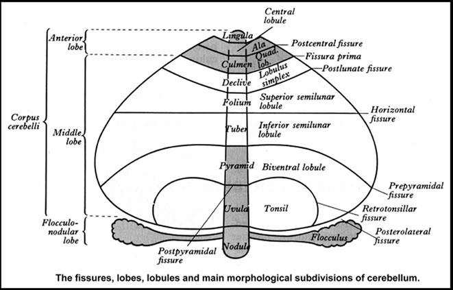
its surface presents many transverse sulci which pass in the same time on hemispheres and vermis; these sulci are generally superficial and separate small rounded folds called folia and only some of them are deep and separate the lobules of cerebellum being called fissures
the horizontal fissure is the greatest and deepest cerebellar fissure, has horizontal direction and separates the cerebellar surface into a sup. surface and an inf. one;
the sup. surface of each hemisphere is related with tentorium cerebelli;
at the level of the inf. cerebellar surface, the cerebellar hemispheres are separated by a notch called vallecula; it is determined by the existence of the medulla oblongata which is pressed on the inf. part of the vermis;
an ant. cerebellar notch is adapted to the pons; it lies at the level of the sup. surface of cerebellum;
a post. cerebellar notch separates dorsally the hemispheres; in this notch enters the falx cerebelli;
lobes and fissures of cerebellum:
the fissures are prolonged both on vermis and hemispheres and bear the same name at the both levels;
the fissures separate lobules of the hemispheres and of the vermis but the lobules bear different names on vermis and on the corresponding part of the hemispheres;
LOBULES ON VERMIS |
FISSURES |
LOBULES ON HEMISPHERES |
|
||
|
Anterior lobe |
Lingula Cerebelli |
Fraenulum Lingulae |
|
||
|
Precentral Fissure | |||||
|
Lobulus Centralis |
Ala Lobuli centralis |
|
|||
|
Postcentral Fissure | |||||
|
Culmen |
Lobulus Quadrangularis |
|
|||
|
Middle Lobe |
Primary Fissure | ||||
|
Declive |
Lobulus Simplex |
|
|||
|
Posterosuperior Fissure (Postlunate Fissure) | |||||
|
Folium |
Lobulus Semilunaris Sup. |
|
|||
HORIZONTAL FISSURE | |||||
|
Tuber |
Lobulus Semilunaris Inf. |
|
|||
|
Prepyramidal Fissure | |||||
|
Pyramis |
Lobulus Biventer |
|
|||
|
Secondary Fissure (Retrotonsilar Fissure) | |||||
|
Uvula |
Tonsilla Cerebelli (Amygdala) |
|
|||
|
Posterolateral Fissure | |||||
|
Flocculo-nodular lobe |
Nodulus |
Flocculus |
|
||
Legend: *** = archicerebellum, = paleocerebellum, = neocerebellum.
Cerebellar grey matter lies principally at the exterior, covering the entire cerebellar surface; thus it forms the cerebellar cortex Cerebellar white matter lies internally forming a cerebellar "white core". Cerebellar grey matter also forms cerebellar nuclei (small masses of grey matter inside the "white core"); there are 4 pairs of such nuclei.
Cerebellar white core
on a sagital section it has the aspect of a "tree" formed by many laminae that diverge radially; these laminae are projected towards the surface where they are covered by the cerebellar cortex; from these primary laminae begin many "secondary laminae" branching at very obtuse angles and is possible that secondary laminae will also branch into "tertiary laminae"; a11 these laminae are covered by the layer of grey matter of the cerebellar cortex; totally, this aspect on a sagital section that looks like a very rich ramified tree was called "arbor vitae" (tree of life):
the white core contains the next types of fibres
proper fibres - they are intrinsic fibres that connect different cerebellar regions; there are more types of proper fibres
association fibres are generally short fibers that connect the cortical folia of the same hemisphere and of the vermis; they do not decussate;
commissural fibres are long fibers that decussate in order to connect both hemispheres; they form 2 commissures: an anterosuperior commissure and a posteroinferior commissure; decussating spinocerebellar and cerebellovestibular fibers are intermingled with these commissural fibres;
projection fibres connect the cerebellum with the other levels of the CNS; they form 3 pairs of large cerebellar peduncles which seem to arise from the ant. cerebellar notch:
inf. cerebellar peduncles are 2 thick masses of white matter like 2 diverging bundles of fibres to the sup. direction; they connect the cerebellum with medulla oblongata; some authors divide each inf. cerebellar peduncle into a small, medial juxtarestiform body and a large, lateral restiform body
afferent cerebellar fibres through the inf. cerebellar peduncle form the next tracts:
dorsal spinocerebellar tract
cuneocerebellar tract
olivocerebellar tract
reticulocerebellar tract
vestibulocerebellar tract
ant. external arcuate fibres
striae medullares of the IVth ventricle (they originate also in the arcuate nuclei, decussate partially and run posterior on both sides of the median raphe to exit in the floor of the IVth ventricle; they run laterally as striae medullares and enter the inf. cerebellar peduncle to project into the fioccu1us (arcuatofloccular tract or the fibers of Picolomini
trigeminocerebellar fibres (from spinal trigeminal and sup. sensory nuclei)
efferent fibres from cerebellum through the inf. cerebellar peduncle form the next tracts:
cerebello-olivary fibres,
cerebellovestibular fibres,
cerebelloreticular fibres
middle cerebellar peduncles (also called brachia pontis) are the greatest cerebellar peduncles; they arise just from the lat. sides of the pontine basis and then they curve dorsally to enter the white core of cerebellum ; they contain the axons of the pontine nuclei neurons which decussate forming the transverse fibers of the pontine basis and then converge laterally to form the middle cerebellar peduncles;
sup. cerebellar peduncles are 2 bundles of fibers that converge in sup. direction and connect the cerebellum with the midbrain;
efferent fibres through the sup. cerebellar peduncle
dentatorubric fibres,
dentatothalamic fibres,
cerebello-olivary fibres for the accessory olivary nuclei,
cerebelloreticular fibres,
cerebellooculomotor fibres.
afferent fibres through the sup cerebellar peduncle
ventral spinocerebellar tract,
tectocerebellar tract,
hypothalamocerebellar tract.
Afferent projection fibres synapse first, as a rule, into cerebellar cortex; they form the so-called mossy fibres and climbing fibers
Efferent fibres from the cerebellar cortex are represented by the myelinated axons of Purkinje cells which, generally, are going to synapse into the cerebellar nuclei and finally the axons of the neurons in these nuclei project the information outside the cerebellum; only a few cerebellovestibular fibers represent an exception from this rule because they run directly from the cerebellar cortex outside the cerebellum;
cerebellar input is somehow distributed after some rules to the phylogenetical subdivisions of cerebellum as it follows:
archicerebellum (vestibulocerebellum) receives generally afferents from the vestibular nuclei;
paleocerebellum (spinocerebellum) receives generally afferents from the spinal cord, medullary reticular formation and accessory olivary nuclei;
neocerebellum receives
pontocerebellar and inf. olivary connections at the level of hemispheres of the middle lobe;
tectocerebellar input at the level of vermian and paravermian regions;
in these cases the terms of pontocerebellum and tectocerebellum may seem appropriate;
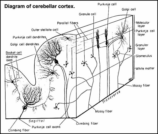
Cerebellar Grey Matter:
Cerebellar Cortex
it consists of 3 layers in which 5 types of neurons can be met; from the surface to the deep we find the next layers
molecular layer contains the next elements:
the rich ramified dendritic trees of Purkinje cells whose bodies lie in the next layer; these dendritic trees are ramified just in one plan of the space which is perpendicular on the long axe of the cerebellar folia;
the rich ramified dendritic trees of Golgi type II neurons whose bodies lie in the granular layer; these dendritic trees are ramified in allthe directions of the space at the level of molecular layer;
neuronal bodies and both types of prolongations of the outer stellate cells which have a superficial localization at the level of molecular layer;
neuronal bodies and prolongations of the basket cells that lie deeper at the level of this molecular layer;
axonal terminals of the granular cells whose neuronal bodies lie in the granular layer; these axons bifurcate in the molecular layer and will extend in opposite direction but both of them remain parallel with the longitudinal axe of the folia and traverse the dendritic trees of molecular layer (they were compared with the telegraph wires through the branches of bushy trees);
terminals of the olivocerebellar climbing fibers that ascend through the other 2 layers to synapse the Purkinje dendrites in molecular layer;
layer of Purkinje cells: it contains only the neuronal bodies of Purkinje cells;
granular layer contains:
the small bodies of the granular neurons and a part of their axons which ascend to the molecular layer; the dendrites of these neurons are also ramified inside this layer and form some terminal expansions;
neuronal bodies, basal dendrites and complex axonal ramifications of Golgi type II neurons;
ramified terminals of the mossy fibers;
climbing fibers ascending to the molecular layer;
cerebellar glomeruli;
neuronal interconnections in the cerebellar cortex
synapses with Purkinje cells are made by:
parallel fascicles of axons from the granular neurons which run through the dendritic trees of Purkinje cells synapsing them;
climbing fibres which are ramified following the ramification of Purkinje dendrites to make very numerous synapses;
axodendritic synapses of the axons of outer stellate cells with the dendrites of Purkinje cells;
collaterals from the axons of basket cells which synapse with the dendrites of Purkinje cells;
complex terminals of basket cells axons surrounding the bodies of the Purkinje cells (they form the so-called baskets
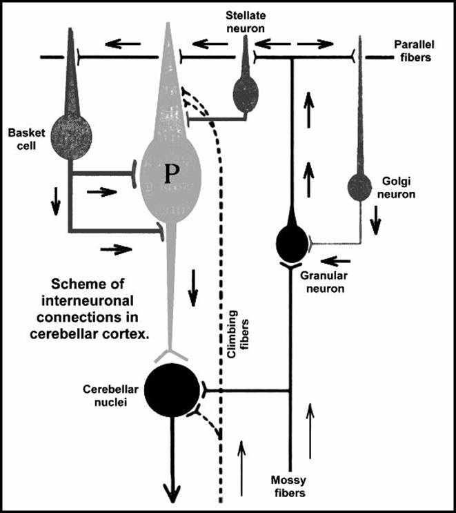
synapses with granular neurons are made by:
terminals of mossy fibres and axonal terminations of Golgi cells upon the dendrites of granular cells; they form totally the so-called "cerebellar glomeruli
synapses with basket neurons are made by:
the parallel fascicles of axons of granular neurons;
the axons of basket cells run in the transverse plan of the folia and synapse with the neuronal bodies (in fact with the preaxon) of the Purkinje cells;
synapses with the outer stellate cells are made by:
the parallel fascicles of axons of the granular neurons;
the axons of outer stellate neurons 1ie also in the transverse plan of the folia and synapse on the dendrites of Purkinje cells or on their preaxon like the basket cells;
synapses with the Golgi neurons are made by:
the parallel fascicles of axons of the granular a1 function:
Neuronal Function of cells in cerebellar cortex:
inhibitory cells are: Golgi neurons, basket cells, and outer stellate neurons;
excitatory cells are: granular neurons, and Purkinje cells.
Cerebellar nuclei
they are masses of grey matter embedded into the cerebellar white core;
from the lateral to medial direction they are: dentate nucleus, emboliform nucleus, globose nucleus and fastigial nucleus; in fact they are 4 pairs of nuclei:
dentate nucleus is the most lateral one and lies near the white core of its hemisphere; it has the shape of a folded bag like the inf. olivary nucleus; the opening of the "bag" is called hillum and it allows the efferent fibers to pass out in order to form an important part of the sup. cerebellar peduncle;
emboliform nucleus - it partially covers the hillum of dentate nucleus which is oriented medially;
globose nucleus - lies medially to the emboliform one; it is elongated in dorsoventral direction;
fastigial nucleus - it lies near the midline in the ventral part of sup. vermis;
afferent connections of the cerebellar nuclei are from intrinsic and extrinsic sources:
intrinsic sources - axons of Purkinje neurons of the cerebellar cortex which make axodendritic and axosomatic synapses on the neurons in these nuclei; all these synapses have inhibitory effect upon the neurons of these cerebellar nuclei
extrinsic sources
rubrocerebellar fibres
fibres from: spinocerebellar tracts, pontocerebellar tracts, olivocerebellar tracts, reticulocerebellar tracts; these fibres are excitatory for the neurons in cerebellar nuclei;
efferent connections of the cerebellar nuclei are, in fact, the efferent connections of cerebellum;
Mechanisms of the cerebellar cortex:
cerebellar cortex has:
2 distinct inputs: mossy fibers and climbing fibres,
just 1 output axons of Purkinje neurons
both inputs convey excitatory impulses from: exteroceptors and proprioceptors, brainstem reticular formation and cerebral cortex;
climbing fibres exert a one-to-one and all-or-none excitation on individual Purkinje neurons;
mossy fibres exert a diffuse excitatory effect on Purkinje neurons via the thousands of synapses made by the axons of granular neurons (parallel fibers) with the dendrites of more Purkinje cells;
axons of Purkinje cells exert inhibitory effect on cerebellar and vestibular nuclei
mossy fibres excite a locus of granular neurons which will excite by their axons (a narrow fascicle of parallel fibers) the dendritic fields of Purkinje, basket and outer stellate neurons; Golgi neurons do not respond because only a small part of their dendritic tree is in contact with parallel fibers; due to the arrangement of Purkinje neurons in regular spaced transverse plans and due to the slow conduction in the parallel fibers, the transverse plans of Purkinje neurons are excited sequentially; the transverse disposition of basket and stellate neurons and their terminals determines by their excitation at the contact with parallel fibers the inhibition of 2 other transverse plans of Purkinje neurons that flank the excited plan of Purkinje neurons on both sides; if the active fascicle of parallel fibers becomes wider than a row of Purkinje cells, the dendrites of Golgi neurons are excited and provide inhibitory impulses in the glomeruli, limiting the excitation provided by the mossy fibers;
|
