Management of Venous Thromboembolism
TABLE OF CONTENTS 2
I. INTRODUCTION: Hemostasis, Thrombosis and VTE 3
1.1 HEMOSTASIS
1.1.1 Primary hemostasis
1.1.2 Secondary hemostasis
1.1.3 Fibrinolysis
1.2 THROMBOSIS
1.2.1 Arterial thrombosis
1.2.2 Venous thrombosis
1.3 What is VTE?
1.3.1 DVT
1.3.2 PE
II. Prevalence and socio-economics of VTE 18
2.1 Prevalences
VTE is an economic burden
III. PATHOPHYSIOLOGY OF VTE 24
3.1 Development of thrombus
3.2 Consequences of VTE
IV. CAUSES OF VTE: Virchow's Triad 27
4.1 Stasis
4.2 Hypercoagulability
4.3 Endothelial injury
V. Risk factors for VTE 29
5.1 Predisposing risk
5.2 Exposing risk
5.2.1 Different risks with different surgeries
5.2.2 Different exposing medical risk factors
5.3 Assessing the risk in patients
VI. Diagnosis of VTE 34
6.1 Diagnosis of DVT
6.1.1 Symptoms of DVT
6.1.2 Physical examination
6.1.3 Diagnostic testing for DVT
6.2 Diagnosis of PE
6.2.1 Symptoms of PE
6.2.2 Diagnostic testing for PE
VII. VTE prophylactic options in medical and surgical patients 38
7.1 The rationale for thromboprophylaxis
7.2 What can be done to reduce the risk of DVT?
VIII. REFERENCES 43
Hemostasis and coagulation
Hemostasis is a complex physiological balance to prevent bleeding and excessive clotting (thrombus formation). It results from the dynamic interaction between the vascular wall, platelets and the processes of coagulation and fibrinolysis.
The coagulation process, called the coagulation cascade, is a series of enzymatic reactions, involving the sequential activation of numerous plasma components, known as coagulation factors. Each reaction leads to the activation of a coagulation factor. One of these, Factor Xa, plays a major role, being at the center of the enzymatic reactions leading to clot formation. It is neutralized by antithrombin, the main endogenous inhibitor of the coagulation cascade.
Thrombosis
Normally, the complex interplay of feedback mechanisms leading to clot formation occurs only when necessary (i.e. in response to vascular injury). Under abnormal circumstances, clots can also form spontaneously in a vessel that has not been breached - a condition called 'thrombosis'. Such clots can block the vessel. While arterial thrombosis is mainly related to an increase in platelet aggregation, venous thrombosis mainly results from disruption of the coagulation cascade.
In arteries, the obstruction resulting from thrombosis may have disastrous consequences, since it leads to infarction of the tissues supplied by the artery: e.g. acute myocardial infarction or stroke.
Venous thrombosis is usually the result of blood stasis. This explains why venous thrombi mostly form in regions of slow or disturbed blood flow, typically the deep veins of the legs where blood flow is slowest. This is deep-vein thrombosis (DVT).
Without treatment, the thrombus generally grows in proximal progression along the vein. The leg becomes red, swollen and painful. Following DVT - i.e. after the clot itself has dissolved - symptoms may persist due to the damage caused by the clot to the vessel structure. This condition is called post-thrombotic syndrome and is characterized by swelling, pain in the limb affected, and sometimes hyperpigmentation or the development of ulcers at the ankle.
The main danger is that part of the thrombus, or the thrombus as a whole, may become detached from the vein lining to form an embolus that migrates in the circulation and can be swept into the lung artery causing lung infarction. This is pulmonary embolism (PE). Large emboli can cause massive blockage of the pulmonary circulation and can lead rapidly to dramatic symptoms and death.
Treatment of DVT and PE is based on anticoagulant therapy. However, best treatment is prophylaxis.
Venous thromboembolism (VTE)
Venous thromboembolism (venous thromboembolic disease - VTE) is a condition where a clot forms in a vein, leading to either DVT and/or its major complication, PE which is sometimes fatal. DVT and PE are actually two manifestations of the same disease, necessitating a similar therapeutic approach. DVT may also predispose patients to long-term morbidity from post-thrombotic syndrome.
VTE is one of the most common disorders of the circulatory system, with an estimated annual incidence of one in every 1000 people in Western countries. It should be noted that in most instances, VTE is clinically silent, sudden death being often the first and only manifestation of the disease. Thus, this incidence probably represents an underestimate. Nevertheless, VTE is the third most common cardiovascular disease after ischemic heart disease and stroke and is the cause of significant morbidity and mortality. It is regarded as a major public health concern and an economic burden.
VTE is a multicausal disease that involves the interaction of both inherited (coagulation factor abnormalities) and acquired risk factors. For instance, DVT is a frequent complication in patients undergoing orthopedic surgery or in patients with cancer or other chronic illnesses, as well as in bed-ridden and immobilized patients. Advancing age is also one of the most important risk factors for VTE. Recent studies indicate that the number of hospitalized patients at high risk for VTE is considerable.
Owing to the often clinically silent nature of the disease, its relatively high prevalence in certain clinical circumstances, and the potentially disabling and life-threatening consequences of unrecognized and consequently untreated VTE, thromboprophylaxis (prevention) has become routine practice for certain groups of patients and is recommended by both North American and European Consensus statements. However, despite thromboprophylaxis, most studies indicate that the incidence of VTE remains too high for a condition that is preventable. In addition, with the continuing aging of populations, the number of cases of VTE is expected to increase.
Hemostasis is a natural physiological process, which serves to maintain blood in a fluid state while being able to stop blood loss quickly in the event of an injury. The initiation and amplification of hemostasis requires an interaction of substances produced by the injured blood vessels, platelets and plasma proteins called clotting factors. Regulatory mechanisms with feedback controls are in place to ensure that the hemostatic response is vigorous and yet controlled.
When damage to a blood vessel occurs through trauma, wear, infection or disease it triggers a natural defence mechanism, known as local vasoconstriction. Vasoconstriction is an immediate reaction to the injury and involves the contraction of vessel walls to reduce the blood flow to the affected area.
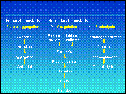 Overview of hemostasis
The process of hemostasis occurs in two stages:
primary hemostasis or platelet aggregation is the formation of a platelet plug or white thrombus at the site of vessel injury to provide an initial seal
secondary hemostasis or coagulation is the formation of an insoluble fibrin plug or red thrombus through a series of enzymatic events known as the coagulation cascade - this clot provides a more permanent patch over the damage to the vessel.
After the damage to the vessel wall has been repaired, it is important that the clot is dissolved to avoid blockage of the vessel. This is the reverse process of fibrin formation and is known as fibrinolysis, sometimes referred to as tertiary hemostasis.
In primary hemostasis, platelets stick to the site of a vessel wall injury and clump together to form aggregates to provide an initial seal. The formation of the initial platelet plug involves adhesive proteins such as collagen and von Willebrand factor (VWF).
Primary hemostasis requires a dynamic change in the behavior of blood platelets so that they are able to bind to the damaged surface of the blood vessel. This process takes place in three phases: platelet adhesion, platelet activation and platelet aggregation.
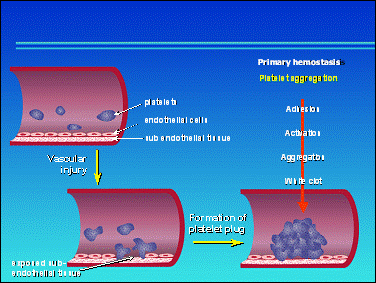
The first phase is the adhesion of platelets to the site of the injury or lesion in the vessel. Platelet adhesion is triggered by exposure to an abnormal surface. Undamaged vessel surfaces have a net negative electrical charge on their surfaces. Since platelets also have a negative surface charge, they are repelled by intact vessels. Damaged vessel surface and the structural protein collagen have a net positive charge that attracts platelets and causes them to stick to the injury site.
Platelet activation and aggregation
The adhesion of platelets to the injured vessel wall activates them. After activation, platelets assemble glycoproteins, which bind fibrinogen and VWF, both cofactors for platelet recruitment and aggregation. Fibrinogen and VWF are mandatory for platelet aggregation.
Activated platelets release two types of procoagulant granules:
dense granules, which contain adenosine diphosphate, ADP, as well as calcium and serotonin
alpha granules which contain specific proteins and certain coagulation factors - in particular, factor V.
On activation, platelets also synthesize and release thromboxane A2 (a prostaglandin) and platelet activating factor (PAF) - both are strong platelet aggregants and vasoconstrictors.
Further factors released by the platelets include the protein platelet factor 4 (PF4). PF4 is implicated in the pathogenic process of heparin-induced thrombocytopenia (a decrease in the number of blood platelets) after heparin administration.
Secondary hemostasis, or coagulation, is initiated at the same time as primary hemostasis but is a slower process involving a cascade of enzymatic reactions that result in formation of a platelet and fibrin clot. The clot is effected by conversion of soluble fibrinogen into insoluble fibrin by the enzyme thrombin.
A lesion in a blood vessel wall activates an enzyme cascade. Additionally, particular pathological situations such as infection, inflammation and atherosclerosis can trigger coagulation.
Many activation and inhibition loops ensure regulated control of coagulation so that formation of the blood clot or thrombus remains within the limits of the vascular lesion and does not grow unnecessarily large and form a blockage.
Coagulation factors
The different components that interact in the coagulation cascade are known as coagulation factors. These are listed in below.
Names, functions and locations of blood coagulation proteins
|
Common name |
Common alternative name |
Infrequent or archaic name |
Function (location) |
|
Tissue factor |
Thromboplastin |
CD142, factor III |
Cofactor, inhibitor (subendothelium) |
|
Factor XII |
Hageman factor |
Protease zymogen (plasma) |
|
|
Factor XI |
Plasma thromboplastin antecedent (PTA) |
Protease zymogen (plasma) |
|
|
Factor X* |
Stuart factor |
Protease zymogen (plasma) |
|
|
Factor IX* |
Antihemophilic factor B |
Christmas factor |
Protease zymogen (plasma) |
|
Factor VIII |
Antihemophilic factor A |
Cofactor for factor IXa in factor X activation (plasma) |
|
|
Factor VII* |
Proconvertin |
Protease zymogen (plasma) |
|
|
Factor V |
Proaccelerin |
Cofactor for factor Xa in prothrombin activation (platelets, plasma) |
|
|
Prothrombin* |
Factor II |
Protease zymogen (plasma) |
|
|
Fibrinogen |
Factor I |
Fibrin precursor (plasma) |
|
|
Factor XIII |
Fibrin-stabilising factor |
Zymogen of transglutaminase (Platelets, plasma) |
|
|
Thrombomodulin |
Cofactor for thrombi 111k103b n in protein C activation (endothelial surface) |
||
|
Protein C* |
Protease zymogen (plasma) |
||
|
Protein S* |
Cofactor for activated protein C (plasma) |
||
|
Antithrombin III |
Antithrombin |
Heparin cofactor |
Protease inhibitor (plasma) |
|
Tissue factor pathway inhibitor (TFPI) |
Extrinsic pathway inhibitor (EPI); lipoprotein-associated coagulation inhibitor (LACI) |
Protease inhibitor (platelets, plasma, endothelial surface) |
* Vitamin K-dependent proteins
Enzyme cascade activation involves two elements:
enzyme precursors, called zymogens, which when stimulated are converted into active enzymes or proteases. Activated enzymes are followed by the letter a. For example, prothrombin (factor II), is a zymogen that when activated is converted into thrombin (factor IIa)
accelerators of enzyme reactions, known as cofactors. A cofactor has no catalytic site but is required by and therefore regulates the activity of an accompanying protease. This is ultimately responsible for the catalysis.
Most enzymes in the coagulation cascade need a cofactor. For example, tissue factor is a cofactor that binds factor VII in initiating the event of clotting and is required for the proteolytic activity of the formed protease factor VIIa.
The liver is the major site of synthesis for probably all the plasma clotting factors. In addition, four factors (II, VII, IX and X) require vitamin K for synthesis. This explains the bleeding tendency associated with liver disease (cirrhosis) and vitamin K deficiency.
The coagulation cascade
Coagulation is activated through two clotting pathways: the initiation (extrinsic) pathway and the propagation (intrinsic) pathway (see graph below). In fact, interactions between the two pathways occur at many levels of the cascade.
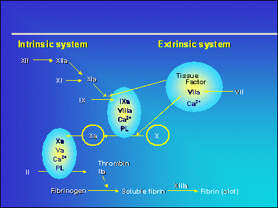
The principal pathway for the initiation of coagulation is the initiation pathway (also termed the extrinsic system). The initiation pathway (which is dependent on tissue factor) is the most important and most rapid pathway, requiring only a few seconds for coagulation and hemostasis.
The propagation pathway (also termed the intrinsic system) is initiated by components entirely contained within the vascular system, such as platelets and the activation of plasma coagulation factors. The pathway involves factors XI, IX, X and VIII. Factor VIII is also a cofactor for the activation of factor X.
The initiation and propagation pathways come together in a cascade mechanism that forms an enzymatic complex, factor Xa. A common pathway then allows the conversion of prothrombin into thrombin (mediated by factor V and calcium), which in turn converts fibrinogen into fibrin.
The initiation pathway
Coagulation is triggered by the activation of tissue factor (factor III or tissue thromboplastin). Injury to the vessel, such as a burn or cut (including surgery), brings blood into contact with tissue factor, which is expressed by the endothelial cells and subendothelial tissue of the damaged vessel. This is a very rapid pathway, in the order of seconds.
Tissue factors act on factor VII (proconvertin) and forms an active enzyme, which in turn activates two plasma zymogens, factors X and XI, producing two alternative pathways for the formation of factor Xa (see graph on previous page).
The propagation pathway
Once coagulation is triggered by the initiation pathway, it is amplified by the propagation pathway. The propagation pathway is the major pathway for the generation of factor Xa.
The pathway is controlled by a positive feedback mechanism; the thrombin produced by the initiation pathway activates factor XI, which converts factor IX to the activated form. This enables the formation of a complex with factor VIII, an enzyme reaction accelerator, which then activates factor X.
The complex formed between factor IXa and factor VIII must bind to a surface in order to activate factor X. This involves a platelet phospholipid, called platelet factor 3 (PF3).
The formation of the factor VIII-IX-phospholipid complex is essential in hemostasis. People who lack factor VIII (hemophilia A) and people who lack factor IX (hemophilia B) suffer with severe bleeding.
Calcium is necessary at all steps of enzyme activation, except for those of coagulation factors XII and X.
The common pathway
At the stage where factor X is activated, the initiation and propagation pathways follow a common pathway. Once formed, factor Xa has its enzymatic activity accelerated by proaccelerin (factor V), an enzyme reaction accelerator, and by calcium.
Both of these compounds bind to the phospholipid (PF3) to form a complex called prothrombinase, which activates prothrombin and splits it into several fragments, one of which is thrombin.
Factor Xa is involved in initiation of the formation of the thrombus following deterioration of the tissue and activation of the initiation pathway. Activated factor X stimulates the conversion of prothrombin (II) into thrombin (IIa), which consequently catalyses the formation of fibrin.
Although the two pathways work in parallel to bring about coagulation, in clinical practice an isolated abnormality in one of these factors results in a bleeding syndrome. For example, in hemophilia A, the initiation pathway does not compensate for the deficiency in factor VIII, and the propagation pathway does not compensate for the congenital deficiency in proconvertin (factor VII).
Positive feedback controls
The coagulation cascade includes many feedback controls. This means that an enzyme formed later in the cascade feeds back and proteolytically activates a zymogen or cofactor precursor earlier in the pathway. The major feedback controls are:
factor Xa activates the tissue-factor VII zymogen complex to tissue factor-VIIa (short amplification loop)
thrombin activates the cofactor precursors VIII and V to their active states (propagation loop)
thrombin activates platelets, providing phospholipid and platelet factor 4 (PF4).
Negative feedback controls
Negative feedback mechanisms are responsible for the inactivation of three activated species generated during clotting. Two major negative feedback mechanisms are known - one inhibitory and one enzymatic: Tissue factor pathway inhibitor and Protein C
The importance of a regulatory cascade
The cascade of reactions which results in coagulation is important. Failure to achieve a clot at an injury site could result in hemorrhage and even death. Failure to clot may result from a lack of one of the factors in the coagulation cascade - for example in hemophilia where even minor injuries can cause uncontrolled bleeding.
On the other hand, spontaneous clot formation at an inappropriate site can produce similarly fatal results by blocking the blood vessel. Or clots may break away from the site at which they are formed and travel through the circulation system, where they can block a smaller vessel (embolism). This can result in a heart attack (if the clot blocks a coronary artery), stroke (if the clot occurs in the brain) or a pulmonary embolism (if a blood vessel in the lungs is blocked).
The cascade of many reaction steps in the coagulation process greatly amplifies the signal from the tissue damage and platelet aggregation. This allows greater control over the process of clotting.
Activation of enzymes in such a cascade mechanism means that reactions are amplified at each step. This ensures that the body is able to respond quickly to injury.
Formation of fibrin
Thrombin plays an essential part in coagulation. The main way that thrombin promotes coagulation is via the generation of fibrin from fibrinogen. But thrombin also activates (positive feedback) the important cofactors, factors V and VIII.
In addition, thrombin is a platelet agonist, activating platelets and promoting aggregation. During fibrin formation, secondary hemostasis and primary hemostasis interact. Platelets - once activated - especially by the first traces of thrombin in primary hemostasis, enable the appearance of receptors for certain coagulation factors and the phospholipid platelet factor. This is essential to development of the propagation pathway.
Thrombin also activates factor XIII, which is important for the crosslinking of the fibrin polymer to form a stable structure. Crosslinking makes fibrin more resistant to the fibrinolytic enzyme plasmin.
The first fibrin network is built around the platelets. The fibrin network forms bridges and consolidates the platelet plug or white thrombus into an insoluble fibrin plug or a red thrombus.
Coagulation inhibitors
Excess coagulation factors are present in the blood. The thrombin in a milliliter of blood can coagulate a volume at least a hundred times greater, hence the existence of potent regulatory mechanisms.
These regulatory mechanisms are important to restrict clot formation to the injury site only and ensure that coagulation stops once the clot has been formed. They normally prevent reactions from causing thrombosis or disseminated intravascular coagulation (DIC).
The excess of activated coagulation factors is diluted by the blood flow and neutralized by plasma inhibitors. Thus a constant balance exists between activated coagulation factors and inhibitors.
There are three coagulation factor inhibitors:
antithrombin III (ATIII), which is the main endogenous inhibitor of coagulation factors
protein C
Tissue Factor Pathway Inhibitor (TFPI).
Antithrombin III (ATIII)
The main inhibitor of coagulation is antithrombin III, which is synthesized by the liver without the assistance of vitamin K. ATIII inhibits thrombin and factor Xa. It also has an inhibitory action against plasmin, which plays a role in fibrinolysis. Because factor Xa occurs earlier than thrombin in the coagulation cascade, when ATIII inhibits one molecule of factor Xa it inhibits the generation of fifty molecules of thrombin. A complete deficiency of ATIII (homozygous defect) is not compatible with life. Deficiency in ATIII is very rare and in these cases 50% of normal plasma ATIII level is available.
Endogenous Inhibitors - The Role of Antithrombin
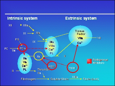
Protein C is another inhibitor. A vitamin K-dependent factor, it is activated by thrombin in the presence of endothelial thrombomodulin. In the presence of its cofactor, protein S, which is also dependent on vitamin K, protein C inactivates factors Va and VIIa.
Although thrombin is a very potent coagulant, it synthesizes protein C, which has an anticoagulant effect. This is the thrombin paradox.
Tissue Factor Pathway Inhibitor (TFPI)
TFPI is another regulator of hemostasis. It inhibits factor VII in the presence of phospholipid and factor Xa. This serves to dampen the coagulation cascade.
Fibrinolysis, sometimes termed tertiary hemostasis, is the process that destroys the fibrinous platelet clot and eliminates fibrinous deposits in the body, wherever they are located. Fibrinolysis corresponds to dissolution of the fibrin clot by plasmin. Thus, fibrinolysis is the reverse of coagulation.
Fibrin is converted into FDP (fibrin and fibrinogen degradation products) under the effect of plasmin. Plasmin is a powerful enzyme that catalyses fibrinolysis.
Plasmin arises from plasminogen under the effect of tissue plasminogen activator (tPA), an endogenous compound secreted by the endothelium. Plasminogen and plasmin are inactivated by antiplasmin. tPa is inactivated by plasminogen activator inhibitor (PAI), which, in turn, is inactivated by protein C. Thus, protein C inhibits coagulation and stimulates fibrinolysis.
Fibrin is broken down first of all into large and then smaller fragments. One specific degradation product released as a result of endogenous fibrinolysis is D-dimer - a useful diagnostic tool for venous thromboembolism. The soluble degradation products are dispersed into the circulatory system.
A decrease in fibrinolytic activity creates a predisposition to venous thrombosis.
Thrombosis results from an imbalance between anticoagulant and prothrombotic activities in which the prothrombotic activities predominate.
Under abnormal circumstances, clots can also form in a vessel that has not been breached - a condition called 'thrombosis'. Such clots can block the vessel.
While arterial thrombosis is mainly related to an increase in platelet aggregation, venous thrombosis mainly results from disruption of the coagulation cascade (see table below). An overactive clotting tendency might be the result of specific hereditary deficiencies. Injury may also result in excessive clotting, particularly if it involves bone fracture. Immobilization, surgery and a number of diseases are associated also with increased risks of clot formation.
Characteristics of the two types of thrombi
|
White thrombi |
Red thrombi |
|
|
Composition |
Mainly platelet and fibrin |
Mainly red cells and fibrin |
|
Location |
Vessels with rapid blood flow |
Vessels with slow blood flow |
|
Clinical complications |
Arterial thrombosis |
Venous thrombosis |
|
Cause |
Hypercoagulation and platelet activation |
Stasis, hypercoagulation and vascular injury (Virchow's triad) |
|
Management |
Antiplatelet agents and anticoagulant agents |
Anticoagulant agents |
In arteries, the obstruction resulting from thrombosis may have disastrous consequences, since it leads to infarction of the tissues supplied by the artery: e.g. acute myocardial infarction, stroke or peripheral arterial disease. Although popular belief holds that myocardial infarction and stroke are caused by vessel narrowing due to atherosclerosis, it appears that the acute symptoms are the consequence of an obstructing blood clot developing on the affected vessel wall. In this perspective, thrombosis is one of the major causes of death in the western world.
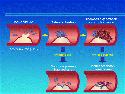
Most acute coronary syndromes (ACS; e.g. unstable angina, myocardial infarction) are triggered by the spontaneous or mechanical rupture of an atherosclerotic plaque, leading to abnormal platelet aggregation and a hypercoagulable state within the arteries. Indeed, when the endothelium is intact, blood constituents do not interact at this site. However, arterial injuries, such as plaque rupture, expose subendothelial tissue proteins, e.g. collagen and von Willebrand Factor (vWF), leading to platelet adhesion and activation. In addition, plaque rupture activates the coagulation cascade leading to thrombin generation and clot formation. Current strategies for the prevention and treatment of acute coronary syndromes are designed to both inhibit platelet activation/aggregation (antiplatelet agents) and control coagulation (anticoagulant agents) (see graph below).
Arterial thrombosis
In contrast to arterial thrombosis, venous thrombosis mainly results from disruption of the coagulation cascade with an excess in fibrin formation. This disequilibrium in the coagulation cascade is mainly due to stasis of blood flow.
In veins, the consequences of thrombosis are less apparent than in arteries and are often not even detected.
Venous thrombosis
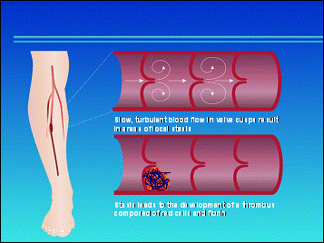
This explains why venous thrombi mostly form in regions of slow or disturbed blood flow, typically the large sinuses and valve cusp pockets in the deep veins of the calf. Indeed, venous thrombosis usually originates in the veins of the legs where blood flow is slowest.
Deep-vein thrombosis (DVT) in the leg
Factors which promote stasis and so can increase the risk of venous thrombosis include immobility, increased blood viscosity, dilatation of the veins (as with varicose veins and pregnancy) and increased venous blood pressure (as in heart failure).
Without treatment, the entire blood content of the vein may clot, forming a large mass behind the original thrombus. The thrombus generally grows in proximal progression along the vein. The leg becomes red, swollen and painful. Following deep-vein thrombosis - i.e. after the clot itself has dissolved - symptoms may persist due to the damage to the vessel structure caused by the clot resulting in venous stasis (reduced blood flow). This condition is called post-thrombotic syndrome and is characterized by swelling, pain in the affected limb, and sometimes hyperpigmentation or the development of ulcers at the ankle. Treatment and prevention of deep-vein thrombosis is based on anticoagulant therapy.
The post-phlebitic syndrome
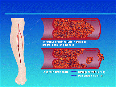
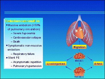
The main danger is that part of the thrombus, or the thrombus as a whole, may become detached from the vein lining to form an embolus that migrates in the circulation and can be swept into the lung artery causing lung infarction. This is pulmonary embolism. If less than 50% of the pulmonary circulation is blocked, symptoms are generally limited to shortness of breath or are absent altogether. Very large emboli, however, can cause massive blockage of the pulmonary circulation and lead rapidly to dramatic symptoms and death. Pulmonary embolism can be silent, without symptoms. However, repetition of asymptomatic pulmonary embolism may lead to pulmonary hypertension in the long term. Once an embolus has formed, immediate treatment with anticoagulant therapy is critical.
Pulmonary embolism (PE)
The term venous thromboembolism (VTE) covers two potentially highly dangerous and closely inter-related medical conditions.
1. The formation of a thrombus (blood clot) within a deep vein of the lower limb. This is termed a deep vein thrombosis and is usually abbreviated to a 'DVT'.
2. Pulmonary embolism (PE). This occurs when a piece of the DVT that detaches from the original thrombus in the leg travels in the venous blood stream to the lungs via the heart. When the PE reaches a vessel in the lungs with a lumen diameter equal to its own diameter, the PE plugs that vessel causing complete blockage. A large PE can block the main vessel leading from the heart to the lungs (pulmonary artery) causing instant death. A smaller PE will block a smaller vessel leading to respiratory dysfunction; the severity of which will depend on the size of the PE. PE is a medical emergency.
DVT and PE are in fact two manifestations of the same disease (VTE) and in many patients they co-exist:
50-70% of patients with proven proximal DVT have asymptomatic PE (Hirsh 96; Turkstra 97; Huisman 89; Meignan 00)
70-90% of patients with PE have asymptomatic DVT (Hirsh 96; Turkstra 97; Girard 99)
in more than 90% of cases, PE originates in the deep veins of the legs (Perrier 96)
1% to 2% of patients with DVT die as a result of PE (Hirsh 96)
DVT and PE co-exist in most patients (Hirsh 96; Turkstra 97; Huisman 89; Meignan 00; Doyle 87; Girard 99; Partsch 96) and share similar risk factors (Hirsh 96; Turkstra 97)
VTE often remains clinically 'silent', not causing any noticeable symptoms. The asymptomatic nature of VTE is the main threat of this disease; a fatal PE can occur without any warning signs.
Finally, the therapeutic approach based on anticoagulant therapy has proved effective in both DVT and PE (Hyers 01)
A patient who has developed VTE is at vastly increased risk of recurrence and so at risk of developing potentially fatal PE despite modern therapeutic interventions. Therefore prevention is the key to reducing the number of deaths from VTE. Accurate identification of patients at risk of VTE enables prophylaxis to be targeted effectively (Hirsh 96; Geerts 01; Nicolaides 01)
VTE is a major medical problem because it can be life-threatening (fatal PE) and may lead to recurrent VTE in the short-term, and chronic problems such as leg ulceration and post-thrombotic syndrome in the long-term.
Many risk factors for VTE are now recognized. These include surgery, major trauma, prolonged bed rest, cancer, pregnancy and increased age. Knowledge of these risk factors is used to target VTE prophylaxis (Hirsh 96; Geerts 01; Nicolaides 01).
VTE is one of the
most common disorders of the circulatory system, with an estimated annual
incidence of one in every 1000 people in
VTE is a multicausal disease that involves the interaction of both inherited and acquired risk factors. For instance, DVT is a frequent complication in patients undergoing orthopedic surgery or in patients with cancer or other chronic illnesses, as well as in bed-ridden and immobilized patients.
Owing to the often clinically silent nature of the disease, its relatively high prevalence in certain clinical circumstances, and the potentially disabling and life-threatening consequences of unrecognized and consequently untreated VTE, thromboprophylaxis has become routine practice for certain groups of surgical or medical patients, according to their level of risk for VTE.
DVT refers to a thrombus occurring in one of the deep veins of the lower limbs. Most thrombi originate in the veins deep within the muscles of the calf, sometimes termed the distal veins. However, some DVTs start in the more proximal veins, such as the popliteal vein (behind the knee) or the deep femoral vein of the thigh. More rarely, the DVT starts even more proximally in a pelvic vein. Proximal DVTs have the potential to be more life-threatening than distal DVTs because the thrombi are more prone to embolize (ie cause a PE).
Once a thrombus has started to develop, it can progress in several ways. It can increase in size so that it completely obstructs all blood flow in the vein. A DVT can also grow along a vein (so-called extension), frequently in a proximal direction, ie up the vein in the leg toward the pelvis. It can also cause damage to valves in the veins. Most seriously, it can give rise to a PE.
As mentioned previously, a piece of the thrombus may break off and travel as a PE to the pulmonary circulation, where its effect can be life-threatening.
PE is a piece of thrombus that has broken away from the original DVT and is carried by the blood stream via the heart to the blood vessels in the lungs. PE is the most serious, but not inevitable, complication of DVT. In 90% of cases, PE is a result of a thrombus that developed in the deep veins of the lower limbs (Anderson 98). PE causes obstruction of the pulmonary artery or of one of its branches. PE is associated with a high mortality rate.
The International Cooperative Pulmonary Registry (ICOPER;
Goldhaber 99) study examined a multicenter registry of patients with PE from 59
hospitals and within seven countries in Europe and
The larger the pulmonary embolus, the larger (and hence the more important) the pulmonary vessel it can obstruct and the more serious the effects on the circulatory system. When a large thrombus blocks the major pulmonary vessels (arteries) it causes cardiogenic shock, which is followed by circulatory failure and death. A smaller thrombus can block a smaller pulmonary vessel, causing symptomatic PE. In chronic cases of PE, often with multiple small pulmonary emboli, pulmonary hypertension (raised blood pressure in the pulmonary arteries) may develop.
In the general
population, the rate of VTE is in the range of two events per 1,000 people
per year. In the
In the
Epidemiological data from Western countries show that the annual frequency of VTE in the general population remains high. The reported annual incidence ranges from approximately 40 to 160 per 100 000 for DVT ( 60% being proximal DVT) and 20 to 70/100 000 for PE (Silverstein 98; Anderson 91; Nordstrom 92).
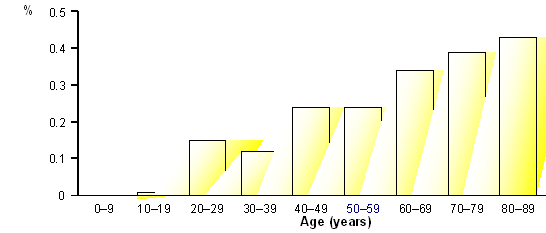
Data from the USA, provided by a retrospective 25-year population-based study (1966 to 1990), indicate that the overall average age- and sex-adjusted annual incidence of a first episode of VTE was 117 in 100 000 (DVT alone: 48/100 000 and PE with or without DVT: 69/100 000), with higher age-adjusted rates among males than females (130 vs 110/100 000). In addition, it was shown that the incidence of VTE rose markedly with increasing age in both sexes, PE accounting for most of the increase. The incidence of VTE in the 65-69 year age range reached 300/100,000 and was more than 800/100,000 among people aged 80 years or older (Silverstein 98).
Both DVT and PE manifest few specific symptoms and the clinical diagnosis is insensitive and unreliable. Indeed, it has been shown that less than half the patients suspected of having VTE actually have the disease. In addition, in most instances, VTE is clinically silent. Almost 80% of DVT are clinically silent and less than 50% of patients with proximal DVT, the more prone to embolize, present with clinical signs.
Sudden death is often the first and only manifestation of the disease and most deaths occur within 30 minutes of the acute event, too soon for anticoagulant therapy to be effective (Sandler 98). Less than 50% of all cases of fatal PE are detected prior to death (Donaldson 63). For instance, among 54 patients identified with anatomically major PE at autopsy, 16 (30%) had a correct antemortem diagnosis (Goldhaber 82). Although accuracy was greater in the postoperative patients of this series, the corresponding figures remained low (64%).
It is also widely acknowledged that DVT and PE co-exist in most patients, one of the two often being silent (Hirsh 96; Turkstra 97). Indeed, 50% to 70% of patients with proven proximal DVT have asymptomatic PE (Hirsh 96; Turkstra 97; Huisman 89; Meignan 00). This is still the case for approximately 30% of patients with proven isolated calf DVT, a condition supposed to be less prone to embolize (Huisman 89; Dole 87). Even when asymtpomatic, DVT can lead to PE. Silent PE has been reported to occur in 35% of patients with asymptomatic (silent) proximal DVT and in 7% to 8% of patients with asymptomatic distal DVT (Partsch 96) . Finally, asymptomatic DVT is found in approximately 70% to 90% of patients with confirmed clinically symptomatic PE (Hirsh 96; Turkstra 97; Girard 99).
PE is associated with very high mortality rates and the risk of recurrence is high in patients who survive PE. Among 2454 North American and European patients with acute PE, mortality at three months was 17% despite the use of state-of-the-art treatment. PE was the cause of death in 45% of cases (Goldhaber 99). After exclusion of 61 patients in whom PE was first discovered at autopsy, the mortality rate at three months was still 15% (see figure below). In 34% of the patients who died from PE, this was silent and only detected at autopsy. Interestingly, 75% of the deaths occurred during the initial hospital admission for PE. In addition, a large proportion of patients who survived the initial episode (17%) had to be readmitted to hospital within three months.

As well as being a frequently occurring and life-threatening disease in the short-term, VTE can lead to long-term complications or sequelae. These sequelae include future episodes of recurrent VTE and chronic medical problems such as phlebitis and venous ulcers in the lower limb that had the original DVT. These long-term problems are part of what is known as the post-thrombotic (or post-phlebitic) syndrome. (Nicolaides 2001)
Surgical patients are at risk of developing VTE both during the operation and after hospital discharge. There are recorded incidences of DVT in many different surgical procedures, as well as in general medical patients (see table 1 below). The incidence of clinical PE is more difficult to obtain because most patients found with proximal DVT are treated with antithrombotics, therefore the true incidence, without screening and intervention, is unknown and is likely to be greater than suggested in Table 2. The incidence of VTE remains too high for a condition that is preventable (Nicoloaides 1992).
Table 1:The incidence of DVT in the absence of prophylaxis
|
Patient group |
No. of studies |
No. of patients |
DVT incidence (%) |
95% CI (%) |
|
Stroke |
8 |
380 |
212 (56) |
51-61 |
|
Elective hip replacement |
17 |
851 |
435 (51) |
48-54 |
|
Multiple trauma |
4 |
536 |
270 (50) |
46-55 |
|
Total knee replacement |
7 |
541 |
252 (47) |
42-51 |
|
Hip fracture |
16 |
836 |
372 (45) |
41-48 |
|
Retropubic prostatectomy |
8 |
335 |
106 (32) |
27-37 |
|
General surgery |
54 |
4310 |
1084 (25) |
24-26 |
|
Spinal cord injury |
9 |
458 |
160 (35) |
31-39 |
|
Neurosurgery |
5 |
280 |
61 (22) |
17-27 |
|
Myocardial infarction |
4 |
180 |
40 (22) |
16-28 |
|
General medical |
2 |
110 |
19 (17) |
10-24 |
|
Gynaecological surgery |
4 |
460 |
63 (14) |
11-17 |
|
Geriatric |
1 |
131 |
12 (9) |
5-15 |
|
Transurethral prostatecotomy |
3 |
150 |
14 (9) |
5-15 |
Diagnosed by surveillance with objective methods: Phlebography or fibrinogen uptake test
Table 2: The frequency of clinical pulmonary embolism in the absence of prophylaxis
|
Patient groups |
No. of studies |
No. of patients |
Incidence (%) |
95% CI (%) |
|
General surgery |
32 |
5091 |
82 (1.6) |
1.3-2 |
|
Elective hip replacement |
25 |
1436 |
57 (4) |
3-5.1 |
|
Traumatic orthopedic surg. |
11 |
494 |
34 (6.9) |
4.8-9.5 |
Frequency of PE in hospitalized patients
A retrospective analysis (Stein 99) considering all cases of PE (n=400) diagnosed in a tertiary care general hospital in Detroit, Michigan, USA, over a 4.75-year period (1993-1997), showed that hospital inpatients are at high risk, with an overal PE incidence of 0.23%, more than three times higher than that observed in the general population. The incidence of PE was linearly correlated with advancing age of the patients.
Annual incidence of PE in a tertiary care general hospital
In addition, it should be noted that this incidence probably represents an underestimate of the true incidence, as records of PE found at autopsy in patients with no diagnosis of PE before death were not taken into account.
Annual Incidence of DVT alone, PE±DVT and all VTE Events among Residents of Omsted County, Minnesota, from 1966 to 1990, by Age (Silverstein 98)
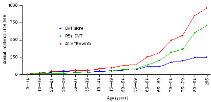
There was a 45% decrease in the incidence of PE between 1966 and 1975, but the incidence then remained stable up to 1990. In contrast, the incidence of DVT remained unchanged in men, but showed a trend to increase in older women. Thus, with the continuing aging of populations, the number of cases of VTE may be expected to increase.
Autopsy studies
Due to the silent nature of the disease and the low rate of autopsy in the USA, the total incidence, prevalence and mortality rates of VTE remain elusive and are probably underestimated (Sandler 98). Data from countries where autopsy is common indicate that PE remains a significant problem (Lindblad 91; Rasmussen 95; Lindblad 91; Bergqvist 85), suggesting the need for even wider application of prophylaxis. In these studies, in-hospital deaths due to PE ranged from 4% to 13%.
In
an early study by Sandler 1989 in the
Data from a general surgical department in
The incidence of VTE in all necropsy reports in Malmö hospital over 30 years (1957 to 1987) is summarized in Tables 1 and 2. It should be noted that calf veins were not routinely examined.
Table 1. Incidence of VTE in
|
% of autopsy findings* |
% of hospital patients** found at autopsy to have had |
||||
|
Year |
VTE |
PE |
Fatal PE |
PE |
Fatal PE |
Incidence of VTE and PE relative to *the number of autopsies and **number of patients in hospital
Table 2. Percentage of Autopsies in which VTE (fatal PE) was Found by Hospital Department
|
Year |
General surgery |
Infectious diseases |
Internal medicine |
Oncology |
Orthopedics |
In addition, one of the studies from Malmö (a 30-year survey of PE in 1274 surgical patients) demonstrated that the risk of postoperative PE continues for at least one month after surgery (Bergqvist 85; Scurr 88; Huber 92). Other reports have confirmed these findings.
Venous thromboembolic events are very expensive to manage, not only in terms of the hospital costs associated with acute episodes, but also when the costs of long-term complications are taken into account. Due to the large number of patients involved, venous thrombosis represents a huge economic burden.
The socio-economic costs of VTE include:
costs associated with the diagnosis and treatment of DVT
costs of managing non-fatal short-term consequences of VTE (eg intensive care costs for managing the respiratory dysfunction caused by PE)
costs associated with the diagnosis, treatment and mortality of PE
costs associated with managing the long-term
consequences (eg recurrence of
The total US inpatient costs for the immediate management of DVT (i.e. without taking into account recurrence and chronicity) have been estimated at US$ 111 millions (based on 1997 Medicare figures). In addition, besides the demand on healthcare resources (primary healthcare providers and hospitals), thrombotic disorders are responsible for considerable loss of productivity due to premature mortality and incapacity. It should also be noted that the long-term healthcare costs of a DVT almost doubles the immediate costs of treatment due to recurrences, development of post-thrombotic syndromes and the need for long-term treatment.
A 15-year
retrospective cohort study in a
Comparison of 15-year Outcomes for DVT and Control patients
|
Outcome at 15 years |
Incidence |
|
|
DVT cohort |
Control cohort |
|
|
Mortality | ||
|
VTE complications or events |
1.3 /patient |
0.13 /patient |
The average cost of primary DVT was estimated at US$ 6083 (see table below). In addition, the long-term costs associated with treating the post-thrombotic complications amounted to approximately 75% of the direct costs of treating the primary DVT.
Costs of post-thrombotic complications or events (US$; 1990-1991 figures)
|
Complication or event |
DVT cohort |
Control cohort |
||
|
Average cost/complication |
Complications (N) |
Average cost/complicat. |
Complicat. (N) |
|
|
Superficial venous thrombosis | ||||
|
Recurrent DVT | ||||
|
Primary DVT | ||||
|
Cellulitis | ||||
|
Venous ulcer | ||||
|
Varicose veins | ||||
|
Stasis dermatitis | ||||
|
Deep venous insufficiency | ||||
|
Pulmonary embolism | ||||
|
Combinations of several conditions | ||||
|
Overall average | ||||
The graph below confirms that the cost incured as a result of DVT is not only due to the acute episode but also to long-term complications, with the first year post-acute episode accounting for 40% of the total cost. (Bergqvist 97)
Distribution of the total cost of a DVT per year following the acute event

VTE represents a huge economic burden, not only because of the cost of treatment of the acute event, but also because of the cost of long-term complications, incapacity and premature death.
Clot formation in the venous lumen
Certain pathological situations interact to initiate the development of a clot (thrombus) within a vein. When this process begins in a deep vein, the resultant thrombus is known as a DVT.
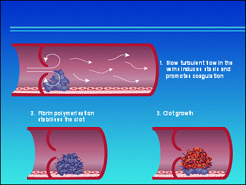
A DVT may begin as a microscopic platelet nidus, often in the cusps of the venous valves or in the large venous sinuses of the lower limb. Abnormal turbulent blood flow near the valves may contribute to the development of these platelet nidi.
At the cellular or subcellular level, clot formation starts when the initiation pathway of thrombosis is triggered by an injury to the endothelial wall of the vein. Substances released from the damaged wall encourage primary hemostasis during which platelet activation and aggregation result in an initial platelet plug or white thrombus at the site of the vessel injury.
In secondary hemostasis, or coagulation, fibrin is formed. This coagulation process is initiated by the membrane protein tissue factor. This process consists of a highly regulated cascade of reactions during which fibrin is produced by the conversion of soluble fibrinogen into insoluble fibrin under the effect of the enzyme thrombin. The production of fibrin results in a red fibrin thrombus around the injured area; this increases in size as it incorporates platelets, and red cells that become trapped in the growing fibrin mesh.
A venous thrombus differs in appearance from an arterial thrombus, being red and less compact, and having a greater number of red blood cells trapped in its fibrin network. Once a thrombus is formed, it may lyse (disintegrate) spontaneously.
This lysis is the result of the actions of the body's fibrinolytic system, which opposes coagulation, attacking the fibrin thrombus and sometimes successfully breaking it down. After the clot dissolves, the affected vein recanalizes within a few weeks. If the fibrinolytic system fails to lyse the thrombus, it stays in the leg and pieces of the thrombus can break off and embolize.
Common sites for thrombus formation
A DVT may begin at one site and extend up the vein to other sites, or there may be multiple DVTs, simultaneously occurring at different sites. Common sites for DVT formation are within the veins that drain the lower limb and pelvis.
These include:
iliac veins - runs through the pelvis to the vena cava
common femoral vein - the main vein that runs from the upper part of the thigh towards the pelvis
deep femoral vein - deep inside the thigh running up to the common femoral vein
popliteal vein - runs behind the knee up towards the deep femoral vein.
posterior tibial vein - runs within the calf towards the knee
deep distal intramuscular veins and sinuses of the calf. These also run up towards, and join, the popliteal vein.
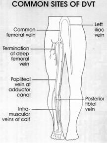
Site of thrombus and risk of PE
The distal veins - the calf muscle veins - are common sites for thrombus formation. A thrombus in the calf can extend (propagates) into the popliteal or thigh veins, and the risk of PE (the major acute complication of DVT) increases greatly.
This is because the location of a thrombus seems to influence whether or not the thrombus will develop into a case of PE. Most cases of PE arise from the proximal veins of the legs - the iliofemoral system - and massive occlusion of the iliofemoral system can be life threatening because of the risk of PE.
These can be categorized into acute and long-term consequences. The most dangerous of the acute consequences, as we have already discussed, is PE. All patients with a DVT are (potentially) at risk of this life-threatening condition.
The long-term consequences of the disease are explored further below, but the fact that a single DVT is the strongest 'predisposing' factor to further events, thus re-exposing the patient to more serious consequences, must not be ignored.
Long-term complications of VTE
Even when treated with current anticoagulation therapy, DVT can lead to the development of long-term clinical complications. Long-term complications include:
recurrent DVT
recurrent PE
post-phlebitic (or post-thrombotic) syndrome
death.
Recurrent venous thrombosis
Patients experiencing their first episode of DVT are at high risk of recurrence. Even under the most effective thromboprophylaxis regimens, recurrence of DVT in the first 3 months is 8% and at one year is 12.9% (Heit 00) .
Recurrent PE
Patients with a history of PE are more prone to have PE recurrences. The risk of recurrence is up to 23% in patients who survive PE in the first year following PE (Oger 00).
Post-phlebitic syndrome
Post-phlebitic syndrome (or post-thrombotic syndrome) is a result of long-term DVT, usually occurring 2-10 years after the development of DVT. The condition affects 50-70% of people with proximal DVT.1 The cumulative incidence of post-phlebitic syndrome is high; 23% at 2 years, 28% at 5 years and up to 29% at 8 years (Prandoni 96). Post-phlebitic syndrome is caused by venous hypertension resulting from damaged calf vein valves and/or persistent obstruction from thrombosis. Damaged valves are incompetent, ie they cease to be one-way valves. Blood flows the wrong way and gets directed to the superficial veins, where edema, ulceration and sclerosis can result. Post-phlebitic syndrome is very disabling for the patient, costly and leads to long-term morbidity.
Death
The mortality rate is much higher in patients with a previous VTE. The percentage of death in patients with a DVT without PE can be up to 30% (Heit 96; Prandoni 96). In patients with a history of PE, 1 year mortality was 39% and 45% at 2 years (compared with 3% at 2 years in the control group) (Kniffin 94). Thus a first event leads to recurrences and exposes the patient to a higher risk of death.
Besides being life-threatening in the short-term, unrecognized and untreated DVT may predispose patients to future episodes of recurrent VTE, and also lead to long-term morbidity from post-thrombotic syndrome (Heit 96; Geerts 01; Nicolaides 01).These two conditions, found even in low-risk patients (Franzeck 96; Prandoni 96; Lagersted 85) have a great impact on quality of life and health care costs.
Before the introduction of anticoagulant therapy, isolated symptomatic calf DVT was associated with an approximately 30% likelihood of recurrent proximal DVT or PE within three months of initial diagnosis. The mortality rate from PE was 20% in hospitalized patients with symptomatic DVT (Hirsh 96). In addition, up to 20% or even 30% of silent calf DVT extended into the popliteal vein and this extension was associated with a 40% to 50% risk of clinically detectable PE. In several studies, the reported incidence of post-thrombotic syndrome ranged from 35% to 69% within three years and from 49% to 100% within 5 to 10 years of initial diagnosis (Nicolaides 01).
Even after the introduction of anticoagulant therapy for the treatment of acute DVT, the risk of recurrent VTE and post-thrombotic syndrome remains high. In an 8-year follow-up study of 528 consecutive patients with first-episode symptomatic DVT who received treatment with anticoagulant therapy, the cumulative incidence of recurrent VTE was 17% after 2 years, 24% after 5 years and 30% after 8 years of follow-up (see table below) The cumulative incidence of post-thrombotic syndrome was 25% after 2 years, 30% after 5 years and 30% after 8 years (Prandoni 96, Prandoni 98; Prandoni 97).
Long-term outcome after a treated first DVT

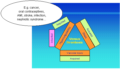
In the nineteenth century, the eminent pathologist Rudolph Virchow proposed a theory (that still holds today) that thrombosis was the result of three interplaying factors known as Virchow's triad:
stasis (slow blood flow)
hypercoagulability (increased tendency of blood to clot)
endothelial injury (injury to inner wall of a blood vessel).
Each of the three factors in Virchow's triad is important in the formation of a DVT, although they do not all need to be present at the same time for a DVT to occur. Stasis and endothelial injury to the endothelial cells are generally the result of acquired factors. In contrast, hypercoagulability may be either inherited - for example, in the form of a coagulation disorder such as antithrombin III deficiency - or acquired, through an underlying disease such as cancer.
The Virchow triad
 6.1
Stasis
6.1
Stasis
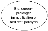
In normal physiological conditions, venous return from the lower limbs is assisted by contraction of the calf muscles - the venous pump - which helps to return blood from the lower part of the body to the heart. Venous stasis describes slow blood flow and may develop as a result of periods of immobility - such as during prolonged bed rest (eg post-surgery) or long journeys in airplanes (resulting in so-called economy class or coach class syndrome).
Immobility causes the blood to pool in the intramuscular calf vein sinuses of the legs. Stasis increases blood viscosity, encourages clotting and prevents small thrombi from being swept away.
As well as those immobilized by bed rest and so on, people at increased risk of venous stasis include those with limb paralysis (such as those who have had a recent stroke) and those patients immobilized with leg casts following fracture. Stasis may also be involved in the development of venous thrombosis in people who are obese.
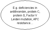
The second aspect of Virchow's triad relates to an underlying disease. Hypercoagulability is an increased tendency of the blood to form clots. Increased coagulability develops naturally with increasing age. Older people are therefore naturally more prone to venous thrombosis. This condition is commonly found in patients who show a history of thromboembolic disease.
Hypercoagulability is also seen in patients with other diseases, particularly cancer. Underlying malignancy appears to accelerate events in the coagulation cascade.
Inflammatory conditions, such as inflammatory bowel disease, systemic lupus erythematosus (SLE), sickle cell disease and sepsis also appear to increase the risk of thrombosis, probably due to hypercoagulability. Thromboembolic disease may also result from inherited disorders of coagulation. Examples include deficiencies of protein S, protein C and antithrombin III. Antiphospholipid syndrome is also a risk factor for VTE because antiphospholipid antibodies accelerate the coagulation process.
Increased levels of estrogen also increase the risk of thrombosis due to a loss of protein S in the urine. This places women who are pregnant, have recently given birth or are taking the combined oral contraceptive pill at increased risk of DVT.
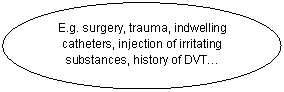
Endothelial injury describes an injury to the venous tunica intima - damage to the inner lining of the vessel wall. Such venous injury occurs in patients undergoing surgical procedures - hip surgery, knee surgery and varicose vein stripping - or those experiencing trauma. Endothelial injury may also be caused by the use of central venous catheters and intravenous drug abuse.
Injury to the endothelium activates both the initiation pathway, resulting in platelet adhesion and aggregation, and tissue factor - triggering coagulation.
Risk factors are present in at least half of patients with DVT. Risk factors are additive in nature, and DVT is usually the result of an interaction between multiple risk factors.
When attempting to confirm the presence of a suspected DVT, or identifying patients suitable for prophylaxis, it is important to take into consideration the presence of the risk factors.
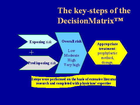
An international taskforce started in 2002 an exiting project to give computer-aided support for approrpriatness of prophylaxis of DVT. This project, called DecisionMatrix , includes one of the most comprehensive collection of predisposing and exposing risk factors.
Predisposing risk factors
= pre-existing risk; those risk factors present before the patient is admitted to the hospital and independent of the procedure the patient is up to undergo
= the predisposing risk may be considered as a risk which cannot be changed
In the DecisionMatrix the predisposing risk factors are grouped into "global patient characteristics", "VTE history and blood factors", "recent clinical conditions" and "chronic clinical conditions". For further details on each single risk factor please refer to the DecisionMatrix .
Following predisposing risk factors have been listed by the DecisionMatrix expert panels:
General patient characteristics:
|
Gender: Male or female |
|
Age (years) <40 40-59 60-74 over 75 |
|
Blood group: non-O group |
|
Obesity: BMI > 30 kg/m2 |
|
Smoking: >15 cigarettes per day |
|
Oral contraceptive pill (OCP): combined oestrogen / progestogen treatment |
|
Hormone replacement therapy (HRT): combined oestrogen/ progestogen menopausal treatment |
|
Specific drug use: protective effect of ASA (aspirin) |
Family history / VTE history / blood factors:
|
Family history of VTE: in first degree, parents or siblings |
|
|
Previous history of VTE Proximal saphenous / superficial venous thrombosis Proximal with or without distal Distal DVT only Pulmonary embolism Clinically idiopathic additional risk |
|
|
Antiphospholipid Syndrome (SLE: Systemic lupus erythematosis; ACL: anticardiolipine; LA: lupus anticoagulant) Primary, without another autoimmune disease (e.g. (SLE)) Secondary with, SLE with LA (with ACL) Secondary with SLE without LA (with ACL) Secondary, with other autoimmune disease or due to drugs |
|
|
Myeloproliferative disorders: including polycythemia Vera, essential thrombocytosis |
|
|
Hyperhomocysteinaemia (fasting homocysteine plasma levels above 40 µmol/L in women; 18 µmol/L in men) |
|
|
Antithrombin deficiency, heterozygote |
|
|
Protein C deficiency, heterozygote |
|
|
Protein S deficiency, heterozygote |
|
|
Factor V Leiden mutation, heterozygote |
|
|
Factor II mutation, heterozygote |
|
|
Factor V / II mutation, homozygote |
|
|
More than one factor (of the previous 6) |
|
Recent clinical conditions (less than 3 month):
|
Recent major surgery with complications without complications |
|
Recent Myocardial infarction |
|
Recent Stroke, disregarding paralysis |
|
Recent prolonged travel (more than 6 hours) |
|
Dehydration: severe dehydration as defined by 10% weight loss |
|
Increased hematocrit: >45% for women; >50% for men |
|
Hyperviscosity: increased blood viscosity |
Chronic clinical conditions:
|
Malignancy Local stage Locally advanced Metastatic cancer Additional risk if specific type is pancreatic, gastrointestinal, ovarian, prostatic, pulmonary, malignant glioma Additional risk if treated with radiotherapy Additional risk if treated with chemotherapy Additional risk if treated with hormonal therapy |
|
Heart failure/Cardiac disease NYHA I or II NYHA III or IV |
|
Chronic respiratory disease (COPD or emphysema) |
|
Nephrotic syndrome, syndrome of proteinuria, hypoalbuminemia of <20g/L |
|
Immobilisation Confinement to bed or chair > 3 days (not wheelchair- or bed bound) Confinement to bed or chair > 3 days (wheelchair- or bed bound) Lower limb paralysis (hemiplegia / paraplegia / neurological disease) |
|
Inflammatory bowel disease (IBD): Crohn's disease and ulcerative colitis |
|
Venous insufficiency Varicose veins, prominence of superficial veins on standing Lower limb swelling, discomfort Ulceration |
|
Lower limb artheropathy: Intermittent claudication |
|
Diabetes Includes both type I and type II, any aetiology |
An international taskforce started in 2002 an exiting project to give computer-aided support for approrpriatness of prophylaxis of DVT. This project, called DecisionMatrix , includes one of the most comprehensive collection of predisposing and exposing risk factors.
In the DecisionMatrix general surgyery, urological and gynecological surgery as well as medical patients are included and evaluated
Exposing risk factors
= risk of medical condition or surgical intervention the patient is exposed to;
the risk incurred or created by the surgical procedure/ medical condition for which the patient is admitted to the hospital;
the reason for hospitalization
Patients who undergo surgery or who suffer major trauma are at risk for developing VTE. Any surgical procedure that lasts more than 30 minutes or involves bone contact (for example, orthopedic or neurological surgery) puts patients at increased risk. In addition, longer surgical procedures require an increased period of immobility for the patient and this also serves to increase the risk.
Orthopedic surgery, including hip and knee replacement and hip fracture surgery, has an especially high incidence of VTE.
Without any prophylaxis, the risk of VTE for example after elective hip surgery is between 45% and 70%, with even higher figures seen after emergency surgery for fractures. Many patients undergoing orthopedic surgery are elderly, which further increases their chance of a VTE. In addition, orthopedic surgery often involves dissection, trauma and torsion of the veins, which again increases the DVT risk. Application of a cast to a leg during orthopedic surgery compounds the problem of venous stasis caused by the immobility of post-operative bed rest. This is because a leg cast impairs the pumping action of the thigh and calf muscles that usually maintain the flow of blood through the veins and back towards the central circulation.
In abdominal, gynecological and urological surgery, cancer is present in more than 50% of cases. Cancer increases the risk of VTE, as tumor cells can activate the clotting system directly, thereby producing thrombin, or indirectly by stimulating mononuclear cells to generate and express procoagulants. Cancer cells can also damage endothelium by direct vascular invasion, resulting in activation of the clotting cascade.
When a patient undergoes surgery that requires them to take prolonged bed rest (>3 days) after the procedure, they are also at increased risk of DVT because of the sustained immobility.
For further details on exposing surgery risk-factors please refer to the DecisionMatrix -program.
Below a summary of the most important medical exposing risk factors, as elaborated by the DecisionMatrix . For further information, specially as far as their weighted risk is concerned, please refer to the DecisionMatrix -program.
Respiratory diseases
|
Asthma | ||
|
COPD (Chronic Obstructive Pulmonary Disease) | ||
|
Lung disease other than COPD | ||
|
Pneumonia |
Cardiovascular diseases
|
Myocardial infarction | ||
|
Acute cardiac heart failure, stages NYHA III or IV | ||
|
Pulmonary oedema | ||
|
Ischaemic stroke without paralysis | ||
|
Ischaemic stroke with paralysis |
Malignancy
|
Malignant disease requiring treatment |
Infectious diseases
|
Septicaemia | ||
|
Severe infections |
| |
|
Infective endocarditis | ||
|
Pneumonia |
Inflammatory diseases
|
Rheumatic diseases | ||
|
IBD (Inflammatory Bowel Disease) |
Others
|
Renal failure without haemodialysis |
|
||||
|
Chronic renal failure |
|
||||
|
Bone marrow transplantation |
|
||||
|
Decompensated liver cirrhosis |
|
||||
|
Psychiatric disorder |
|
||||
|
Patient with shock |
|
||||
|
Other general medical patient admitted to intensive care unit (ICU) without mechanical ventilation | |||||
|
Other general medical patient admitted to ICU with mechanical ventilation | |||||
|
Other general medical patient with acute severe disease | |||||
Risk assessment of VTE is based on the exposing risk (type of surgery/ acute medical illness) and the predisposing risk (patient risk factors). Patients suffering major trauma and patients undergoing orthopedic surgery are at greatest risk. Predisposing factors, which further increase risk, include previous history of VTE, increasing age, malignancy, varicose veins, etc. Furthermore, these factors are modified by general care, including duration and type of anesthesia, pre- and post-operative immobilization, level of hydration and the presence of sepsis.
For further details on the combination of exposing and predisposing risk-factors please refer to the DecisionMatrix program.
Risk categories for VTE in surgical patients have been
defined at consensus conferences in Europe and the
In the US, four risk categories were defined at the Sixth American College of Chest Physicians Consensus (2001) (Table 1), whereas in the International Consensus Statement (2001), which is based on the European Consensus (1991), three categories are defined (Table 2).
Table 1: Classification of level of risk
|
Calf vein thrombosis (%) |
Proximal vein thrombosis (%) |
Clinical PE (%) |
Fatal PE (%) |
|
|
Low risk Minor surgery in patients <40 years with no additional risk factors |
2.0 |
0.4 |
0.2 |
0.002 |
|
Moderate risk Minor surgery in patients with additional risk factors; non-major surgery in patients aged 40-60 years with no additional risk factors; major surgery in patients <40 years with no additional risk factors ; Non-major surgery in patients >60 years or with additional risk factors; major surgery in patients >40 years or with additional risk factors |
10-20 |
2-4 |
1-2 |
0.1-0.4 |
|
High risk Non-major surgery in patients >60 years or with additional risk factors; major surgery in patients >40 years or with additional risk factors |
20-40 |
4-8 |
2-4 |
0.4-1.0 |
|
Highest risk Major surgery in patients >40 years plus prior VTE, cancer, or molecular hypercoagulable state; hip or knee arthroplasty, hip fracture surgery, major trauma; spinal cord injury |
40-80 |
10-20 |
4-10 |
0.2-5.0 |
Adapted from the
Table 2: Classification of level of risk
|
Calf vein thrombosis (%) |
Proximal vein thrombosis (%) |
Fatal PE (%) |
|
|
Low risk Uncomplicated surgery in patients <40 years without additional risk factors |
<10 |
<1 |
<0.1 |
|
Moderate risk General surgery in patients >40 years lasting 30 minutes and in patients <40 years on oral contraceptives |
10-40 |
1-10 |
0.1-1 |
|
High risk General and urological surgery in patients >40 years with recent history of DVT or PE; extensive pelvic or abdominal surgery for malignant disease; major orthopedic surgery of lower limbs |
40-80 |
10-30 |
>1 |
Adapted from the European Consensus Statement 1997 and the International Consensus Statement 2002
While it must be emphasized that the key to the management of VTE is prevention, there is still a need to be able to diagnose the condition promptly and accurately.
DVT is frequently described as 'silent' because it often produces little or nothing in the way of symptoms, which can be recognized by the patient, and signs, which the doctor can find by physical examination. There may be little or no pain, swelling or leg tenderness when the doctor examines the patient. One study in patients after major lower limb surgery found that DVT was asymptomatic in 85% of patients. For these reasons the key to diagnosis is that the doctor must have a high level of suspicion of DVT, particularly when confronted with the known risk factors. The doctor must then promptly organize the necessary diagnostic tests, without which diagnosis cannot be made in most cases.
As has been said, the signs and symptoms of DVT may be absent or minimal. However, when present they may have developed over hours, days or even weeks, and they may include:
calf pain or calf, popliteal or femoral vein tenderness
warmth caused by increased skin temperature - the affected limb is often warmer than the unaffected limb
erythema or redness
acute leg swelling - calf swelling greater than 3 cm
pitting edema
superficial venous dilatation.
While a DVT may cause localized pain, redness, warmth and swelling in the leg, it may also present with non-specific symptoms and the first sign of the disease may even be a fatal PE.
A DVT is usually unilateral. Patients with symptoms in both legs are usually not suffering from bilateral DVT; however, bilateral involvement can occur. In fact, in cases of DVT found in orthopedic patients, 40% are shown to be bilateral.
The likelihood of DVT is extremely high in orthopedic surgery. After orthopedic surgery, patients are bed-ridden and they often have a swollen leg or calf that is entirely attributable to their orthopedic problem (eg swelling around the site of knee replacement surgery). This further illustrates why clinical symptoms of DVT are unreliable especially in orthopedic patients. Clinical prediction models aim to make diagnosis more objective. In such models, the scores for every risk factor present in a patient are tallied to give a final score to indicate the patient's level of risk.
Due to the non-specific nature of the clinical symptoms and signs of venous thrombosis, the majority of suspected cases are proved negative by objective testing.
Clinical diagnosis of DVT is unreliable when used alone without objective testing. Physical examination alone confirms fewer than one in three cases of DVT. The consequences of an untreated DVT can be potentially grave, thus in all cases of suspected DVT (where the possibility of DVT cannot be eliminated), treatment should commence immediately.
The whole leg should be inspected for swelling, erythema (redness), warmth and ulceration (a possible sign of previous DVT). Swelling with edema (fluid in the tissues
of the foot and ankle) may be seen, sometimes with the presence of pitting (the skin pits if gently pressed with a finger). The swelling may simply be noted as an asymmetry of calf size, which can often be significant. A limb affected by DVT may often be warmer than the opposite limb.
Physical assessment also includes palpation for calf muscle tenderness (discomfort or pain on gentle pressure). A thrombus behind the knee may be felt as a 'cord'. Superficial veins may be dilated or tender and inflamed (superficial thrombophlebitis).
A patient with suspected DVT should also be examined for signs of PE, such as tachycardia, chest pain and tachypnea (rapid, shallow respiration).
Suggested diagnostic approach in patients with suspected DVT. PTP= pretest probability
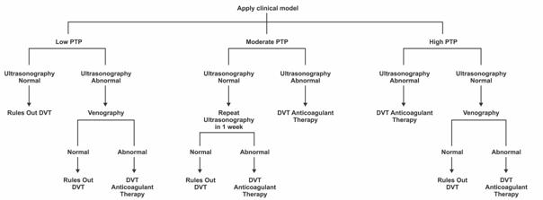
Since physical examination is unreliable, the diagnosis of DVT must be pursued further in any patient who presents with unexplained pain or swelling of the lower limbs. For patients with risk factors for DVT, the threshold for obtaining imaging studies is lower. Accordingly, almost all patients with symptoms compatible with venous thrombosis, require an imaging study. Patients with suspected DVT who are suffering from chest pain or shortness of breath should also be investigated for PE.
Imaging studies for DVT
Imaging studies include both invasive (venography and radiolabeled fibrinogen) and non-invasive (ultrasound, plethysmography and magnetic resonance imaging [MRI]) techniques.
Ultrasonography
Venous compression ultrasonography is the most frequently used diagnostic test, being used in approximately 95% of cases. It is a non-invasive test and is very accurate for proximal DVT. It involves compressing the proximal veins with an ultrasound transducer and watching the venous compression on the machines monitor; an inability to compress the veins is suggestive of DVT.
Color-coded Doppler methods are now available and are the imaging method of choice for detecting DVT. These methods are highly accurate and effective in detecting thrombi above the knee, but are less reliable for detecting thrombi in the calf veins.
The color-flow duplex scan also uses ultrasound to image the leg veins. Although excellent at detecting proximal thrombi, it is limited in its ability to identify thrombi in the calf and pelvic veins.
Impedance plethysmography
Impedance plethysmography (IPG) is a non-invasive imaging method that can detect changes in blood volume in the leg as a function of venous outflow. It works by detecting changes in calf circumference or cutaneous blood flow that may occur in the presence of DVT. Unfortunately, any impairment of venous outflow, which can occur in pregnancy and heart failure, affects the results of IPG, and therefore many false positives can occur. IPG is useful for detecting proximal thrombi, but is less helpful for detecting thrombi in the calf veins.
Magnetic resonance imaging
Another non-invasive testing method, MRI, is able to detect thrombi in the legs, pelvis and lungs; however, it is expensive. This method is non-radioactive and therefore is useful in pregnancy and when ultrasound is not helpful.
Contrast venography
For the most part, ultrasonography has replaced venography. This is because venography is an invasive procedure and thus itself carries a risk of causing thrombosis. Consequently, its use tends to be limited to particular circumstances, such as if the ultrasound is inconclusive, or if clear imaging of the calf veins is required.
Contrast venography involves the injection of a radio-opaque contrast medium into the veins of both feet. X-rays are then taken following the contrast medium through the lower limbs. This makes it possible to detect thrombi in the calves, thighs and vena cava.
Venography is the most accurate method for the diagnosis of any thrombi (symptomatic or asymptomatic) and is therefore used in clinical trials.
Blood tests: D-dimer blood test
Blood testing can check concentrations of D-dimer, a degradation product of crosslinked fibrin. Patients with VTE have high concentrations of D-dimer in the blood. D-dimer concentration may also rise as a result of serious illness - so false negatives can occur.
The test has 98% sensitivity for DVT, moderate specificity and a high negative predictive value. Sensitivity is lower for smaller distal thrombi in the calf veins. A normal D-dimer level in a patient with no risk factors excludes the need for other diagnostic investigations.
The clinical characteristics of PE can range from asymptomatic to sudden death. The symptoms, when present, will vary depending on the extent of pulmonary arterial occlusion: a large single PE causes major problems whereas multiple small emboli cause a different pattern of symptoms.
Symptoms include dyspnea, pleuritic chest pain (pain that is worse on taking a deep breath), cough, tachycardia and tachypnea.
The diagnosis of PE by clinical assessment is often unreliable; therefore, it is essential to confirm the diagnosis with appropriate diagnostic tests.
Angiography
Angiography is the gold standard for PE diagnosis, but this invasive procedure is not without risk, therefore a ventilation/perfusion lung scan has been the favored, non-invasive test.
Angiography involves the injection of a contrast agent (X-ray dye) into the blood vessels of the lungs via the heart. This makes the vessels visible on the X-ray and thus blockages or narrowings in the vessels can be detected.
Lung scan
The lung scan is usually conducted to diagnose PE. The lung scan consists of two parts: a perfusion and a ventilation portion. Perfusion refers to the blood flow through the lungs and ventilation refers to the air flow into and out of the lungs.
Perfusion lung scan
The purpose of the perfusion portion is to detect any blockage of blood flow through the vessels of the lungs. An intravenous injection of the radioactive isotope Technetium-99m is given. These radioactive particles are transported through the circulatory system to the vessels of the lungs. The lungs are then scanned with a special radioactive sensitive camera.
This perfusion scan creates an image showing the distribution of the radioactive Technetium-99m in the vessels of the lungs. If a PE is present causing blockage of blood flow to a part of the lung, the image reveals a reduced or absent distribution of Technetium-99m to the involved area. This causes a perfusion defect on the scan image.
Unfortunately, other diseases can result in similar perfusion defects; therefore, in order to improve the specificity of the perfusion lung scan, a ventilation scan is normally done as well.
Ventilation lung scan
In the ventilation portion of the lung scan a radioactive gas is inhaled. An image is taken with a special camera that shows the distribution of the inhaled radioactive gas in the airways. By cross-matching the information from both scans, a more accurate diagnosis of PE can be made.
Owing to the often clinically silent nature of the disease, the potentially harmful consequences of unrecognized and consequently untreated VTE, including the risk of sudden death, and its high prevalence, particularly in certain certain groups of surgical or medical patients, the use of systematic thromboprophylaxis has been advocated as the best answer to this threat and is now recommended by both the North American and European Consensus statements (Nicolaides 2001; Geerts 2001).
Indeed,
an alternative to systematic prophylaxis would be to use serial surveillance
tests such as impedance plethysmography or duplex ultrasonography in high-risk
patients (Geerts 2001). However, this approach is expensive and can
be applied to only limited numbers of patients at risk. In addition, available
screening methods suffer from various limitations. Impedance plethysmography,
duplex ultrasonography and color doppler ultrasound have been shown to have
only moderate sensitivity and
positive predictive value when used in asymptomatic medical or high-risk
surgical patients (Keller 95; Harenberg 96; Wells 95; Davidson 92; Agnelli 91).
Leg scanning with labeled fibrinogen was widely used to diagnose DVT in
both asymptomatic surgical and medical patients (Geerts 01; Nicolaides 01)
until this product was taken off the market owing to the risk of
potential viral contamination. Venography remains the most accurate and
reliable and thus reference method to screen for DVT in asymptomatic patients.
However, this procedure is invasive, cumbersome and potentially harmful and
cannot be used as a routine screening method.
Importantly, broad application of effective methods of prevention has been shown to be more cost-effective and is probably safer than selective, intensive surveillance (Geerts 01). Various methods are recommended according to the levels of patient risk (Geerts 01; Nicolaides 01) (see table below).
Preventive Strategies According to VTE Risk
|
Low Risk |
No
specific measures |
|
Moderate Risk |
Anticoagulant
agents |
|
High Risk |
Anticoagulant
agents in higher dose regimens |
|
Highest Risk |
Combination of various pharmacological and physical methods |
Despite these clear-cut recommendations based on solid scientific evidence, recent surveys among surgeons suggest that thromboprophylaxis is underused.
In the US, no more than 26% of major abdominothoracic surgery patients, who are at very high risk for VTE, were found to receive appropriate prophylaxis (Bratzler 98), a rate which increased to 50% only in a more recent survey (Stratton 00).Other surveys in New Zealand revealed that only 37% of patients undergoing total hip replacement (THR) or total knee replacement (TKR) (Francis 97), conditions in the highest risk category for developing VTE, and only 20% of high-risk medical patients (Egermayr 99) received some kind of VTE prophylaxis. A French survey found that 36% of THR and TKR patients received a lower anticoagulation dosage than the recommended one (Villemur 98), a rate comparable to that found in the overall at-risk population in a French hospital (Di Sandro 99)
The most frequent reasons found to explain this underuse of thromboprophylaxis are fear of adverse effects, treatment costs, and lack of awareness regarding the high incidence of postoperative VTE (Geerts 01). Many surveys have also demonstrated that clinicians may have difficulties in identifying patients at risk for VTE, whether surgical or medical patients, as they may not be aware of all common risk factors or may not fully grasp their impact on the VTE risk (Caprine 94; Turton 99). It has been suggested that the preventive management of VTE could be improved by the implementation of training sessions for physicians on the apropriate use of anticoagulant therapy (Di Sandro 99).
All modern strategies for the prevention of VTE are based on assigning individual patients to risk categories on the basis of the presence of risk factors. The risk category is then used to guide the choice of prophylactic therapy. It should be noted that, as already discussed, a wide variety of conditions and situations as well as genetic predispositions can contribute to an increased risk of VTE, so that a large number of patients may have multiple risk factors for VTE simultaneously. Thus, even a situation intrinsically associated with a low VTE risk may trigger a sudden, unexpected VTE event in an apparently healthy individual when superimposed on other chronic and/or transient risk factors. Improved identification of VTE risk profiles could lead to much better prophylaxis and a substantial reduction in the incidence of VTE (Rosendaal 99).
A recent project, the DecisionMatrix for prophylaxis of venous thromboembolism, tries to give computerised support for this difficult decision. The physician has to created a very detailed patient profile and then gets feedback on the calculated overall risk and the recommended appropriate method of prophylaxis. For further details please refer to the DecisionMatrix-program.
Another area of probable underuse is in the duration of propylaxis. There is convincing data, specially for orthopedic patients, that prolonged prophylaxis up to 35 days significantly decreases the DVT rate (Eikelboom 01).
The risk of postoperative VTE is often delayed.
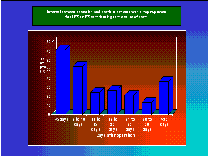
Simple measures that are helpful for patients undergoing surgery are avoiding dehydration, ensuring early mobilization and undertaking leg exercises. Early ambulation after surgery helps to activate the calf muscle pump to encourage venous return. Early ambulation is usually sufficient to prevent DVT in low-risk patients.
In addition to these measures, mechanical devices - surgical stockings and mechanical pumps - can reduce venous stasis in the legs.
Antithrombotic agents
In the past, antithrombotic agents were described as anticoagulants, but now the term 'antithrombotic agent' is more widely used. Essentially, the two terms are synonymous.
Prevention of thrombus formation with prophylaxis agents can reduce morbidity and mortality in surgical patients at risk of developing VTE. Antithrombotic agents prevent thrombus formation after surgery by interfering with the coagulation process.
It is evident from the way that older antithrombotics interact in the coagulation process that they differ in the way they act at non-specific multiple targets and the unwanted side effects that they cause. Nevertheless, they have been very useful and the systematic use of therapeutic prevention has strongly reduced the number of VTE events after surgery.
Oral anticoagulants
Oral anticoagulants used as prophylaxis include warfarin and aspirin.
Warfarin is a vitamin K antagonist that causes reductions in the concentrations of vitamin K-dependent factors IIa, VIIa, IXa and Xa in the blood. Prophylaxis using warfarin can be given before, during and after surgery.
The advantages of warfarin are that it can be given by mouth and it is inexpensive. However, regular laboratory monitoring of coagulation is needed to ensure that the patient is receiving the correct dose, because there is a high risk of bleeding complications when doses are too high - adding further costs to the treatment protocol. Warfarin also has important interactions with many drugs routinely given to surgical patients. As it is an oral agent it cannot be given to many surgical patients in the immediate post-operative period, because they are 'Nil by mouth'. Additionally, warfarin does not produce its maximum effect for 3 or more days after it is started.
Aspirin inhibits platelet aggregation and reduces levels of thromboxane A2, an inducer of platelet aggregation and a vasoconstrictor. It is inexpensive, can be given orally and, unlike warfarin, does not require laboratory monitoring. In arterial thrombotic disorders, aspirin remains one of the main drugs of choice. However, aspirin is much less effective than low molecular weight heparins (LMWHs) or warfarin in preventing VTE, and as such is not recommended for the prophylaxis of VTE .
Unfractionated heparins (UFH)
Low-dose heparin prevents DVT by inhibiting thrombin and coagulation factors IIa and Xa, as well as factors XIIa, XIa, and IXa. Low-dose heparin is given by subcutaneous injection, usually after surgery, although the first dose can be given pre-operatively. Administration continues for as long as the risk persists from about one week or until the patient is fully mobile.
Low-dose heparin works quickly and does not require laboratory monitoring. There is a risk of bleeding during and after surgery, and also a risk of heparin-induced thrombocytopenia (HIT).
Unfractionated heparins (UFHs) are also used at higher doses. Administration of this so-called adjusted-dose heparin begins 2 days before surgery and is subcutaneous in dose-adjusted steps and requires close monitoring. This monitoring, known as activated prothrombin time monitoring, can only be carried out on inpatients; therefore the use of UFHs is not always convenient.
Heparin binds to numerous blood components, endothelial vessels, proteins, macrophages and osteoblasts. Such interactions decrease its activity in an unpredictable manner. Heparin has decreased bioavailability when administered subcutaneously in low doses, but has a higher bioavailability when administered in high doses - these pharmacokinetic properties account for the 24-hour delay before steady state levels are reached. Because of this, constant monitoring of the antithrombotic activity is required. Osteoporosis remains a serious, if rare, complication of heparin therapy.
Low molecular weight heparins (LMWH)
LMWHs are given once- or twice-daily by injection for the treatment or prevention of DVT.
LMWHs are an effective and safe form of prophylaxis for DVT. They have been recommended for use after major orthopedic surgery and as a preventative measure in surgical patients at moderate risk, high risk and highest risk of thromboembolism. They are simple to administer and can be given before or after surgery. They also have an improved efficacy and bleeding profile as well as a reduced risk of HIT compared with UFHs.
A number of LMWHs, each with different properties, have been developed.
Hirudin agents
Hirudin is an anticoagulant originally derived from the salivary glands of the leech. It is a direct thrombin inhibitor that appears to be very effective and does not require any monitoring. Two recombinant hirudins have recently been introduced, Lepirudin and Desirubin. The disadvantages with hirudins are that they are costly, require twice-daily parenteral administration and carry a high risk of bleeding.
Surgical stockings
For an at-risk patient undergoing surgery, the use of elastic graduated compression stockings in the peri-operative period may help reduce the risk of DVT, although their use is often seen as complementary or secondary to other treatments. The stockings, theoretically, reduce the venous cross-sectional area, which increases venous flow and avoids stasis. They prevent blood from pooling in the distal veins by progressively applying more pressure as they descend towards the ankles.
To date, the stockings are virtually free of side effects and are of low cost, but they do need to be fitted carefully to be effective. Additionally, they are limited in use as they are dependent on the limb being free of bandages or casts. The recommended use of surgical stockings in high-risk patients is in conjunction with prophylaxis. However, use of compression stockings is often considered as a basic preventative measure in all at-risk patients undergoing surgery.
LMWH in combination with graduated compression stockings has been shown to be significantly more effective than stockings alone in the prevention of DVT in patients who have undergone hip replacement under spinal anesthesia (Samama 97).
Mechanical compression devices
Intermittent pneumatic compression devices applied to the leg - usually to the calf, but occasionally to the thigh or foot - work to reduce venous stasis by generating intermittent pressure for about 10 seconds in every minute. They can be used alone or with elastic stockings. This method is virtually free of side-effects, but is quite inconvenient, dependent on the limb being free of bandages or orthopedic casts, and the patient must comply with instructions of use.
It is not recommended as first choice in patients undergoing major orthopedic surgery. However, it may be used in patients undergoing surgery with a high risk of bleeding, such as neurosurgery and prostate surgery.
Mechanical compression devices may lower the risk of DVT but they do not remove the risk. Direct comparison between mechanical compression devices and the best available pharmacological prophylaxis available (LMWH) demonstrates the clear superiority of LMWH.
Comparative efficacy of treatments
Mechanical methods of prophylaxis - graduated compression stockings and intermittent pneumatic compression - appear to complement the effects of drug prophylaxis in general surgery, but are not relied upon as the sole method of prophylaxis.
Warfarin is effective in preventing post-operative DVT in all at-risk patients, but its maximum effect is not seen for several days after initiation. In addition, warfarin requires very careful monitoring because of its inhibitory activity against vitamin K (leading to vitamin-K deficiency) and its known adverse interactions with many commonly used drugs. Heparins, therefore, are much more widely used for the prophylaxis of VTE in surgical patients. Warfarin's key role in VTE is for the long-term treatment, rather than the prevention, of DVT. Aspirin is not as effective at preventing DVT as any of the other agents.
Currently, the heparins (LMWH and UFH) are the drugs of choice for the prophylaxis of VTE.
LMWH is an agent of choice for moderate-to-high risk general surgical and medical patients, as it can reduce the risk of VTE by 50-70% (Anderson 98).
An overview of more than 70 studies investigating the prophylactic effects of LMWH in general and orthopedic surgery found that DVT would be reduced from approximately (Anderson 98; Mismetti 00):
22% to 9% in general surgery, ie 59% risk reduction
48% to 24% in orthopedic surgery, ie 50% risk reduction
41% to 14% in urological surgery, ie 68% risk reduction.
LMWHs are to date the most effective form of prophylaxis for patients undergoing hip and knee surgery, hip fracture and after major trauma. They appear to be more effective than low-dose heparin and seem to produce less bleeding than the UFHs for a similar antithrombotic effect with no need to monitor antithrombotic effect and a reduced number of injections.
Virchow R. Gesammelte Abhandlungen zur
Wissenschaftlichen Medizin. 1856;
Rosendaal FR. Venous thrombosis: a multicausal disease. Lancet 1999;353:1167-73.
Mattson JC, Crisan D. Inherited
Thrombophilia due to Factor V
Molecular Diagnostics 1998; 3:55-61.
Ageno W. Applying risk assessment models in general surgery: overview of our clinical experience. Blood Coagulation Fibrinolysis 1999;10 (Suppl 2):S71-S78.
Perrier A, Bounameaux H, Morabia A, et al. Diagnosis of pulmonary embolism by a decision analysis-based strategy including clinical probability, D-dimer levels, and ultrasonography: a management study. Arch Int Med 1996;156:531-6.
Hirsh J, Hoak J. Management of deep vein thrombosis and pulmonary embolism. A statement for healthcare professional. Circulation 1996;93:2212-45.
Turkstra F, Koopman MMW, Büller HR. The treatment of deep vein thrombosis and pulmonary embolism. Thromb Haemost 1997;78:489-96.
Huisman MV, Büller HR, ten Cate JW, van Royen EA, Vreeken J, Mersten MJ, Bakx R. Unexpected high prevalence of silent pulmonary embolism in patients with deep venous thrombosis. Chest 1989;95:498-502.
Meignan M, Rosso J, Gauthier H, Brunengo F, Claudel S, Sagnard L, d'Azemar P, Simonneau G, Charbonnier B. Systematic lung scans reveal a high frequency of silent pulmonary embolism in patients with proximal deep venous thrombosis. Arch Intern Med 2000;160:159-64.
Doyle DJ, Turpie AGG, Hirsh J, Best C, Kinch D, Levine MN, Gent M. Adjusted subcutaneous heparin or continuous intravenous heparin in patients with acute deep vein thrombosis. Ann Intern Med 1987;107:441-5.
Girard P, Musset D, Parent F, et al. High prevalence of detectable deep venous thrombosis in patients with acute pulmonary embolism. Chest 1999; 116:903-8.
Partsch H, Kechavarz B, Mostbeck A, Kohn H, Lipp C. Frequency of pulmonary embolism in patients who have iliofemoral deep vein thrombosis and are treated with once- or twice-daily low-molecular-weight heparin. J Vasc Surg 1996; 24:774-82.
Goldhaber SZ, Visani L, De Rosa M. Acute pulmonary embolism: clinical outcomes in the International Cooperative Pulmonary registry (ICOPER). Lancet 1999;353:1386-9
Stein PD, Huang H, Afzal A, et al. Incidence of acute pulmonary embolism in a general hospital. Chest 1999; 116:909-13.
Hyers TM, Agnelli G,
Geerts WH, Heit JA, Clagett GP, et al. Sixth ACCP Consensus Conference on antithrombotic therapy. Prevention of venous thromboembolism. Chest 2001;119 (Suppl):132S-175S.
Nicolaides AN. Prevention of venous thromboembolism. International Consensus Statement. Guidelines compiled in accordance with the scientific evidence. Int Angiol 2001;20:1-37.
Haeger K. Problems of acute deep venous thrombosis. Angiology 1969;20:219-23.
Lensing AWA, Prandoni P, Prins MH et al. Deep-vein thrombosis. Lancet 1999; 353:479-85.
Lethen H, Flachskampf FA, Schneider R, Sliwka U, Kohn G, Noth J, Hanrath P. Frequency of deep vein thrombosis in patients with patent foramen ovale and ischemic stroke or transient ischemic attack. Am J Cardiol 1997;80:1066-9.
McLachlin J,
Richards T,
Sandler DA, Martin JF. Autopsy proven pulmonary embolism in hospital patients. Are we detecting enough deep vein thrombosis. J Royal Soc Med 1989;82:203-5.
Goldhaber SZ,
Hennekens H, Evans DA,
Franzeck UK, Schalch I, Jäger KA, Grimm J, Bollinger A. Prospective 12-year follow-up study of clinical and hemodynamic sequelae after deep vein thrombosis in low-risk patients (Zürich study). Circulation 1996;93:74-9.
Prandoni P, Lensing AWA, Cogo A, Cuppini S, Villalta S, Carta M, Cattelan AM, Polistena P, Bernardi E, Prins MH. The long-term clinical course of acute DVT. Ann Intern Med 1996:125:1-7.
Lagersted CI, Olsson CG, Fagher BO, Öqvist BW, Albrechtsson U. Lancet 1985;2:515-8.
Prandoni P, Lensing AWA, Prins MR. Long-term outcomes after deep venous thrombosis of the lower extremities. Vasc Med 1998; 3:57-60.
Prandoni P, Villalta S, Bagatella P, et al. The clinical course of deep vein thrombosis. Prospective long-term follow-up of 528 symptomatic patients. Haematologica 1997;82:423-8.
NIH Consensus Conference. Prevention of venous thrombosis and pulmonary embolism. JAMA 1986;256:744-9.
Silverstein MD, Heit JA, Mohr DN, et al. Trends in the incidence of deep vein thrombosis and pulmonary embolism: a 25-year population-based study. Arch Intern Med 1998; 158:585-93.
Nordstrom M, Lindblad B, Bergqvist D, Kjellstrom T. A prospective study of the incidences of deep-vein thrombosis in a defined urban population. J Intern Med 1992;232:155-60.
Lindblad B,
Rasmussen MS, Wille-Jorgensen P, Jorgensen LN. Postoperative fatal pulmonary embolism in a general surgical department. Am J Surg 1995; 169:214-6.
Lindblad B, Eriksson A, Bergqvist D. Autopsy-verified pulmonary embolism in a surgical department: analysis of the period from 1951 to 1988. Br J Surg 1991; 78:849 -52.
Bergqvist D, Lindblad B. A 30-year survey of pulmonary embolism verified at autopsy: an analysis of 1,274 surgical patients. Br J Surg 1985; 72:105-8.
Scurr JH, Coleridge-Smith PD, Hasty JH. Deep venous thrombosis: a continuous problem. Br Med J 1988;297:28.
Huber O, Bounameaux H, Borst F, Rohner A. Postoperative pulmonary embolism after hospital discharge: an underestimated risk. Arch Surg 1992;127:310-3.
Thromboembolism Risk Factors (THRIFT) Consensus Group. Risk and prophylaxis for venous thromboembolism in hospital patients. Br Med J 1992;305:567-74.
Bergqvist D, Jendteg S, Johansen L, et al. Cost of long-term complications of deep vein thrombosis of the lower extremities: an analysis of a defined patient population in Sweden. Ann Intern Med 1997; 126:454-7.
Keller F, Flosbach CW and the PRIME study group. A randomized multicenter study investigating the efficacy and safety of the low molecular weight heparin enoxaparin versus unfractionated heparin in the prevention of thromboembolism in immobilized medical patients. Thromb Haemostas 1995;73:1106 (ABS 788).
Harenberg J, Roebruck P, Heene DL on behalf of the Heparin Study in Internal Medicine Group. Subcutaneous low-molecular-weight heparin versus standard heparin and the prevention of thromboembolism in medical inpatients. Haemostasis 1996;26:127-39.
Wells PS, Lensing AWA, Davidson BL, Prins MH, Hirsh J. Accuracy of ultrasound for the diagnosis of deep venous thrombosis in asymptomatic patients after orthopedic surgery. A meta-analysis. Ann Intern Med 1995;122:47-53.
Davidson BL, Elliot CG, Lensing AWA. Low accuracy of color doppler ultrasound in the detection of proximal leg vein thrombosis in asymptomatic high-risk patients. Ann Intern Med 1992;117:735-8.
Agnelli G, Cosni B, Ranucci V, Renga C, Mosca S, Lupatelli L, Di Filippo P, Rinonapoli E, Nenci GG. Impedance plethysmography in the diagnosis of asymptomatic deep vein thrombosis in hip surgery. Arch Intern Med 1991;151:2167-71.
Jongbloets LMM, Lensing AWA, Koopman MMW, Buller HR, ten Cate JW. Limitations of compression ultrasound for the detection of symptomless postoperative deep vein thrombosis. Lancet 1994;343:1142-4.
Bratzler DW, Raskob GE, Murray CK, Bumpus LJ, Piatt DS. Underuse of venous thromboembolism prophylaxis for general surgery patients: physician practices in the community hospital setting. Arch Intern Med 1998;158:1909-12.
Stratton MA, Anderson FA, Bussey HI, et al. Prevention
of venous thromboembolism: adherence to the 1995 American
Francis CW, Felcher AH, White J, Braaten JV, Goss R. Thrombin activity associated with indwelling central venous catheters. Thromb Haemost 1997;77:48-52.
Egermayer P, Robins C, Town GI. Survey of the use of thromboprophylaxis for medical patients at Christchurch Hospital. N Z Med Journal 1999;112:246-8.
Villemur B, Bosson JL, Diamand JM. Deep venous thrombosis (DVT) after hip or knee prosthesis. Evaluation of practices for prevention and prevalence of DVT on doppler ultrasonography. J Mal Vasc 1998;23:257-62.
Di Sandro I, et al. Le Pharmacien Hospitalier 1999;34:19-25.
Caprini JA, Arcelus JI, Hoffman K, et al. Prevention of venous thromboembolism in North America: results of a survey among general surgeons. J Vasc Surg 1994;20:751-8.
Turton EP, Whiteley MS, Berridge DC, Scott DJ. Calman, venous surgery and the vascular trainee. J R Coll Surg Edinb 1999;44:172-6.
Collins R, Scrimgeour A, Yusuf S, Peto R. Reduction in fatal pulmonary embolism and venous thrombosis by perioperative administration of subcutaneous heparin. Overview of results of randomized trials in general, orthopedic, and urologic surgery. N Engl J Med 1988;318:1162-73.
Hirsh J, Warkentin TE, Shaughnessy SG, et al. Heparin and low-molecular-weight heparin. Mechanisms of action, pharmacokinetics, dosing, monitoring, efficacy, and safety. Chest 2001;119:64S-94S.
Hirsh J, MD, Dalen JE, David R, Anderson DR, et al. Oral Anticoagulants: mechanism of action, clinical effectiveness, and optimal therapeutic range. Chest 2001;119:8S-21S.
Ansell J, Hirsh J, Dalen J et al. Managing Oral Anticoagulant Therapy. Chest 2001;119:22S-38S.
Eriksson BI, Eriksson E, Gyzander E, Teger-Nilsson A-C, Risberg B. Thrombosis after hip replacement. Relationship to the fibrinolytic system. Acta Orthop Scand 1989;60:159-63.
Hoek JA, Nurmohamed MT, ten Cate JW, et al. Thrombin-antithrombin III complexes in the prediction of deep vein thrombosis following total hip replacement. Thromb Haemost 1989;62:1050-2.
Ginsberg JS, Brill-Edwards P, Panju A, et al. Pre-operative plasma levels of thrombin-antithrombin III complexes correlate with the development of venous thrombosis after major hip or knee surgery. Thromb Haemost 1995;74:602-5.
Cofrancesco E, Cortellaro M, Corradi A, Ravasi F, Bertocchi F. Coagulation activation markers in the prediction of venous thrombosis after elective hip surgery. Thromb Haemost 1997;77:267-9.
Gallus AS. Applying risk assessment models in orthopaedic surgery: overview of our clinical experience. Blood Coagul Fibrinolysis. 1999; 10:S53-S61.
Seagroatt V, Tan HS, Goldacre M, Bulstrode C, Nugent I, Gill L. Elective total hip replacement: incidence, emergency readmission rate, and postoperative mortality. Br Med J 1991;303:1431-5.
Kelton JG. Heparin-induced thrombocytopenia.Haemostasis 1986;16:173-86.
Davidson BL. What are the most reliable detection methods for DVT and PE to be used as endpoints in trials of venous thromboprophylaxis. Haemostasis 1998;28 (Suppl 3):113-9.
Kearon C, Julian JA, Math M, Newman TE, Ginsberg JS for the Mc Master Diagnostic Imaging Practice Guidelines Initiative. Noninvasive diagnosis of deep venous thrombosis Ann Intern Med 1998;128:663-
Anderson F, Audet A. Best practices. Preventing deep vein thrombosis and pulmonary embolism. Worcester: Center for Outcomes Research, University of Massachusetts Medical Center, 1998.
Goldhaber SZ, Visani L, De Rosa M et al. Acute pulmonary embolism: clinical outcomes in the International Pulmonary Embolism Registry (ICOPER). Lancet 1999; 353: 1386-1389.
Ferris EJ. Deep venous thrombosis and pulmonary embolism: correlative evaluation and therapeutic implications. Am J Roentgenol 1992; 159: 1149-1155.
Linblad B, Sternby WH, Bergquist D. Incidence of venous thromboembolism verified by necropsy over 30 years. BMJ 1991; 302: 709-711.
Heit JA, Silverstein MD, Mohr DN et al. The epidemiology of venous thromboembolism in the community. Thromb Haemost 2001; 86: 452-463.
Nicolaides AN, Arcelus J, Belcaro G et al. Prevention of venous thromboembolism. European Consensus Statement. Int Angiol 1992; 11: 151-159.
Heit JA, Mohr DN, Silverstein MD et al. Predictors of recurrence after deep vein thrombosis and pulmonary embolism: a population-based cohort study. Arch Intern Med 2000; 160: 761-768.
Oger E for EPI-GETBO Study Group. Incidence of venous thromboembolism: a community-based study in western France. Thromb and Hemostasis 2000; 83: 657-660.
Prandoni P, Lensing AW, Cogo A et al. The long-term clinical course of acute deep venous thrombosis. Ann Intern Med 1996; 125(1): 1-7.
Kniffin, WD Jr, Baron JA, Barrett J et al. The epidemiology of diagnosed pulmonary embolism and deep vein thrombosis in the elderly. Arch Intern Med 1994; 154: 861-866.
Rosendaal FR. Risk factors for venous thrombotic disease. Thromb Haemost 1999; 82: 610-619.
Coon WW, Willis PWI, Keller JB. Venous thromboembolism and other venous disease in the Tecumseh: Community health study. Circulation 1973; 48: 839.
Prandoni P, Piccioli A. Venous thromboembolism and cancer: a two-way clinical association. Frontiers in Bioscience 1997; 2: e12-21.
de Thomasson E, Strauss C, Girard P et al. Detection of asymptomatic venous thrombosis after lower limb prosthetic surgery. Retrospective evaluation of a systematic approach using Doppler ultrasonography: 400 cases. Presse Med 2000; 29: 351-357.
Wells PS, Anderson DR, Bormanis J et al. Value of assessment of pretest probability of deep-vein thrombosis in clinical management. Lancet 1997; 350: 1795-1798.
Hull RD, Raskob GE, Leclerc J et al. The diagnosis of clinically suspected venous thrombosis. Clin Chest Med 1984: 5: 439-462.
O'Donnell T, Abbot W, Athanasoulis CM et al. Diagnosis of deep venous thrombosis in the outpatient by venography. Surg Gynecol Obstet 1980; 150: 69-74.
Anand S, Wells P, Hunt D et al. Does this patient have DVT? JAMA 1998; 279(14): 1094-1099.
Collins R, Scrimgeour A, Yusuf S et al. Reduction in fatal pulmonary embolism and venous thrombosis by perioperative administration of subcutaneous heparin. Overview of results of randomized trials in general, orthopedic and urologic surgery. N Engl J Med 1988; 318: 1162-1173.
Hirsh J. Guidelines for antithrombotic therapy, 4th edition. London: Hamilton. 2001.
Samama CM, Clergue F, Barre J et al. Low molecular weight heparin associated with spinal anaesthesia and gradual compression stockings in total hip replacement surgery. Br J Anaesthesia 1997: 78: 660-665.
Blanchard J, Meuwly JY, Leyvraz PF et al. Prevention of deep-vein thrombosis after total knee replacement. Randomised comparison between a low-molecular-weight heparin (nadroparin) and mechanical prophylaxis with a foot-pump system. J Bone Joint Surg 1999; 81: 654-659.
|
