RESTORATIVE TREATMENT OF CROWDED TEETH - Geoffrey W.
Sheen, DDS, MS, Ronald E. Goldstein, DDS, Steven T. Hackman, DDS
INTRODUCTION
Orthodontics is generally the first consideration when the patient presents
with crowded teeth.3,13,19,34 If a patient is unable to accept
comprehensive orthodontic procedures, the general practitioner must determine
whether the patient can be treated with minor tooth movement, restorations,
extraction, or a combination of these procedures.
To analyze the treatment of crowded teeth, this chapter has been organized into
the following sections: Treatment Considerations, Treatment Strategy, Treatment
Options, and Unusual or Rare Clinical Presentations.
TREATMENT CONSIDERATIONS
Many patients have slightly crowded or overlapping anterior teeth that are not
an esthetic problem. However, when an individual who finds this situation
unesthetic seeks treatment, it may present a challenge for the dentist.
Choosing the correct approach is the most important aspect of the treatment.36
Prior to properly developing a plan of treatment, the dentist should consider a
number of preoperative conditions. A thorough evaluation of the patient will
establish the basis for potential treatment options.37 The areas to
be evaluated include arch space, gingival architecture, influence of root
proximity, smile line, emergence profile, and oral hygiene (Table 24-1
Arch Space
The most significant factor in the treatment of crowded teeth is the available
arch space, as well as how that space is occupied by the dentition. Locating
space deficiencies and their degree will determine which teeth will require
modification.
Berliner presented a classic formulation and clinical rule in his text that
helps to make treatment of crowding more predictable. He stated:
When the sum of the mesiodistal widths (at contact-point level from distal or
right lateral to distal of left lateral) in any given segment measures more
than the available arch space, when measured between the two points (obtained
by dropping perpendicular lines from the mesial contact-point levels of the
right and the left cuspids to the gingival line), the central and the lateral
teeth will be buckled (displaced labially or lingually) or overlapped;
conversely, when the sum of the combined mesiodistal widths of the central and
the lateral teeth measure less than the available arch space (as indicated
above), the involved teeth present diastemas.2
This formula can aid in the correction of crowded or spaced teeth by measuring
the amount to be added or subtracted for the desired objective (Figures 24-1A to
C
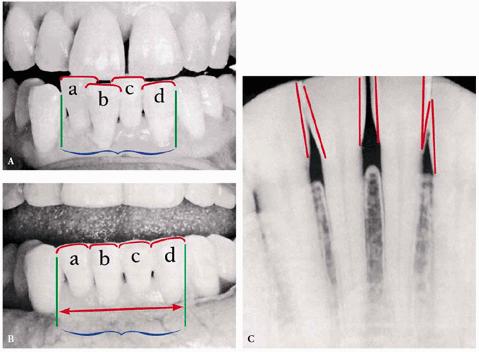
Figure 24-1A to C: (A) Preoperative view: note the relationship of the combined widths of the lower central and lateral teeth (A, B, C, D) to the extent of available arch space. Sum of combined tooth widths > available arch space, resulting in buckling. (B) Postoperative measurement, after remodeling of the lower central and lateral incisor teeth: the combined tooth widths equal the available arch space, and repositioning of the teeth became a feasible clinical procedure. (C) The proximal thickness of the enamel "caps" of the teeth on a lower anterior segment of the dental arch is indicated in outline. (Reproduced with permission from Berliner A. Ligatures, splints, bite pla 737j923h nes and pyramids. Philadelphia: JB Lippincott, 1964:65.)
Gingival Architecture
An often overlooked component of an esthetic smile is the gingival
architecture. When there is crowding in the anterior region, certain teeth will
be forced facially or lingually. In a Class II, Division 2 occlusion, for
example, the maxillary lateral incisors may be positioned labially, and the
gingival tissue will be forced more apically. This creates a discontinuity in
the overall smile of the patient. Treatment considerations in this situation
may require slight modification to the gingival architecture around the central
incisors to create a more harmonious smile. If the patient's lip line hides the
gingival discrepancy, then surgical intervention may not be necessary (Figures 24-2A 24-2B 24-2C, and 24-2D
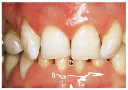
Figure 24-2A: This patient was dissatisfied with her crowded anterior teeth. Note how the gingival height differs between the central and lateral incisors.
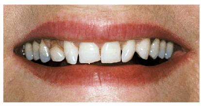
Figure 24-2B: The dissimilar gingival heights did not bother the patient because her natural smile line concealed these irregularities.
Figure
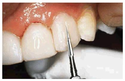
24-2C: After a slight reproportioning of the six anterior teeth, direct
composite resin was placed and contoured (6-mm ET [Brasseler,
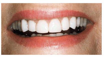
Figure 24-2D: The final result shows improved proportion in tooth size and form.
Likewise,
crowding in the mandibular anterior often results in the rotation or
lingualization of the central or lateral incisors. The gingival tissue will
therefore be positioned more incisally. It may be necessary to perform a
crown-lengthening procedure prior to esthetic restoration of these teeth.30,43
Influence of Root Proximity
Root proximity may complicate the restoration of crowded mandibular anterior
teeth. Root structures may be so close to one another that a separation is not
possible. This creates gingival impingement that can be almost impossible to
treat.
It may be necessary to extract one of the crowded teeth, leaving three incisors
in place of four.24 The decision should be based on radiographs and
a study of the periodontium to determine the amount of bone present. If bone
loss exists due to crowding, then extraction and repositioning are generally
the treatment of choice. This can be successful when the teeth are properly
proportioned. It is seldom noticeable to the patient. The tooth is extracted,
and the remaining anterior teeth are repositioned, providing additional bone
support. When small diastemas remain, the teeth can be bonded with composite
resin or splinted to prevent further tooth movement.
In most cases, some form of retainer is advisable. The patient should be
informed that he or she can reduce the wearing time of the retainer provided
that it does not fit too tightly each time it is placed. A tight fit might
indicate some relapse. The patient should return for possible further treatment
either to relax retentive gingival fibers or to adjust the occlusion to help
equilibrate the stressful occlusal forces.
Smile Line
It is important to study the patient's smile line. The extent to which
incisogingival tooth structure will show in the widest smile and in other
expressions should be noted. If the patient will be embarrassed by a small
metal collar or opaque margin of a porcelain-fused-to-metal crown, but there
are occlusal demands for the strongest restoration possible, a porcelain butt
margin on the labial surface of the crown is used. If occlusion is not a
problem, an all-ceramic crown or other esthetic restoration, such as bonding or
porcelain laminates, should be considered.1
Emergence Profile and Oral Hygiene
Many of the nonorthodontic treatment options discussed in this chapter are
meant to "camouflage" malposed or malaligned teeth. Consideration
must be given to the contours that will be created by the restorative process.18,22,42
Often, these contours are unnatural and create areas around the teeth that the
patient will find difficult to maintain with good oral hygiene. If these
esthetic restorations are to survive, the dentist must consider the final
contours being created, and the patient must be given the necessary
instructions to maintain them.
TREATMENT STRATEGY
Development of an appropriate treatment plan for the correction of crowded
teeth should follow a strategy. First, it is necessary to identify what type of
correction and how much correction of tooth contours are required to achieve
the desired esthetic results.23,34 Then it becomes necessary to
evaluate the dentition, identify clinical limitations to treatment, and select
appropriate restorative options that will accomplish the desired esthetic
outcome.
Identifying the Degree of Esthetic
Correction Required
Following a process of evaluation and development of a problem list, the
dentist may use this information to determine the degree of corrections
required. Esthetic computer imaging can assist the dentist and the patient to
visualize the proposed treatment.5,17 The development of a
diagnostic wax-up is a necessary procedure and may be used to confirm the
viability of the proposed treatment developed by computer imaging.
Two sets of diagnostic casts should be made. One set will serve as a historical
record of the patient's preoperative condition and should never be modified.
The second set of casts should be used for the diagnostic wax-up.
Developing a diagnostic wax-up involves both the addition of wax to deficient
areas of the dentition and the removal of stone as necessary to achieve the
desired esthetic results. The diagnostic wax-up should be accomplished with
attention to detail. Line angles, embrasure spaces, incisal lengths, and
gingival contours must represent the desired results if this effort is to be an
effective tool in the esthetic treatment of a patient.25 Through
this process, arch space deficiencies can be worked out, and specific
modifications to each tooth involved can be identified.
Once completed, the diagnostic wax-up is used to develop additional clinical
aids for the accurate and successful completion of the esthetic treatment plan.
Polyvinylsiloxane interocclusal record material, such as Regisil 2x
(DENTSPLY/Caulk,
A provisional matrix can also be fabricated by using the same materials and
technique. In addition to the palate, lingual surfaces, and incisal edges, the
interocclusal record material also covers the facial surfaces and extends
several millimeters onto the gingival tissue. The matrix formed will accurately
duplicate the subtleties of the diagnostic wax-up. With proper embrasure form
and gingival contours accurately duplicated in the provisional restorations,
chairside adjustment will be significantly reduced.
A well-planned and executed diagnostic wax-up is an essential communications
tool for both the patient and the laboratory.8,21,41 Dental
esthetics is truly in the eyes of the beholder. Everyone has a certain concept
of how the teeth should look. Since provisional restorations are closely
fabricated to the contours of the diagnostic wax-up,10 the patient
will have a chance to observe and identify any changes in contour and function
he or she may desire. If necessary, changes can be made in the provisional restorations,
and an impression of these newly contoured restorations can be made. The new
cast will serve as a clinically evaluated diagnostic tool used to communicate
this vital information to the laboratory.
In summary, a diagnostic wax-up will identify to what degree corrective
contours must be made to idealize a crowded dentition. With knowledge of the
specific modifications required for each tooth, the dentist can begin to select
the proper treatment options.35 This process should be undertaken
whether the treatment is minor esthetic contouring or as comprehensive as a
complete restoration of the anterior region with full-coverage crowns.
Identifying the Type of Restoration
Required
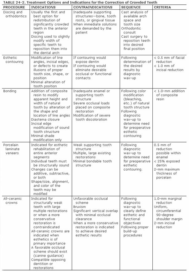
There are many treatment options available to the dentist for correcting
crowded teeth,20,26,29,33 including esthetic contouring, bonding,
porcelain laminates, and crowns (Table 24-2). The condition of the existing
dentition is a factor in determining which restorative option is ideal.
Teeth without any restorations or caries should be treated as conservatively as
possible. If only minor modifications to tooth contours are required to achieve
the desired esthetic result, esthetic contouring and bonding provide the least
invasive treatment options. Small existing restorations are easily incorporated
into other restorative treatments.
Caries in
the teeth to be treated may require that more extensive restorations be
considered, such as porcelain laminates or crowns. The size and location of the
caries may dictate the design of these restorations.
The presence of a root canal-treated tooth,14 with or without a post
and core, may require a crown. In the crowded tooth scenario, this may be
beneficial. The ability to reposition the crown into an ideal esthetic location
can more easily be accomplished when the tooth has been treated with a root
canal. Care must be taken, however, not to overextend or overcontour the final
restoration, which would result in possible gingival irritation.
TREATMENT OPTIONS
Conservative treatments such as esthetic contouring,11 discing
combined with minor tooth movement, and bonding are available for minor
corrections of crowded teeth. When corrections that are more substantial are
required, porcelain laminates and crowns become the treatment of choice.18
Correction by Discing
If the evaluation of arch space, as previously described by Berliner,2
shows that the combined addition of ABCD in the lower arch equals 21 mm (see Figure 24-1A) but the available space is 20 mm,
the amount of crowding is 1 mm. Therefore, if minor movement or repositioning
is attempted to realign the teeth, 1 mm of combined mesiodistal width can be
sacrificed through discing. However, not all of the tooth surface will have to
be lost from the central and lateral incisors. The mesial surfaces of the
cuspids are also available and, under certain rare conditions, the distal
surfaces of cuspids as well.
One
limitation on reducing tooth structure through discing is the thickness of
enamel on the teeth. Radiographs must be accurate enough to measure the
available enamel. A measurement can be made of the proximal surfaces on each of
the anterior teeth to predict the maximum reduction possible without
perforating the dentin.
For example, if 0.25 mm is found to be the amount that can be reduced per
proximal surface, then 0.5 mm can be reduced per tooth. Therefore, 3.0 mm could
theoretically be reduced from the six anterior teeth by discing to increase the
available arch space. In applying this to the earlier example of 1-mm crowding,
there should be no repositioning problem.
The procedure for applying the above principle is as follows:
1. Measure the mesiodistal width of the individual teeth and the available arch
space with a dental dial caliper.
2. Measure the enamel thickness by studying the radiographs of the involved
anterior teeth (see Figure 24-1C). Peck and Peck caution that
accessing enamel thickness from radiographs alone is subject to possible
distortion.32 Instead, they offer an arbitrary, but safe, guideline
of 50% of the mesiodistal enamel thickness as the maximum limit of
reproximation.
3. After determining that the amount of space necessary to realign the teeth is
attainable without perforating dentin, disc the teeth accordingly. This can be
done at one time, if the space is minimal, or over a period of time, depending
on the conditions present. The patient can be instructed to return weekly or
biweekly for stripping. Diamond separating strips (Compo-Strip, Premier Dental
Products,
4. Repositioning can now be accomplished by any of several different methods
shown in Chapter 25: Esthetics in Adult Orthodontics.
Correction by Bonding
The success of composite resin bonding has made immediate restorative
correction of crowded teeth possible.9,12 In most cases, it will be
necessary to combine the treatments of composite resin bonding with esthetic
contouring (see Chapter 11, Esthetics in Dentistry, Volume 1, 2nd
Edition) to produce the greatest effect. As with all bonding techniques, the
patient must be apprised of not only the esthetic life expectancy and
limitations of the bonded restoration but also an estimate of how much
maintenance may be required. The following example will show just how much can
be accomplished through composite resin bonding.
Bonding and Esthetic Contouring to Eliminate Crowding
PROBLEM: This 30-year-old horse trainer had extremely large central
incisors (Figure 24-3A). In addition, the mandibular
centrals were crowded and rotated lingually (see Figures 24-3A, and 24-3B
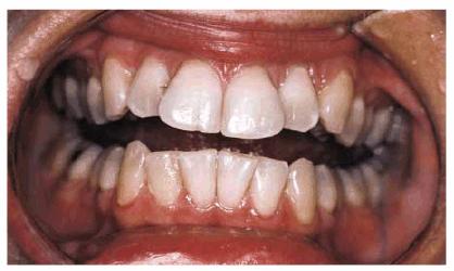
Figure 24-3A: This 30-year-old woman had extremely large central incisors with overlapping maxillary and mandibular teeth.
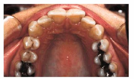
Figure 24-3B: An occlusal view showing the overlapping central incisors.
TREATMENT: Although the patient was informed about the advantage of
orthodontics as the ideal treatment, she nevertheless chose an immediate
correction in the form of esthetic contouring and esthetic resin bonding. The
central incisors were first contoured on the study cast to approximate the
desired tooth size. Esthetic contouring was accomplished in both the maxillary
(Figure 24-3C) and mandibular arches (Figure 24-3D). Next, the lateral incisors were
bonded labially to round out a more symmetric arch (Figures 24-3E, and 24-3F
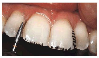
Figure 24-3C: Cosmetic contouring was first performed on the maxillary central incisors and mandibular anterior.
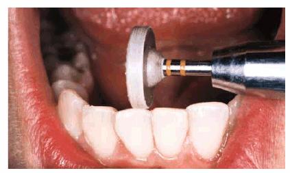
Figure
24-3D: After contouring the mandibular anterior teeth, they were polished with
an impregnated aluminum oxide wheel (Cosmetic Contouring Kit, Shofu,
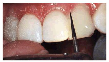
Figure 24-3E: Next, the maxillary laterals were bonded with composite resin and contoured and finished with ET carbide finishing burs (Brasseler).
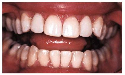
Figure 24-3F: Better tooth proportion and straighter- appearing teeth can be seen here before the final polishing.
RESULT: By comparing occlusal views, one can see how effective the two
procedures were in creating the illusion of straightness (see Figures 24-3F, and 24-3G). A more proportional smile can be
seen by comparing Figures 24-3H, and 24-3I
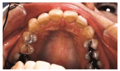
Figure 24-3G: Final result shows a straighter arc with more proportional teeth.
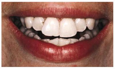
Figure 24-3H: Pretreatment view of the smile.
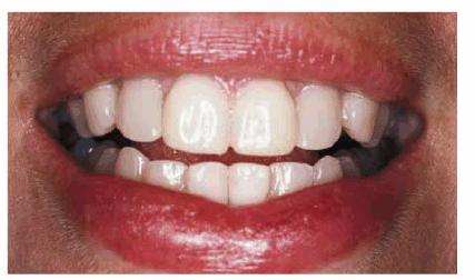
Figure 24-3I: Post-treatment view showing better harmony with the smile.
Correction by Porcelain Laminates
The advantage in selecting porcelain laminates to correct crowded teeth is the
ability for the laboratory to properly proportion the new restorations. This
permits a conservative solution to be used that will need less maintenance.15,18
When selecting porcelain laminates, determine arch space deficiencies on each
side. Both lateral incisors may be rotated in a similar fashion, resulting in
an equal amount of space on both sides of the arch. However, if one lateral is
overlapped more than the other, the available space may be asymmetric.
Correction may require shifting of line angles in the final restorations to
create an illusion of equal dimension in the final restorations.
After reproportioning the anterior space, if the total space will result in
teeth that would look much too narrow, building out the teeth in a slight
labioversion should be considered. The more the buccal surface is positioned
anteriorly, the wider the teeth will become. The added thickness will go
unnoticed if it occurs throughout the restored teeth and results in an entire
arc that is positioned labially from bicuspid to bicuspid. A curve that will
look good from an occlusal and a labial view should be selected. It is usually
possible to compromise by building out the other teeth slightly and
lingualizing the most labially positioned teeth. How much and where the
existing teeth will need reducing during the preparation of the teeth should be
determined. This is easily accomplished by using a reduction matrix fabricated
from a diagnostic wax-up. A good example of the above principles can be seen in
Figures 24-4A 24-4B 24-4C 24-4D 24-4E 24-4F 24-4G, and 24-4H
Usually,
the most severe reduction would resemble a similar amount of dentin loss, as in
a full crown. Even if a tooth will have to be severely prepared for a laminate,
the total loss would be much less with a laminate than for the crown. The worst
scenario would be to require endodontic therapy by doing a vital extirpation of
the pulp.
Following are several examples of the use of porcelain laminates to correct
crowding in the anterior region.
Use of Diagnostic Wax-up and Matrices in the Correction of Crowding with
Porcelain Laminates
PROBLEM: A 56-year-old businessman presented with a desire to straighten
his anterior teeth. In addition, he was concerned about the wear and
irregularity of the incisal edges of his teeth (see Figures 24-4A 24-4B, and 24-4C). Orthodontics was presented as a
first option, but the patient declined this treatment option because of the
lengthy treatment time that would be required.
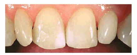
Figure 24-4A: Pretreatment-anterior view. Note the prominence of tooth #7 due to significant rotation and the slightly shortened appearance of tooth #10.
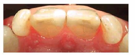
Figure 24-4B: Pretreatment-incisal view. Variations in available arch space for the lateral incisors are evident.
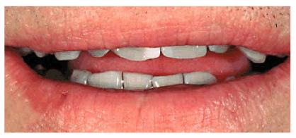
Figure 24-4C: Pretreatment-view of normal smile. Irregular incisal edges and incisal embrasures create an unbalanced esthetic appearance.
TREATMENT: Because of uncertainty about the ability to achieve a
satisfactory result using restorative treatment options to correct the
significant rotation of the lateral incisors, diagnostic casts were made. A
diagnostic wax-up was used to determine if appropriate modifications could be
made that would correct this crowding.
Using a diagnostic cast, it was noted that the space available (see Figures 24-4D, and 24-4E) for tooth #7 was 4.5 mm and for
tooth #10 was 6 mm. The central incisors were evaluated, and tooth #8 was found
to be 8.75 mm and tooth #9 was 8 mm. The crowding problem was then summarized
as the deficiency in arch space of 1.5 mm for tooth #7 with an excessive width
of 0.75 mm for tooth #8. If the distal of tooth #8 was reduced by 0.75 mm, then
the space needed to be created for tooth #7 was 0.75 mm if both sides of the
arch were to have a symmetric appearance.
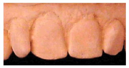
Figure 24-4D: Diagnostic cast-facial view.
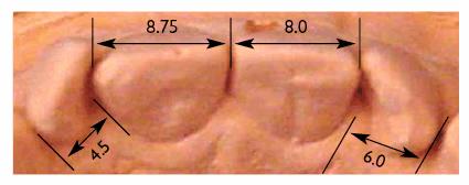
Figure 24-4E: Diagnostic cast-incisal view. By measuring the available arch space for each incisor, the source of the crowding problem was determined. The results of this analysis demonstrated a 1.5-mm deficiency in the area of tooth #7 and an excessive amount of arch space with tooth #8.
Stone was removed from the prominent mesial line angles, and wax was added to
the distal half of the lateral incisors to reduce the appearance of rotation
(see Figures 24-4F 24-4G, and 24-4H). The central incisors were
contoured to create a symmetric appearance and uniform width. Wax was added to
the incisal edges to create a uniform length. Incisal embrasures were developed
to define the width of each of the incisors. The facial surface and mesial edge
of tooth #7 were allowed to remain slightly facial and overlapped with tooth
#8. This facial positioning of the tooth (increasing the arch form in this
area) provided the additional space needed to create an appropriately sized
tooth.
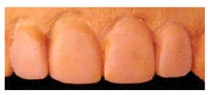
Figure 24-4F: Completed diagnostic wax-up-anterior view. Proposed reduction to the distal tooth #8 and additional width to the mesial of tooth #7 create a more harmonious distribution of tooth width in the arch.
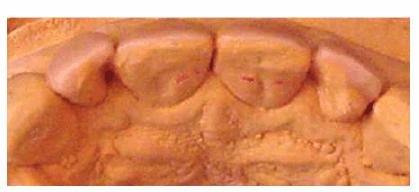
Figure 24-4G: Completed diagnostic wax-up-incisal view. The additional width required for tooth #7 was achieved by allowing the facial surface to remain slightly facial from the ideal arch form. This slight overlap was unnoticeable from an anterior view.
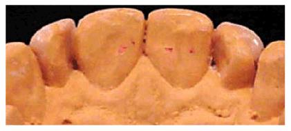
Figure 24-4H: Completed diagnostic wax-up-lingual view. The transition from the actual tooth position to the final tooth position created an unusual lingual embrasure on the mesial of both lateral incisors. Special instructions were given to the patient regarding oral hygiene in these areas.
Once it was determined that restorative treatment could adequately camouflage
this patient's crowding, the decision was made to use porcelain laminates to
accomplish this goal. The patient was then presented with this option, and he
elected to proceed with treatment. A reduction matrix was fabricated over the
diagnostic wax-up and trimmed to the facioincisal line angle. Figures 24-4I 24-4J, and 24-4K illustrate its use. Figure 24-4I shows the matrix in position prior
to tooth reduction. This identified the amount of facial reduction necessary to
bring the natural tooth back into the desired final contours. Figure 24-4J demonstrates the initial tooth
reduction to achieve this. The teeth were then prepared for porcelain
laminates, with the matrix being used to determine the proper incisal and
facial reduction (see Figure 24-4K).
Provisional restorations were fabricated from a matrix formed over the
diagnostic wax-up. The intended results of treatment were shown to the patient
(Figure 24-4L). Following the patient's approval
of esthetics, phonetics, and function, the laminates were completed (Figures 24-4M, and 24-4N
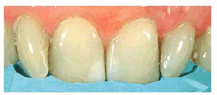
Figure 24-4I: Matrix of diagnostic wax-up used to identify areas of tooth structure beyond the desired final contours.
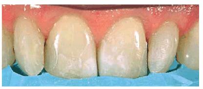
Figure 24-4J: Prior to final preparation of the teeth, only the excessive tooth structure was reduced using the diagnostic wax-up matrix as a guide.
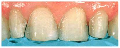
Figure 24-4K: Use of the diagnostic matrix to ensure proper incisal and facial reduction of the final preparations for porcelain laminate veneers.
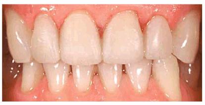
Figure 24-4L: Idealized provisional restorations fabricated using a second diagnostic wax-up matrix that remained untrimmed. From these restorations, the patient can accurately visualize the proposed results of the treatment.
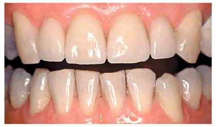
Figure 24-4M: Completed restorations-facial view. Note accurate reproduction of the diagnostic wax-up.
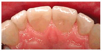
Figure 24-4N: Completed restorations-incisal view.
RESULTS: Porcelain laminates were used to
correct significant crowding of four maxillary incisors with satisfactory
results. The importance of the diagnostic casts, wax-up, and matrices in the
overall planning, communication, and implementation of treatment was
demonstrated.
Esthetic Contouring and Porcelain Laminates to Eliminate Crowding
PROBLEM: This 58-year-old housewife was concerned about her eroded,
crowded, and stained front teeth (Figures 24-5A, and 24-5B). Measurement with a dental dial
caliper helped to accurately determine available space for reproportioning tooth
size (Figure 24-5C). Although orthodontics was
mentioned as a first step to an ideal solution, the patient preferred to accept
a compromise treatment of porcelain laminates and cosmetic contouring. Although
crowding was less of a concern to the patient (Figures 24-5D, and 24-5E), she nevertheless decided to have
straighter-looking teeth through a compromise treatment of porcelain laminates
that would also esthetically correct the erosion and discoloration.
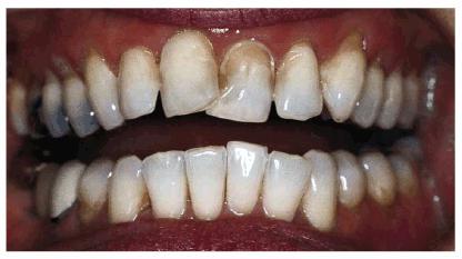
Figure 24-5A: This 58-year-old woman was dissatisfied with her crowded, eroded, and discolored teeth.
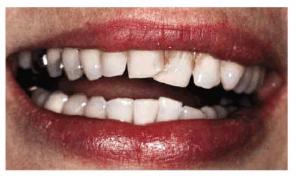
Figure 24-5B: Preoperative view.
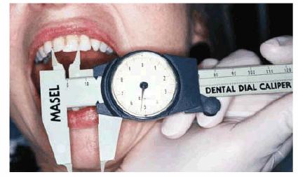
Figure
24-5C: Each tooth and the available space for the laminated teeth are measured
with Dental Dial Calipers (Masel Enterprises,
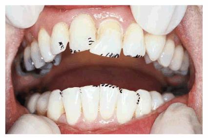
Figure 24-5D: The teeth are then esthetically contoured to begin creating the illusion of straighter teeth.
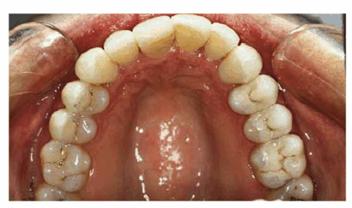
Figure 24-5E: Porcelain laminates were cemented from the right cuspid to the left second bicuspid; the laminate onlays were inserted on the maxillary right bicuspids and first molar.
TREATMENT: Figure 24-5D shows the areas that will be
esthetically contoured. Following contouring to reproportion spaces, Figure 24-5F demonstrates the gingival chamfer
margin being placed with a two-grit LVS diamond bur (Brasseler). The occlusal
view (Figure 24-5G) reveals just how much overlapping
existed. Figure 24-5H shows the teeth after esthetic
contouring and tooth preparation have occurred, as well as how defective
amalgam restorations were removed and glass ionomer bases were placed. Figure 24-5E shows finished laminates in place.
Note the newly proportioned, straighter, and lighter-looking teeth. The final
occlusal view also shows a new arch created by the laminates, building out
teeth #9 and #10 and the new posterior laminate onlays on the upper right side.
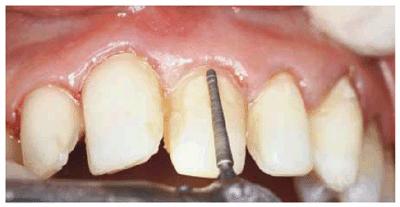
Figure 24-5F: The anterior teeth are prepared for porcelain laminates. A special two-grit diamond bur ([Brasseler]; see Esthetics in Dentistry, Volume 1, 2nd Edition, p. 346-7) helps prepare both the body and margin of the tooth.
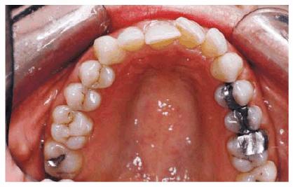
Figure 24-5G: The occlusal view shows the extent of overlapping in the central incisors.
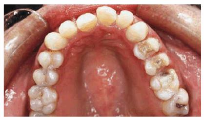
Figure 24-5H: This occlusal view shows the final tooth preparation. Note the patient's right posterior quadrant. The teeth were prepared for combination laminate/onlays.
RESULT: Before and after smiles can be seen
by comparing Figures 24-5B, and 24-5I. Esthetic contouring has also
improved the alignment of the lower anteriors. Constructing the porcelain
laminates indirectly allows the laboratory to better proportion tooth size.
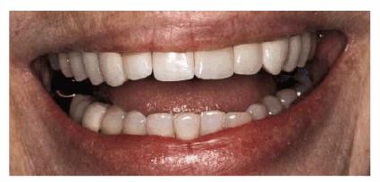
Figure 24-5I: Postoperative view of improved smile created by use of porcelain laminate veneers and esthetic contouring.
Correction by Crowning
As in bonding, the first problem in restoring crowded teeth by crowning is
tooth size. Each tooth needs to be or appear to be proportional. The more teeth
that are crowned, the less distortion there is.4,27,31 This means
that if only one or two teeth are crowned, there may be a noticeable difference
between the crowned teeth (which would be smaller) and the natural ones,
depending on the space involved. However, it is possible to accomplish this by
carefully shaping both the tooth to be crowned and the adjacent teeth to appear
harmonious in size. Figures 24-6A 24-6B, and 24-6C provide an example of how one tooth
can be crowned to relieve anterior crowding. After the adjacent central and
lateral incisors were reshaped, a full porcelain crown was placed on the left
central (see Figure 24-6B
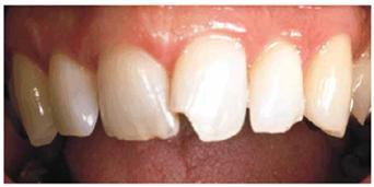
Figure 24-6A: This patient presented with chipped and crowded maxillary anterior teeth.
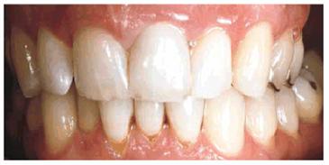
Figure 24-6B: A full porcelain crown was placed on the left central incisor; the right central and lateral incisors were esthetically contoured.
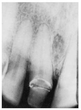
Figure 24-6C: The post-treatment radiograph shows the change in axial inclination for esthetic result.
An
alternative solution is to reshape the existing teeth.40 For
example, in a maxillary anterior crowded condition, instead of merely crowning
two central incisors, the lateral incisors can be reshaped by reducing the
mesial surface slightly so that the adjacent central incisors can be enlarged.
This same principle can be applied in other areas of the mouth. The teeth
adjacent to crowns are always reduced to recover some of the space lost because
of crowding. The more the adjacent teeth are reduced, the less noticeable is
the distortion. An example of crowning six incisors to
eliminate the crowding of anteriors follows.
Crowning to Eliminate Crowding
PROBLEM: This 38-year-old store owner presented with crowded and
discolored maxillary and mandibular teeth (Figures 24-7A, and 24-7B). Although orthodontic treatment was suggested as ideal
treatment, he elected a compromise that consisted of bonding the mandibular and
crowning the maxillary teeth.
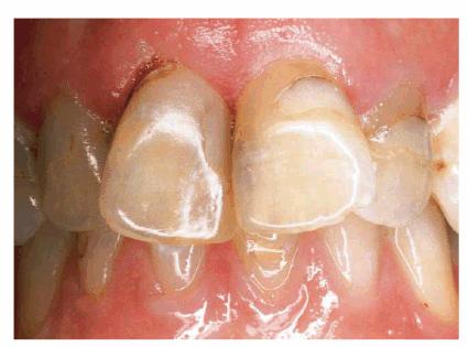
Figure 24-7A: This 38-year-old man wanted to improve his crowded maxillary and mandibular teeth.
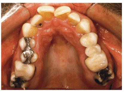
Figure 24-7B: This occlusal view shows why full orthodontic treatment was originally presented as the ideal treatment. The patient insisted on a "quick fix" solution.
TREATMENT: When teeth are as crowded as this, it is sometimes necessary
to do a vital pulp extirpation to prepare the teeth for adequate porcelain
thickness. Thus, tooth preparation and diagnostic wax-up were first completed
on the study casts (Figures 24-7C, and 24-7D). The patient was fully informed of the possibility of
endodontic therapy. The actual tooth preparation can be seen in Figures 24-7E, and 24-7F. Fortunately, the pulp had receded, so extirpation was
not necessary. Electrosurgery was completed prior to impressions to improve
access to the preparation margins. Six full porcelain crowns restored the
esthetics of the maxillary arch (Figure 24-7G), whereas composite resin bonding helped restore
mandibular esthetics. A maxillary occlusal night appliance was constructed for
the patient to wear since the patient had a history of clenching while
sleeping.
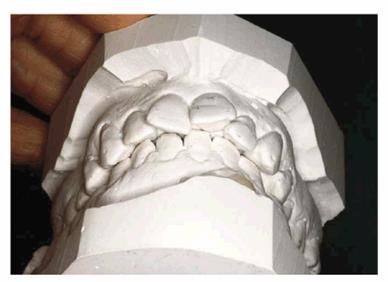
Figure 24-7C: Diagnostic casts show the extent of crowding in the maxillary anterior teeth.
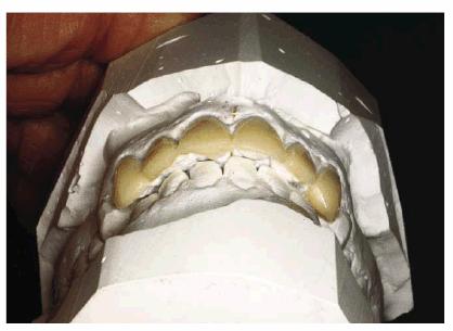
Figure 24-7D: A wax-up was completed to demonstrate to the patient and dental team how crowns could be used to accomplish the esthetic goal.
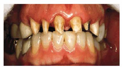
Figure 24-7E: Although the patient was warned that endodontic therapy might be necessary on the maxillary incisors, the teeth were prepared without pulpal exposures.
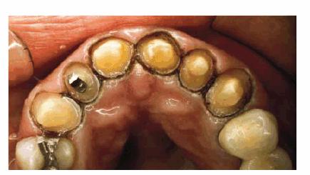
Figure 24-7F: The occlusal view shows the patient ready for impressions after electrosurgery for effective tissue displacement.
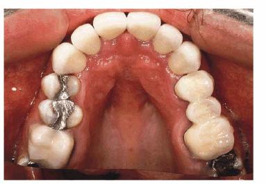
Figure 24-7G: The final six crowns show improved proportion and symmetry in the arch.
RESULT: The resulting smile with straighter and lighter teeth (Figures 24-7H, and 24-7I) was most appreciated by the patient.
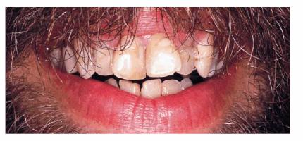
Figure 24-7H: Pretreatment smile.
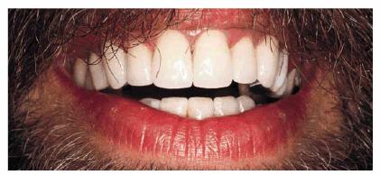
Figure 24-7I: Post-treatment smile with six maxillary full porcelain crowns and four mandibular incisors with bonded composite resins.
If crowded teeth are to be corrected with bonding, laminating, or full crowns,
the central incisors must be proportioned correctly. This can be accomplished
by either discing the mesial surfaces of adjacent uninvolved teeth or reducing
the size of adjacent crowned teeth. For final esthetics, contouring of adjacent
teeth should be considered.
The decision whether to laminate or crown should be made primarily on the
position of the overlapping (protruding) teeth. To restore the arch on or near
the labial-most position of the teeth, porcelain laminates can be used.
However, if the choice is to use maximum position (including vital pulp
extirpation), then crowns will probably be the best choice.
Crowning and Repositioning of Mandibular Anteriors. Although
crowding can occur in both arches, it is more common in the mandibular anterior
teeth. Treatment for these teeth is usually repositioning. There may be
occasions when the orthodontist will choose not to reposition, and the patient
may want these teeth bonded, laminated, or crowned. For teeth that are badly
broken down or have significant gingival recession that has made them unattractive,
bonding, laminating, or crowning can accomplish two things: it can restore and
straighten each tooth to its proper form. How much correction can be achieved
by repositioning is governed partially by root structure and crown inclination.
If the axial alignments of the teeth are divergent, there is a limit to how
much they can be straightened. If one of the teeth is in extreme labioversion,
it is difficult to do much straightening without building the adjacent tooth
somewhat thicker. This may create a gingival impingement on the tooth that is
being overcontoured. Excessive labial reduction could cause pulp damage, so
some compromise has to be reached. For this reason, repositioning is generally
the better solution. Sometimes, a combination approach is the best solution. If
the teeth are broken down and discolored and have unattractive, large
restorations, partial repositioning can be attempted, and crowning may take
care of the remainder of the problem. This way, the patient might not mind
wearing an appliance for a short while. One of the main objections that
patients have to orthodontic treatment is the length of time the appliances
have to be worn.
UNUSUAL OR RARE CLINICAL PRESENTATIONS
Occasionally, the dentist is presented with unusual dental problems associated
with crowded dentitions. Some of these may include malposed and misaligned
teeth, a protruding tooth, a retruded tooth, or the "lingually
locked" tooth.6,7,16,28,38,39
Malposed and Misaligned Teeth
The method of choice for correcting malposition or misalignment of a tooth or
teeth is orthodontia. For adults, consideration should be given to removable
appliances, plastic or ceramic brackets, or lingual braces. However, it is
sometimes possible to treat malposed or misaligned teeth without constructing
special appliances. Provisional acrylic splints can be adapted to accomplish
necessary repositioning.
An effective technique for repositioning crowded lower incisors is use of the
provisional splint with small hooks. A composite resin stop that is
mechanically bonded to the teeth helps keep the elastic from slipping. Some
discing in the interproximal surfaces of the anterior teeth is necessary to
create space for the incisors to move lingually. Finally, it is necessary to
plan some sort of retention; either an A-splint or direct bonding with
composite resin splint can be used.
Adult patients who come to the dentist for correction of malposed or misaligned
teeth may think that they are too old for orthodontic therapy. The dentist will
have to judge the importance of immediate facial esthetics to the patient. If
repositioning is the best solution to the esthetic problem, then the patient
should be advised and motivated to accept this therapy. When the patient will
accept only those procedures that offer an immediate solution, then bonding,
laminating, or crowning may be the only feasible compromises. Esthetic
contouring should also be considered. Rather than no treatment, esthetic
contouring may provide some compromise benefit.
The Protruding Tooth
In restoring a crowded labially positioned incisor, careful preparation can
make the protruding tooth appear to be in a more lingual position. Care must be
taken to avoid a short preparation. The labial surface is reduced as far as
possible without damaging the pulp; very little tooth structure is removed from
the linguoincisal surface (Figures 24-8A, and 24-8B). It is extremely important not to reduce the
incisogingival height until the preparation is essentially complete. This will
help avoid a short preparation. If the labial protrusion is so extreme that the
pulp may become involved, the dentist may perform vital pulp extirpation.2
Such radical procedures should be undertaken only when appearance is extremely
important and the patient is completely aware of the possible consequences and
has signed an informed consent for treatment.
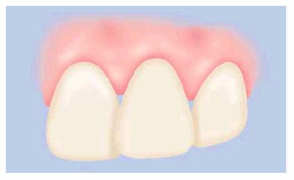
Figure 24-8A: The labial surface is reduced as far as possible without damaging the pulp. It is extremely important not to reduce the incisogingival height until the preparation is essentially complete.
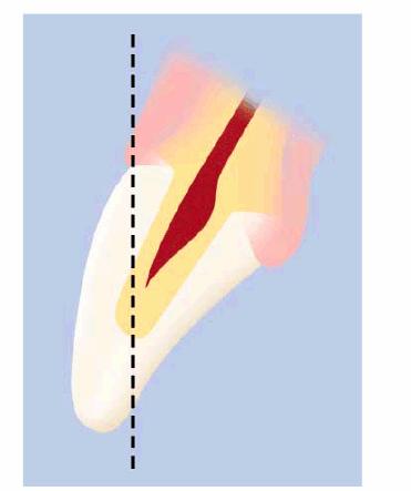
Figure 24-8B: The crowded labially positioned incisor requires a careful preparation to make it appear to be in a more lingual position.
The Retruded Tooth
This esthetic problem is similar to that of the protruded tooth. However,
realignment of the tooth in linguoversion frequently necessitates reduction and
recontouring of the opposing teeth to allow for clearance of the newly crowned,
bonded, or laminated tooth. To achieve the desired result, a large bulk of the
tooth structure may have to be removed from the linguoincisal surface of the
opposing tooth. Esthetic results can then be achieved by crowning or
laminating. If a porcelain-fused-to-metal restoration is desired, the lingual
side can be covered with a thin layer of metal, and the labial porcelain may be
built out to correct alignment. Laminating can be especially useful since
virtually no enamel needs to be reduced on the labial surface, with only
linguoincisal enamel being reshaped to mask the amount of retrusion present.
The Lingually Locked Tooth
If a lingually locked tooth is fully erupted, it can be restored to correct
position by pulp extirpation and placement of an off-center endodontic post and
porcelain crown. However, if the tooth is too short and in moderate
linguoversion, this treatment may be impractical. A porcelain shoulder is
prepared on the labial, and the porcelain is built up butted against the labial
gingiva. The patient must be warned that oral hygiene must be scrupulous. It is
far better to try to convince the patient of the advantages of repositioning
the lingually locked tooth than to try to restore it in place. In severe
situations, a third choice involving extraction and moving the remaining teeth
together with elastic thread ligature or bonded brackets may be indicated.
TO BOND, LAMINATE, OR CROWN?
The following questions should be considered:
1. What is the size of the crowded tooth? Will bonding make the tooth too
bulky? Bonded lower anteriors are more susceptible to this problem.
2. How much enamel is left for bonding? Are there very large restorations that,
once removed, will lessen the retention for a bonded restoration?
3. What is the appearance of the enamel? Is it badly stained or discolored so
that a large amount of opaquer plus several layers of composite resin will be
required to mask the defects? If so, then lamination and crowning are the
better choices.
4. Does the patient have a bad habit that may stain a bonded restoration? Heavy
smokers or coffee/tea drinkers may choose laminating or crowning to lessen the
amount of postoperative discoloration.
5. Is there an economic problem? Frequently, patients may wish to have their
teeth laminated or crowned, but finances enter into the decision since bonding
is less expensive than either crowning or laminating.
6. How long does the patient expect the restoration to last? Chances are that
both laminating and crowning can provide much longer life than direct bonding
with composite resin.
REFERENCES
1. Bello A, Jarvis RH. A review of esthetic alternatives for the restoration of
anterior teeth. J Prosthet Dent 1997;78:437-40.
2. Berlinger A. Ligatures, splints, bite planes, and pyramids. Philadelphia: JB
Lippincott, 1964.
3. Burstone CJ, Marcotte MR. Problem solving in orthodontics: goal-oriented
treatment strategies. Chicago: Quintessence, 2000.
4. Chiche GJ, Pinault A. Esthetics of anterior fixed prosthodontics. Chicago:
Quintessence, 1993.
5. Crispin B. Contemporary esthetic dentistry: practice fundamentals. Chicago:
Quintessence, 1994.
6. Curry FT. Restorative alternative to orthodontic treatment: a clinical
report. J Prosthet Dent 1999; 82:127-9.
7. Cutbirth ST. Treatment planning for porcelain veneer restoration of crowded
teeth by modifying stone models. J Esthet Restor Dent 2001;13:29-39.
8. Derbabian K, Marzola R, Arcidiacono A. The science of communicating the art
of dentistry. J Calif Dent Assoc 1998;26:101-6.
9. Dietschi D. Free-hand composite resin restorations: a key to anterior
esthetics. Pract Periodont Aesthet Dent 1995;7(7):15-25.
10. Donovan TE, Cho C. Diagnostic provisional restorations in restorative
dentistry: the blueprint for success. J Can Dent Assoc 1999;65:272-5.
11. Epstein MB, Mantzikos T, Shamus IL. Esthetic recontouring: a team approach.
N Y State Dent J 1997;63(10):35-40.
12. Fahl N Jr, Denehy GE, Jackson RD. Protocol for predictable restoration of
anterior teeth with composite resins. Pract Periodont Aesthet Dent 1995;7(8):13-21.
13. Foster TD. A textbook of orthodontics. 2nd edn. Oxford: Blackwell
Scientific, 1982.
14. Fradeani M, Aquilino A, Barducci G. Aesthetic restoration of endodontically
treated teeth. Pract Periodont Aesthet Dent 1999;11:761-8.
15. Garber DA, Goldstein RE, Feinman RA. Porcelain laminate veneers. Chicago:
Quintessence, 1988.
16. Gleghorn TA. Use of bonded porcelain restorations for nonorthodontic
realignment of the anterior maxilla. Pract Periodont Aesthet Dent 1998;10:563-5.
17. Goldstein CE, Goldstein RE, Garber DA. Imaging in esthetic dentistry.
Chicago: Quintessence, 1998.
18. Goldstein RE. Esthetics in dentistry. Vol. 1. 2nd edn. Hamilton, ON: BC
Decker, 1998.
19. Graber TM, Vanarsdall RL. Orthodontics: current principles and techniques.
St. Louis: Mosby, 2000.
20. Heyman HO. Conservative concepts to achieving anterior esthetics. J Calif Dent Assoc 1997;25:437-43.
21. Jun S. Communication is vital to produce natural looking metal ceramic
crowns. J Dent Technol 1997; 14(8):15-20.
22. Kokich VG. Esthetics: the orthodontic-periodontic restorative connection. Semin Orthodont 1996;2(1): 21-30.
23. Levine JB. Esthetic diagnosis. Curr Opin Cosmet Dent 1995;3:9-17.
24. Lieberman MA, Gazit E. Lower incisor extraction-a method of orthodontic
treatment in selected cases in the adult dentition. Isr J Dent Med 1973;22:80.
25. Magne P, Magne M, Belsor V. The diagnostic template: a key element to the
comprehensive esthetic treatment concept. Int J Periodont Restor Dent 1996;16: 560-9.
26. Margolis MJ. Esthetic considerations in orthodontic treatment of adults. Dent Clin North Am 1997;41:29-48.
27. Narcisi EM, Culp L. Diagnosis and treatment planning for ceramic
restorations. Dent Clin North Am 2001;45:117-42.
28. Narcisi EM, DiPerna JA. Multidisciplinary full-mouth restoration with
porcelain veneers and laboratory-fabricated resin inlays. Pract Periodont Aesthet Dent 1999;11:721-8.
29. Okuda WH. Creating facial harmony with cosmetic dentistry. Curr Opin Cosmet Dent 1997;4:69-75.
30. Oringer RJ, Iacono VJ. Periodontal cosmetic surgery. J Int Acad Periodontol 1999;1:83-90.
31. Paul SJ, Pietrobon N. Aesthetic evolution of anterior maxillary crowns: a
literature review. Pract Periodont Aesthet Dent 1998;10(1):87-94.
32. Peck H, Peck S. Reproximation (enamel stripping) as an essential
orthodontic treatment ingredient. London: Transactions of the Third
International Orthodontics Congress, 1975.
33. Portalier L. Composite smile designs: the key to dental artistry. Curr Opin Cosmet Dent 1997;4:81-5.
34. Proffit W, Fields HW. Contemporary orthodontics. 3rd edn. St. Louis: Mosby,
2000.
35. Rada RE. Interdisciplinary management of a common esthetic complaint. Gen Dent 1999;47:387-9.
36. Roblee RD. Interdisciplinary dentofacial therapy. Chicago: Quintessence,
1994.
37. Rufenacht CR. Principles of esthetic integration. Chicago: Quintessence,
2000.
38. Salama M. Orthodontics vs. restorative materials in treatment plans.
Contemp Esthet Restor Pract 2001; 5:20-30.
39. Shannon A. Reconstruction of the maxillary dentition utilizing a
nonorthodontic technique. Pract Periodont Aesthet Dent 1999;11:973-8.
40. Singer BA. Esthetic dentistry: a clinical approach to techniques and
materials. Philadelphia: Lea & Febiger, 1993.
41. Small BW. Laboratory communications for esthetic success. Gen Dent 1998;46:566-8, 572-4.
42. Studer S, Zellweger V, Scharer P. The aesthetic guidelines of the
mucogingival complex for fixed prosthodontics. Pract Periodont Aesthet Dent 1996;8: 333-41.
43. Wilson TG, Korman KS. Esthetic periodontics (periodontal plastic surgery).
Chicago: Quintessence, 1996.
|
