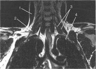
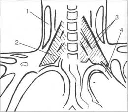
![]()
![]() лизации
плечевого
сплетения и
его ветвей. С
помощью
современных
томографов и
программного
обеспечения
удается
получить
ин 10510f510k формацию
не только об
окружающих
плечевое сплетение
тканях, но и о
самих
нервных
структурах.
лизации
плечевого
сплетения и
его ветвей. С
помощью
современных
томографов и
программного
обеспечения
удается
получить
ин 10510f510k формацию
не только об
окружающих
плечевое сплетение
тканях, но и о
самих
нервных
структурах.
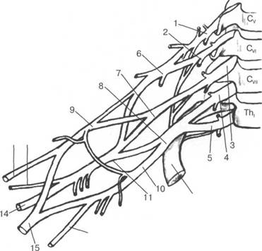
V, VI, VII, VIII I, II Cv CVI Cv
CVIII CIV,
Cv CV] CV]] CVIII Thj I
![]()
![]()
![]()
![]()
Cv; CVI; CVII; Thr |
|
|
|
I |
|
|
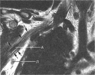
грудино-ключичного ключичной мышцы. Медиальный I
I
CV-CVII II
Cv-Th,
![]()
|
|
|
б
|
|

б
|
|
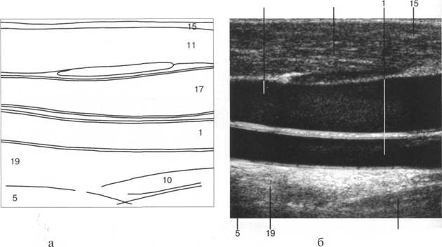
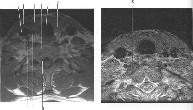
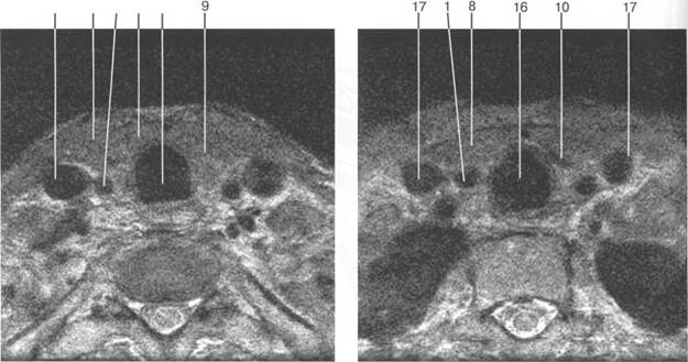
T2-BH(FATSAT).
![]()
|
|
|
a
![]()
|
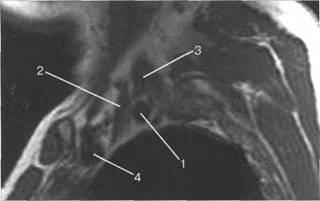
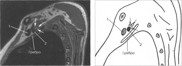
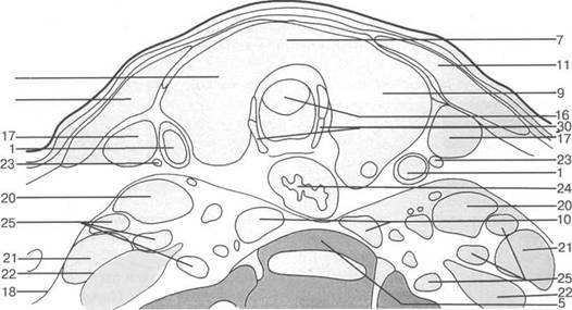
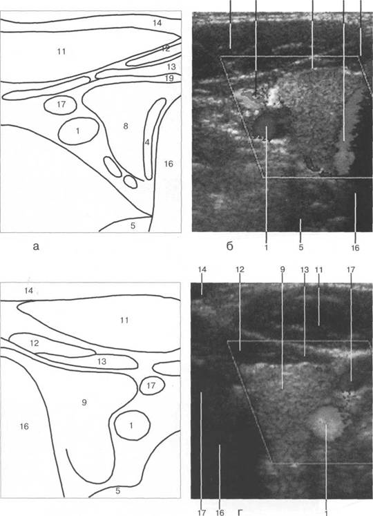
I располагается позади. Иногда удается хорошо различить ин 10510f510k дивидуальное деление пучков. Ла-
![]()
|
б
|
|
![]()
![]()
Blair
D.N., Rappoport S. et al.
Bonnel F., Rabischong P. Anatomy and
systematization of the brachial plexus in adult An. of
Clin.- 1981, N2.- P.
Filler A.G., Howe F.A. et al. Magnetic resonance neurography Lancet.- Vol. P.
Higgins
Steinbach
L.S. The brachial plexus Higgins
C.B, Hricak H., Helms C.A. (Eds)
Magnetic resonance imaging of the body.-
KellmanG.M., KneelandJ.B., MiddletonW.D.etal. MR imaging of the
supraclavicular region:
normal anatomy//Am. J. Roentgenol.- Vol. P.
Posniak H. V., Olson M.C. et al. MR imaging of the brachial plexus AJR.- Vol. P.
Seeger
L.L., Ruszkowski J. Bassett
L. W. et al. MR imaging of the normal shoulder: anatomic
correlation //AJR.- Vol.
P.
Sherrier R.H., Sostman H.D. Magnetic resonance imaging of the brachial plexus J. Thorac. Imaging.- Vol. P.
Van Es H. W.,
Witkamp T.D., Feldberg M.A.M. MRI of the brachial plexus and its region:
anatomy
and pathology Eur. Radiol.- Vol. P.
![]()
![]()
![]()
|
|
lig. thyrohyoideum;
![]()
![]()
![]()
|
|
[W.Swobodnic, M. Herrmann].
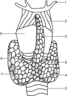
a. carotis dextra; a. carotis sinistra; cartilago thy-roidea; a. thyroidea superior; v. thyroidea superior; v. jugularis interna; a. subclavia dextra; v. subclavia dextra; a. subclavia sinistra; v. thyroidea inferior; v. thyroidea superior im-par; trancusbrachiocephalicus; v. brachiocephalica dextra; v. brachiocephalica sinister; a. subclavia; a. pulmonalis;
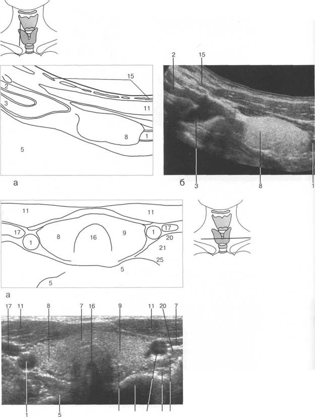
![]() стно
располагается
тонкий пласт
подкожной
мышцы и кожа.
стно
располагается
тонкий пласт
подкожной
мышцы и кожа.
![]()
![]()
|
железы
I II III IV
![]()

![]()

b d
|
|
|
|
|
|
|
|
|
|
|
|
|
|
|
|
|
|
|
|
![]() железы
должны быть
ровными и
четкими, с
четко
дифференциру
железы
должны быть
ровными и
четкими, с
четко
дифференциру
V V (RI) (PI) (Vo6),
Vo6
![]()
правой и левой долей. Ширина в норме в среднем составляет 10 мм, длина - 20 мм, толщина
F.Delange
|
|

![]()
|
|
|
|
|
|
17 13 |
|
|
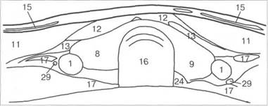
m. longus coli; 1 sternocleidomastoideus; sternohyoideus; sterno-thiroideus; thyreoideus; platysma; fascia coli profunda; m. longus capitis; m. scalenius anterior; m scalenius medius; m. scalenius posterior; a. facialis; a. lingualis; a. thyreoideus superior; cartilago cricoidea; cartilago thiroidea.
![]()
![]()
|
|
|
|
|
|
|
|
|
|
|
|
|
|
|
|
|
|
|
|
|
|
|
|
|
|
|
|
|
|
|
|
|
|
|
|
|
|
|
|
|
|
|
|
|
|
|
|
|
|
|
|
|
|
|
|
|
|
|
|
|
|
|
|
|
|
|
|
|
|
|
|
|
|
|
|
|
|
|
|
|
|
|
|
|
|
![]()
 17
17
12
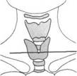
железы при поперечном сканировании.
![]()
|
|
|
|
8 |
|
|
|
|
|
|
|
|
|
|
|
. |
|
|
|
|
|
|
|
|
|
![]()
|
|
|

8
![]()
-■ |
|
|
|
15 28 29 |
|
|
2 28 29
![]()
![]()
|
|
|
2'4 5 1 25 21 |
![]()
![]()
![]()
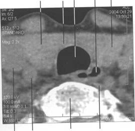
Cv.
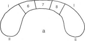
Поперечные размеры составляют 30x20 мм, высота - 30-40 мм.
HU.
![]()
|
|
|

[W. Swobodnic, M.Herrmann].
1Z3
![]()
![]()
|
|
|
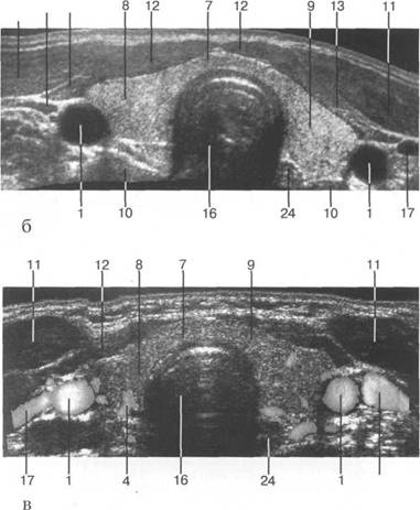
 13 26 4.1 16
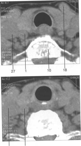
27 1
8 15 13 26 41 18 16
железы
хряща.
1
10 26 41
![]()
|
|
8 24 16 |
|
|
MPT
|
|
|
17 8 1 7 16 |
STIR
|