Decreased
In
- Secondary hypogonadism
- Kallmann's syndrome
(inherited autosomal isolated deficiency of hypothalamic
gonadotropin-releasing hormone; occurs in both sexes): Found in ~5% of
patients with primary amenorrhea. Causes failure of both gametogenic
function and sex steroid production (LH and FSH are "normal" or
undetectable but rise in response to prolonged gonadotropin-releasing
hormone stimulation).
- Pituitary LH or FSH
deficiency.
- Gonadotropin deficiency.
Müllerian
Inhibiting Substance, Serum
(Gonadal hormone
produced by prepubertal testes to 919p1523j promote involution of müllerian ducts during
normal male sexual differentiation. Detectable in normal boys from birth to
puberty, when concentration declines.)
Use
- Differentiate anorchia
from nonpalpable undescended testes in boys with bilateral cryptorchidism.
- Presence indicates
testicular integrity in children with intersexual anomalies.
- Supplements or replaces
measurement of response of serum testosterone to administration of hCG for
gonadal evaluation in prepubertal children.
Decreased
or Absent In
- Anorchia
- Negligible concentration
in girls until puberty
- Female
pseudohermaphroditism
Interpretation
- In prepubertal children,
normal value in boys is sensitive and specific test predictive of
testicular tissue (98%) and undetectable value predicts anorchia or
ovaries (89%).
- Values are better than those
for serum testosterone alone; combined with serum testosterone,
sensitivity = 62% and specificity = 100% for absence of testes.23
Progesterone,
Serum
Increased
In
- Luteal phase of menstrual
cycle
- Luteal cysts of ovary
P.666
- Ovarian tumors (e.g., arrhenoblastoma)
- Adrenal tumors
Decreased
In
- Amenorrhea
- Threatened abortion (some
patients)
- Fetal death
- Toxemia of pregnancy
- Gonadal agenesis
17-Hydroxycorticosteroids
(17-Ohks), Urine
(Derived from
cortisol and cortisone. Measure approximately one-half to two-thirds of
cortisol and its metabolites.)
Use
- Evaluation of
adrenocortical function
- Screening and diagnostic
test of glucocorticoid hypo- or hypersecretory disorders. Often replaced
by measurement of urine free cortisol or serum cortisol, which it parallels.
Increased
In
- Cushing's syndrome
- Adrenal tumors
- Marked stress (e.g.,
burns, surgery, infections)
- Use of certain drugs
(e.g., acetazolamide, chloral hydrate, chlordiazepoxide, chlorpromazine,
colchicine, erythromycin, estrogens, etryptamine, glucocorticoids,
meprobamate, oleandomycin, paraldehyde, quinine and quinidine,
spironolactone)
Decreased
In
- Addison's disease
- ACTH deficiency
- Hypothyroidism
- Fasting
- Use of certain drugs
(e.g., high-potency steroids [dexamethasone], narcotics, oral
contraceptives, phenothiazines, phenytoin, reserpine)
17-Ketogenic
Steroids (17-Kgs) (Corticosteroids), Blood and Urine
Use
- Evaluation of excessive
or deficient glucocorticoid secretion
- Evaluation of
21-hydroxylase deficiency type of CAH
- Increasingly supplanted
by measurements of serum cortisol, urine free cortisol, serum
17-hydroxyprogesterone, urine pregnanetriol
Increased
In
- Adrenal hyperplasia
- Adrenal adenoma
- Adrenal carcinoma
- ACTH therapy
- Stress
- Other conditions (e.g.,
obesity, smoking)
- Use of certain drugs
(e.g., glucocorticoids, ampicillin)
Decreased
In
- Addison's disease
- Panhypopituitarism
P.667
- Cessation of
corticosteroid therapy
- General wasting disease
- Use of certain drugs
(e.g., estrogens and oral contraceptives, dexamethasone)
17-Ketosteroids
(17-Ks), Urine
(Metabolites of
adrenal and gonadal androgenic steroids)
Use
- Indication of adrenal
rather than testicular status; two-thirds are of adrenal origin in men;
almost all are of adrenal origin in women.
- Diagnosis of ovarian and
adrenal tumors. Supplanted by more specific RIA of DHEA and DHEA-S.
- May
show daily variation of 100% in same individual
Increased
In
- Interstitial cell tumor
of testicle
- Virilizing ovarian tumors
(e.g., adrenal rest tumor, granulosa cell tumor, hilar cell tumor, Brenner
tumor, and, most frequently, arrhenoblastoma); increased in 50% of
patients and normal in 50% of patients
- Adrenocortical
hyperplasia (causing Cushing's syndrome, adrenogenital syndrome,
precocious puberty)
- Adrenocortical adenoma or
carcinoma
- Severe stress (e.g.,
burns, surgery, infections); exercise
- Pituitary tumor or
hyperplasia
- ACTH or testosterone
administration
- Third trimester of
pregnancy
- Nonspecific chromogens in
urine
- Use of certain drugs
(e.g., ampicillin, cephaloridine, cephalothin, chloramphenicol,
chlorpromazine, cloxacillin, danazol, dexamethasone, erythromycin,
ethinamate, nalidixic acid, oleandomycin, penicillin, phenaglycodol,
phenazopyridine, phenothiazines, quinidine, secobarbital, spironolactone)
Decreased
In
- Primary hypogonadism
(e.g., primary ovarian agenesis)
- Secondary hypogonadism
- Addison's disease
- Panhypopituitarism
- Nephrosis
- Generalized wasting
disease
- Use of certain drugs
(e.g., chlordiazepoxide, estrogens and oral contraceptives, metyrapone,
opiates, phenytoin, probenecid, promazine, reserpine)
Testicle,
Biopsy
Use
- Infertility workup
- Diagnosis of tumor
Interpretation
Normal spermatogenesis and normal endocrine
findings in patient with aspermia and infertility suggests a mechanical
obstruction to sperm transport that may be correctable.
Testosterone,
Free, Plasma
Use
Evaluation of gonadal hormonal function
P.668
Decreased
In (Men)
- Primary hypogonadism
(e.g., orchiectomy)
- Secondary hypogonadism
(e.g., hypopituitarism)
- Testicular feminization
- Klinefelter's syndrome
levels lower than in normal male but higher than in normal female and
orchiectomized male
- Estrogen therapy
- Total testosterone
decreased due to decreased sex hormone-binding globulin (e.g., cirrhosis,
chronic renal disease)
Increased
In
- Adrenal virilizing tumor
causing premature puberty in boys or masculinization in women
- CAH
- Idiopathic
hirsutism-inconclusive
- Stein-Leventhal
syndrome-variable; increased when virilization is present.
- Ovarian stromal
hyperthecosis
- Drugs that alter T -binding
globulins may also affect testosterone-binding globulins; however, free
testosterone level is not affected.
Gonadal
Disorders
Ambiguous
Genitalia
(Sexual
ambiguity occurs in 1 in 1000 live-born infants.)
Females24
|
Condition
|
Laboratory Finding
|
|
CAH with or without salt losing
|
|
|
Iatrogenic virilization
|
No diagnostic test. History of maternal
ingestion of virilizing agents (i.e., progestins).
|
|
Maternal virilization
|
Increased androgens in maternal serum
|
|
Idiopathic virilization
|
Normal plasma and urine steroids. 46 XX
karyotype. Gonadal biopsy may show Leydig's tissue.
|
|
Gonadal dysgenesis
|
No specific laboratory test. Laparotomy
usually shows a streak gonad on one side and testicular tissue on other
side. Karyotype may be nondiagnostic (46 XX or 46 XY), multiple mosaic (44
XO/46 XX/47 XXY), or typical (45 XO/46 XY).
|
|
True hermaphrodite
|
No specific laboratory test. Biopsy of
gonad shows ovarian follicles and testicular tubules. Karyotype may be 46
XX, 46 XY, or any mosaic included under gonadal dysgenesis. H-Y antigen is
present.
|
|
Males24
|
Condition
|
Laboratory Finding
|
|
Absent müllerian inhibiting factor
|
46 XY karyotype. Normal steroid levels.
No specific laboratory tests. Testes and uterii inguinali present.
|
|
Undescended testes
|
Normal gonadotropin and hormone levels.
|
|
Anorchia
|
May have low plasma testosterone and
very high plasma FSH and LH. Later hCG stimulation is negative.
|
|
Leydig's cell agenesis or hypoplasia
|
Plasma
testosterone is very low and fails to rise after hCG stimulation. High LD.
Normal FSH. Biopsy of testicle is diagnostic.P.669
|
|
Unknown cause for unresponsiveness to
androgens
|
Karyotype 46 XY. Normal testosterone
and dihydrotestosterone levels. No specific laboratory test.
|
|
Faulty androgen action
|
46 XY karyotype. Normal testosterone
level. Sex-linked defect type is diagnosed by in vitro binding
study. LH may be high. Dihydrotestosterone level is normal. Autosomal
recessive type has normal LH and FSH levels. Dihydrotestosterone level is
low.
|
|
Abnormal testosterone synthesis, no
salt loss
|
Increased 17-KS in urine and low plasma
testosterone in one type. Decreased 17-KS in urine in other types.
|
|
Abnormal testosterone synthesis, with
salt loss
|
Decreased 17-KS in one type. Increased
plasma pregnenolone in other type.
|
|
Hypopituitarism
|
Decreased GH levels. Other tropic
hormones may be deficient. Neonatal hypoglycemia is usual.
|
|
Microphallus
|
No specific laboratory test.
|
|
Congenital malformations
|
No specific laboratory test.
|
|
Laboratory
Differential Diagnosis
Gonads Palpable
- Buccal smear chromatin
positive and 17-KS normal
- True hermaphroditism
- Klinefelter's syndrome
variant
- Buccal smear chromatin
negative and 17-KS normal
- True hermaphroditism
- Anatomic defect
- Inherited enzyme
deficiency syndrome affecting testosterone synthesis, metabolism, or
action on target tissues
- Buccal smear chromatin
negative and 17-KS increased
- CAH (3-beta-hydroxysteroid
dehydrogenase deficiency)
Gonads Not Palpable
- Buccal smear chromatin
positive and 17-KS normal
- True hermaphroditism
- Ovarian tumor (maternal
17-KS increased)
- Maternal exposure to
androgens (history)
- Buccal smear chromatin
positive and 17-KS increased
- CAH
- 11-beta-hydroxylase
deficiency
- 21-hydroxylase
deficiency
- 3-beta-hydroxysteroid
dehydrogenase deficiency
- Buccal smear chromatin
negative and 17-KS normal
- True hermaphroditism
- Gonadal dysgenesis (45
X/46 XY, 46 XY)
Precautions
in Workup of Neonate with Ambiguous Genitalia
- Buccal mucosal smear for
nuclear sex chromatin determination may show false-negative patterns
during first 2 days of life so all chromatin-negative smears should be
repeated after the third day. Sex chromatin in >25% of cells from the
buccal mucosa indicates presence of at least two X chromosomes. A
leukocyte culture for karyotype preparation should begin immediately
whenever possible to confirm the sex chromosome constitution. The Y
chromosome fluorescence test may also be valuable.
- A chromatin-positive
newborn is almost always female.
- External
genitalia are normal in Klinefelter's and most cases of Turner's syndrome
Amenorrhea/Delayed
Menarche (Primary)
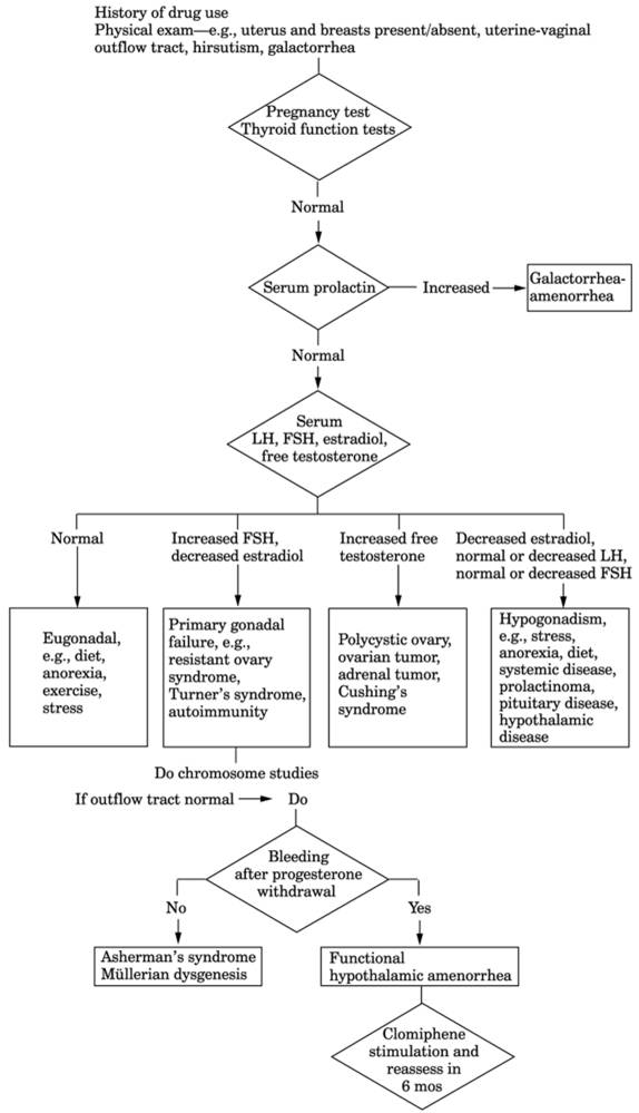
See Fig. 13-21.
P.670
|
|
|
Fig. 13-21. Algorithm for workup of
amenorrhea. Asherman's syndrome is the obliteration of endometrial lining by
adhesions due to pelvic inflammatory disease, tuberculosis, postabortal or
puerperal endometritis, etc. Normal blood steroid levels that do not respond
to progesterone administration by bleeding. Müllerian dysgenesis is a
congenital deformity or absence of tubes, uterus, or vagina; karyotype and
hormone levels are normal. (LH = luteinizing hormone; FSH =
follicle-stimulating hormone.)
|
P.671
Due To
- Gonadal disorders (60% of
all causes)
- Gonadal dysgenesis (75%
of gonadal disorders)
- Testicular feminization
syndrome (most common form of male hermaphroditism; female phenotype
with male 46 XY karyotype, testosterone in male range; testes are
present)
- Polycystic ovaries
- Resistant-ovary syndrome
- Structural genital tract
disorders (35-40% of all causes)
- Imperforate hymen
- Uterine agenesis
- Vaginal agenesis
- Transverse vaginal
septum
- Pituitary disorders
(rare)
- Hypopituitarism
- Adenomas (prolactin
secreting)
- Hypothalamic disorders
(rare)
- Anatomic lesions (e.g.,
craniopharyngioma)
- Functional disturbance
of hypothalamic-pituitary axis (e.g., anorexia nervosa, emotional stress)
- Systemic disorders
- Hypothyroidism
- CAH
- Debilitating chronic
diseases (e.g., malnutrition, congenital heart disease, renal failure,
collagen diseases)
Hormone
Profiles
- Normal LH, FSH,
prolactin, estradiol, testosterone, T , and TSH (eugonadal)
- Drugs
- Diet, anorexia
- Exercise
- Stress, illness
- Structural genital tract
disorders (see previous section)
- Increased LH and normal
FSH
- Early pregnancy
- Polycystic ovarian
disease (Stein-Leventhal syndrome)
- Ectopic gonadotropin
production by neoplasm (e.g., lung, GI tract)
- Increased LH and FSH
(>30 mIU/mL), decreased estrogen (<50 pg/mL)
- Primary ovarian
hypofunction
- Normal or low LH and FSH,
decreased estrogen
- Hyperprolactinemia
- Isolated gonadotropin
deficiency due to pituitary or hypothalamic impairment.
- Administer clomiphene
citrate for 5-10 days; if gonadotropin level increases or menses return,
cause is probably hypothalamic.
- Administer hypothalamic
LH-releasing factor; normal or exaggerated response in hypothalamic
amenorrhea (cause in 80% of patients); smaller or no response in
pituitary tumor or dysfunction.
- Increased androgen
- Polycystic ovarian
disease (testosterone level usually <200 ng/dL)
- Tumor of adrenal or
ovary (testosterone level may be >200 ng/dL)
- Testicular feminization
- Use of anabolic steroids
(e.g., in athletes)
Androgen
Abuse
- (By
athletes who use synthetic androgens to enhance performance or body
building; effects depend on type and dose of drug used.)
- When exogenous testosterone is used, urine
testosterone/epitestosterone ratio >6:1 is often considered indicative
of steroid abuse (normal ratio is ~1:1 in men and women)
- Synthetic androgen or its metabolites are identified in urine.
- Erythrocytosis may occur.
P.672
- Serum testosterone may be
low.
- Decreased or normal LH
and FSH
- Plasma HDL may be
decreased and LDL may be increased.
- Platelet counts and
platelet aggregation may be increased.
- Laboratory findings due
to infertility and testicular atrophy
Androgen
Deficiency (Hypogonadism)
See Tables 13-23 and
.
Due To
- Secondary
hypogonadism (hypogonadotropic)
- Secondary to
pituitary-hypothalamic disorders
- Hyperprolactinemia
- Panhypopituitarism
(pituitary or hypothalamus)
- Tumor
- Granulomatous disease
- Hemochromatosis
- Trauma
- Infarction, vasculitis
- Isolated gonadotropin
deficiency
- Isolated FSH or LH
deficiency
- Idiopathic hypothalamic
hypogonadism
- Kallmann's syndrome
- Genetic disorders (e.g.,
Prader-Willi, Laurence-Moon-Biedl syndromes)
- Systemic (e.g., chronic
disease, nutritional deficiency, massive obesity)
- Drugs (e.g.,
glucocorticoids)
- Constitutional (delayed
puberty)
- Decreased serum testosterone (<100 ng/dL) with low or normal LH and FSH
- Decreased gonadotropin-releasing hormone
- Administration of gonadotropin-releasing hormone increases serum gonadotropin,
testosterone, FSH, and LH
- Primary
hypogonadism (hypergonadotropic)
- Gonadal
- Genetic
- Klinefelter's syndrome
- True hermaphroditism
- Defects in synthesis of
androgens due to deficiency of various enzymes (e.g.,
20-alpha-hydroxylase, 17,20-desmolase, etc.)
- Agenesis of testicles
- Miscellaneous (e.g.,
Noonan's syndrome, streak gonads, myotonia dystrophica, cystic fibrosis)
- Acquired (e.g.,
chemotherapy, irradiation, castration, drugs, alcohol, viral orchitis
[especially mumps], cryptorchidism, chronic liver or kidney disease)
- Hormonal
- Hormonal insensitivity
(e.g., androgen or LH insensitivity)
- Defects in action of
androgens (pseudohermaphroditism)
- Complete (testicular
feminization)
- Incomplete
- Type I (defects in testosterone receptors)
- Type II (5-alpha-reductase deficiency)
Climacteric,
Male
- ○ Decreased
testosterone level in blood (<300 ng/mL) and urine (<100 µg/24 hrs)
- ○ Urinary
gonadotropin level is elevated. (Gonadotropin is decreased when low
testosterone level is due to pituitary tumor, gout, or diabetes.)
P.673
|
|
|
Fig. 13-22. Algorithm for evaluation of
nonazoospermic infertility. (FSH = follicle-stimulating hormone.)
|
P.674
|
|
|
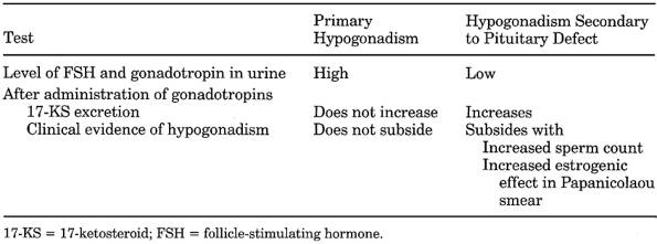
Table 13-23. Laboratory Differentiation
of Primary and Secondary (to Pituitary Defect) Hypogonadism
|
Corpus
Luteum Deficiency
(Corpus luteum
produces insufficient progesterone for development of endometrium receptive for
pregnancy.)
Due To
- Any condition that interferes
with follicle growth and development
- Severe systemic illness
including liver, kidney, or heart dysfunction
- Hyperprolactinemia
- X-chromosome
abnormalities
- Polycystic ovarian
disease or other causes of inadequate FSH level early in cycle
- Deficient LH receptors
on corpus luteum cells
- Inadequate LH level or
deficient ovulatory surge
- Findings of endometrial
biopsy on 26th day of cycle show less development than those of biopsy on
menstrual day.
- Serum progesterone measured on three different days during midluteal
phase totals <15 ng/mL and random level is <5 ng/mL.
Germinal
Aplasia
- Biopsy of testicle shows that Sertoli's and Leydig's cells are intact and germinal
cells are absent.
- ○ Azoospermia
- ○ Buccal smears are
normal (negative for Barr bodies).
- ○ Chromosomal
pattern is normal.
- Urinary gonadotropin is
normal.
- Urinary pituitary
gonadotropin is increased.
- 17-KS is decreased.
|
|
|
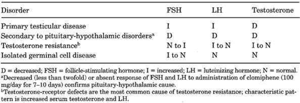
Table 13-24. Serum Hormone Levels in
Various Types of Androgen Deficiency
|
P.675
Gynecomastia
See Fig. 13-23.
Due To
- Neonatality
- Puberty (25%)
- Drugs (10-20%) (e.g.,
spironolactone, estrogens, cimetidine)
- Cirrhosis or malnutrition
(8%)
- Testicular tumors (3%)
(e.g., Leydig's cell, Sertoli's cell, germ cell tumors containing
trophoblastic tissue)
- Ectopic production of hCG
by tumors (e.g., lung, liver, kidney)
- Primary gonadism (8%)
- Secondary gonadism (2%)
- Hyperthyroidism (1.5%)
- Renal disease (1%)
- Klinefelter's syndrome
- Feminizing adrenal
cortical tumors
- Idiopathic (25%)
- Conditions usually
associated with ambiguous genitalia or deficient virilization
- Androgen-insensitivity
syndromes
- True hermaphroditism
- Enzymatic defects of
testosterone production
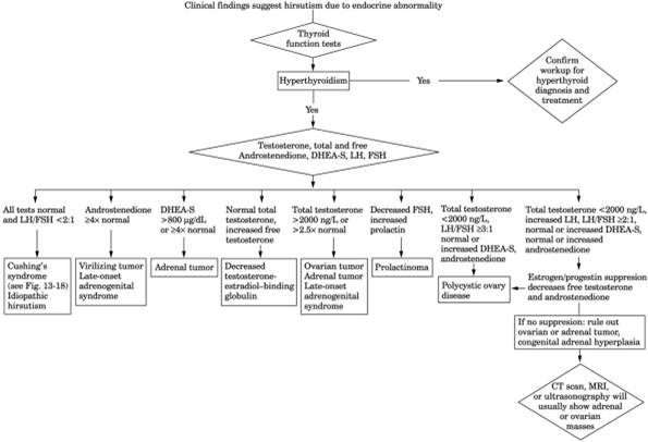
Hirsutism
See Fig. 13-24.
Due To
- Ovarian
- Polycystic ovary
syndrome
- Hyperthecosis syndrome
- Tumors (e.g.,
arrhenoblastoma, gonadoblastoma, dysgerminoma; Brenner cell, granulosa-theca
cell, lipoid cell tumors)
- Adrenal
- Adenoma, carcinoma
- Cushing's syndrome
- CAH (21-hydroxylase
deficiency, 11-hydroxylase deficiency, 3-beta-hydroxysteroid
dehydrogenase deficiency)
- Drugs (e.g., anabolic
steroids, androgens)
- Idiopathic (e.g.,
increased 5-alpha-reductase activity)
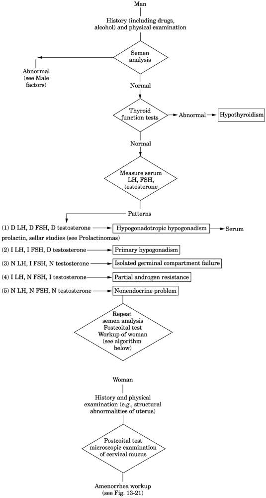
Infertility
- See Figs.
13-22, and .
- 85% of couples conceive
after 12 mos of unprotected intercourse.
- Remaining 15% warrant
investigation for infertility.
Due To
- Male factors (identified
in ~40% of couples) (see Fig. 13-25)
- Testicular abnormalities
(e.g., cryptorchidism, torsion, trauma, infection, varicocele)
- Coital factors (e.g.,
impotence)
- Toxins (e.g., anabolic
steroids, marijuana, alcohol, medications [cyclosporine, spironolactone,
cimetidine, nitrofurantoin])
- Others, e.g.,
- Sperm antibodies
(numerous assay methods)
- Clinical significance of serum antibodies in men and women is
controversial.
- Present in 10% of infertile men
- Present in infertile women in cervical mucus in 25% and in serum
in 13%
- Irradiation
P.676
|
|
|
Fig. 13-23. Algorithm for evaluation of
patients with gynecomastia. (CT = computed tomography; E = estradiol; FSH = follicle-stimulating
hormone; hCG = human chorionic gonadotropin; LH = luteinizing hormone; MRI =
magnetic resonance imaging; T = testosterone.) (Data from
Braunstein GD.
Gynecomastia. N Engl J Med
|
P.677
|
|
|
Fig. 13-24. Algorithm for hirsutism. (CT
= computed tomography; DHEA-S = dehydroepiandrosterone sulfate [preferable to
urinary 17-ketosteroid]; FSH = follicle-stimulating hormone; LH = luteinizing
hormone; MRI = magnetic resonance imaging.)
|
P.678
|
|
|
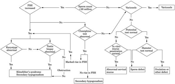
Fig. 13-25. Algorithm for evaluation of
azoospermia.
|
- Hyperthermia
- Heavy metals, e.g.,
lead, cadmium, manganese
- Pesticides
- Hypothalamic/pituitary
disorders (e.g., hyperprolactinemia, deficiency of gonadotropin-releasing
hormone)
- Chromosome abnormalities
(e.g., Klinefelter's syndrome, Down syndrome)
- Female factors
(identified in ~40% of couples) (see Fig. 13-22)
- Uterine factors
- Cervical (e.g.,
decreased cervical mucus quality or quantity, sperm antibodies)
- Uterine (e.g.,
endometriosis)
- Tube (e.g.,
salpingitis)
- Hypothalamic/pituitary
disorders (e.g., hyperprolactinemia)
- Disorders of ovulation
(e.g., polycystic ovaries)
- Chromosome abnormalities
(e.g., Turner's syndrome)
- Others (e.g.,
irradiation)
- Combined male and female
or unidentified factors in 20%.
Semen
Analysis25, ,
- Use
- Infertility studies
- Absence of sperm to
confirm vasectomy
- DNA test to confirm rape
assailant
P.679
|
|
|
Fig. 13-26. Algorithm for investigation
of the infertile couple. (D = decreased; FSH = follicle-stimulating hormone;
I = increased; LH = luteinizing hormone; N = normal.) (Male portion of figure
from
Swerdloff RS.
Infertility in the male. Ann Intern Med
|
P.680
|
Volume
|
>2 mL
|
|
pH
|
|
|
Color
|
Translucent, gray-white, or opalescent
|
|
Liquefaction
|
<30 mins
|
|
Viability
|
>65%
|
|
Motility
|
>50% viable sperm with forward
progression Progressive motility 3+ to 4+
|
|
Sperm density (count)
|
>20 million/mL
|
|
Morphology
|
>30% normal forms
|
|
Total motile functional sperm (= volume
× % motility × sperm density × % normal morphology)
|
>40 million
|
|
RBCs
|
0-5/HPF
|
|
WBCs
|
0-5/HPF (<10 /mL)
|
|
Crystals
|
None
|
|
Clumping
|
None
|
|
Mixed antiglobulin reaction
|
Negative
|
|
Bovine cervical mucus penetration
|
>30 mm
|
|
Cultures for bacteria (Ureaplasma,
Chlamydia)
|
No pathogens
|
|
Antisperm antibodies
|
Negative
|
|
- Sterile males usually
show
- Volume of <3 mL
- <20 million sperm/mL;
only a count <5 million sperm/mL seems to reduce chance for pregnancy
- <25% motility
- Abnormal motility or
morphology can occur with normal sperm counts but are usually seen with
decreased counts. Abnormal forms indicate impaired spermatogenesis.
Decreased motility may reflect defects in cilia structure elsewhere (in
respiratory and reproductive tracts). Agglutination may indicate antisperm
antibodies (which can be measured but relationship to infertility is not
established).
- Normal morphology is
>60% normal oval forms, <6% tapered forms, <0.5% immature forms,
<8% amorphous forms. Tapered forms and spermatids are often increased
in infertility associated with varicocele.
- Inflammatory cells may
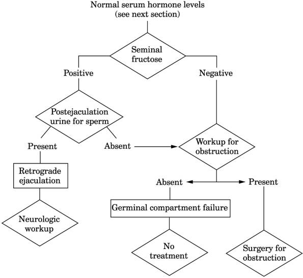
indicate infection of GU tract.
- Absent fructose (normally
produced by seminal vesicles) may indicate absence or obstruction of vas
deferens and seminal vesicles. Azoospermia, normal semen fructose, and
normal serum FSH suggest obstruction proximal to entry of ejaculatory
ducts.
- Large numbers of sperm in
postejaculation urine in these patients suggests retrograde ejaculation.
- Repeated semen analysis
(specimens collected 7 days apart) are necessary to characterize average
spermatogenesis.
- Specimens should not be
collected within 24 hrs of, or >5-7 days later than, previous
ejaculation. Should be received in laboratory within 1 hr.
- ≤40% variability
between different semen samples
- Comparison of split
ejaculate specimens is useful in patients with abnormal semen analysis
associated with a high volume; specimens may show marked differences.
- Antisperm antibodies (may
test male serum or seminal fluid or female serum or cervical mucus) may
occur in
- Testicular trauma (even
minor)
- Almost all vasectomized
patients
- Viral orchitis
(permanent)
- Bacterial infections of
GU tract (usually transient)
- Cervical mucus
penetration test measures greatest distance traveled by an individual
sperm from a small aliquot of semen incubated 90 mins in a capillary tube
of bovine cervical mucus. 68% of infertile men had penetration scores
<20 mm whereas 79% of fertile men had scores >30 mm.
- Hamster egg penetration
assay: hamster oocytes enzymatically treated to remove outer layers of egg
(which prevent cross-species fertilization) are incubated with human sperm
selected for their motile ability. Penetration rates of <15% (number of
eggs penetrated) indicate reduced fertility. May also be reported as
number of sperm penetrations
P.681
per egg (normal = ≥5). Positive test results indicate ability of sperm to
propel itself to oocyte, bind to oocyte, and penetrate oocyte.
- After vasectomy,
spermatozoa are present for some time. To confirm efficacy, two
centrifuged specimens properly collected 1 mo apart should be sperm free
and fructose should be absent.
Klinefelter's
Syndrome
- (Patients
have 2 or more X chromosomes)
- Azoospermia
- Plasma LH and FSH are increased; high FSH is best demarcator between normal men and
those with Klinefelter's syndrome.
- Urinary gonadotropin
level is elevated.
- Plasma testosterone
levels are decreased to normal.
- Buccal smears are helpful
if positive for Barr bodies but a negative result does not rule out
mosaicism. If negative, chromosome analysis should be performed, but in
70% of patients mosaic pattern may occur only in testes, so that
chromosomal analysis of testicular cells is required for definite
diagnosis.
- Abnormal chromosomal pattern. XY males have an extra X; 47 XXY is the classic type; 10% of
patients have the mosaic form (46 XY/47 XXY); may have additional X (e.g.,
XXXY, XXXXY).
- Biopsy of testicle shows
atrophy, with hyalinized tubules lined only by Sertoli's cells, clumped
Leydig's cells, and absent spermatogenesis.
- Laboratory findings due
to associated conditions, e.g., breast cancer, diabetes mellitus, thyroid
dysfunction.
Menopause
(Female Climacteric)
- Serum estradiol <5 ng/dL and FSH >40 mU/mL confirms primary ovarian failure;
progesterone is <0.5 ng/mL.
- Urinary estrogens are
decreased.
- Urinary 17-KS are
decreased.
- Plasma and urinary
gonadotropin are increased.
- Vaginal cytology shows
menopausal pattern.
Ovarian
Insufficiency, Secondary
Due To
- Deficient estrogen
production, e.g., diseases of the pituitary or hypothalamus (see separate
sections)
- Normal or increased
estrogen production, e.g., ovarian tumors, functional cysts of ovary that
suppress LH and FSH secretion
- Disorders of adrenal
function (increased production of cortisol or androgens) or thyroid
function.
- Urinary gonadotropin is decreased or absent
- Plasma LH is <0.5 mU/mL.
Ovarian
Tumors
Feminizing
Ovarian Tumors
- (E.g.,
granulosa cell tumor, thecoma, luteoma)
- ○ Pap smear of
vagina and endometrial biopsy show high estrogen effect and no
progestational activity; no signs of ovulation during reproductive phase.
- ○ Urinary FSH is
decreased (inhibited by increased estrogen).
- Urine 17-KS and 17-OHKS
are normal.
- ○ Pregnanediol is
absent.
Masculinizing
Ovarian Tumors
- (E.g.,
arrhenoblastoma, hilar cell tumors, adrenal rest tumors)
- Androgen-secreting tumor of ovary or adrenal gland is highly likely if serum total
testosterone is >200 ng/dL or DHEA-S is >800 µg/dL. Localization may
require androgen measurement in blood from adrenal and ovarian veins.
P.682
- ○ Pap smear of
vagina shows decreased estrogen effect.
- ○ Endometrial
biopsy shows moderate atrophy of endometrium.
- Urine FSH (gonadotropins)
is low.
- Urine 17-KS level is normal or may be slightly increased in arrhenoblastoma. Level
may be markedly increased in adrenal tumors of ovary
("masculinovoblastoma"). The higher the urine 17-KS level, the greater the
likelihood of adrenocortical carcinoma; value of >100 mg/24 hrs is
virtually diagnostic. Level may be moderately increased in Leydig's cell
tumors.
- In
arrhenoblastoma an increased amount of androsterone, testosterone, etc.,
may be excreted in urine even though the 17-KS level is not much
increased. Normal or slightly increased urine 17-KS level in association
with plasma testosterone in male range is almost certainly due to ovarian
tumor.
- In
adrenal cell tumors of ovary, laboratory findings may be the same as in
hyperfunction of adrenal cortex with Cushing's syndrome, etc
- In
some cases no endocrine effects are seen from these tumors. Some cases of
arrhenoblastoma with masculinization also show evidence of increased
estrogen formation
Struma
Ovarii
~5-10% of cases are hormone producing. Classic findings of hyperthyroidism
may occur. These tumors take up radioactive iodine. (Simple
follicle cysts may also take up radioactive iodine.)
Primary
Chorionepithelioma of Ovary
- Urinary chorionic gonadotropins are markedly increased
- Estrogen and progesterone
secretion may be much increased.
Nonfunctioning
Ovarian Tumors
Only effect may be hypogonadism due to
replacement of functioning ovarian parenchyma.
Tumor Markers
- Serum CA-125 is useful
for
- Postoperative monitoring
for persistent or recurrent disease; poorer prognosis if elevated 3-6 wks
after surgery. Lower levels in patients with no residual tumor or <2
cm of residual tumor. But a negative test does not exclude residual
disease.
- Rising level during chemotherapy
is associated with tumor progression and fall to normal is associated
with response. Remains elevated in stable or progressive disease.
- Rising level may be
indication for second-look operation even in presence of normal clinical
examination. Specificity = 99%, sensitivity = 46%, positive predictive
value = 97% for second-look cases.
- Higher levels are seen
in less differentiated tumors (grade 2 and 3) and in serous
cystadenocarcinoma. Not increased in mucinous adenocarcinoma.
- Sequential determinations
are more useful than a single test because levels in benign disease do
not show significant change but progressive rise occurs in malignant
disease.
- Rising level may precede
clinical evidence of recurrence by up to 11 mos.
- Not used for screening
because it is negative in 20% of cases at time of diagnosis; normal level
does not exclude tumor; greater elevation roughly related to poorer
survival.
- CA-125 is positive in
80% of cases of common epithelial tumors, 50% of early-stage disease,
0.6% of healthy women older than age 50 yrs.
- Beta-hCG is positive in
almost all cases of choriocarcinoma, 10-30% of cases of seminomas, and
5-35% of cases of dysgerminoma. See Trophoblastic
Neoplasms section.
- AFP is present in 80-90%
of cases of endodermal sinus tumors or immature teratomas.
- CEA is present in 50-70%
of cases of serous carcinoma. CA-125/CEA ratio is much higher in serous
carcinoma (>10 and often >100) than in carcinomas of breast, lung,
colon, or pancreas (usually <10), which may also cause increased levels
of these markers.
P.683
Germ
Cell Tumors of the Ovary
|
Tumor
|
AFP
|
hCG
|
|
Seminoma
|
|
|
|
Seminoma with syncytiotrophoblastic
giant cells (STGC)
|
|
|
|
Embryonal carcinoma
|
|
|
|
Embryonal carcinoma with STGC
|
|
|
|
Yolk sac tumor
|
|
|
|
Yolk sac tumor with STGC
|
|
|
|
Choriocarcinoma
|
|
|
|
Mature teratoma
|
|
|
|
See Chapter 16.
When both markers are positive, both should be assayed after therapy, as
recurrence or metastases may be reflected by increase of only one marker.
|
|
Stein-Leventhal
Syndrome (Polycystic Ovarian Disease)
- Serum increased ~3× normal (>35 mU/mL) in ~60% of patients in association
with normal or slightly low FSH level. Abnormally high LH/FSH ratio
(>2) is more consistently abnormal than is either measurement alone.
Ratio ≥ 2 is considered highly suggestive; ratio ≥ 3 is
considered diagnostic.
- Increased serum LH, LH/FSH ratio of >2, and mild increase of ovarian
androgen level are sufficient for diagnosis in presence of the symptoms
and clinical signs. Because of erratic daily
fluctuations of LH and androgens, obtaining daily plasma specimens for 3-5
days may be necessary.
- Plasma free testosterone is increased ≤200 µg/dL in 40-60%
of cases
(>200 µg/dL usually indicates an androgen-producing tumor); not
suppressed by dexamethasone.
- Plasma androstenedione (DHEA) is increased in ≤50% of cases.
- Serum 3-alpha-androstanediol glucuronide (metabolite of dihydrotestosterone)
is markedly increased in this and in idiopathic hirsutism.
- Synthetic estrogens and
progestins (as in oral contraceptives) for 21 days, with before and after
measurement of free testosterone and androstenedione:
- Free testosterone and
androstenedione decrease by 50% or become normal in LH-dependent
hyperandrogenism, e.g., polycystic ovaries.
- No suppression occurs in
patients with ovarian tumors or adrenal disorders.
- Change in free
testosterone accounts for estrogen-caused increase in sex hormone-binding
globulin, which could result in unchanged or increased total testosterone
level.
- ~85% of these patients have one or more abnormalities of serum LH/FSH ratio,
testosterone, or androstenedione. Hyperandrogenism does not differentiate
condition from CAH but CAH is more likely if LH/FSH ratio is <2:1 and
ovaries are normal in size.
- Urinary 17-KS are
somewhat increased (higher values occur in congenital virilizing adrenal
hyperplasia and hyperadrenalism due to Cushing's syndrome). (Measurement
of DHEA-S is preferable to evaluate adrenal disease.) Dexamethasone
administration (0.5 mg four times a day for 5-7 days) causes partial
suppression in cases of ovarian origin, but complete suppression suggests
adrenal origin (e.g., late-onset CAH). Administration of gonadotropin
increases urinary 17-KS.
- Biopsy of ovary is
consistent with increased androgen effect but is not specific; ovarian
visualization and biopsy are not routine part of diagnosis.
- Plasma cortisol, urinary
17-OHKS, and 17-KGS are normal.
- Plasma prolactin is
increased in ~30% of patients.
- Hyperinsulinemia occurs
for unknown reason; correlates with degree of increased androgens.
- 10-13% of these patients
have partial 21-hydroxylase defects.
- ○ If testosterone
is >2 ng/mL or DHEA is >7000 ng/mL, ovarian or adrenal tumor should
be ruled out.
- Laboratory tests may be
helpful in defining pathogenesis, following course of treatment, or ruling
out adrenal or ovarian tumors.
- Increased
serum LH with normal or decreased FSH may occur in simple obesity,
hyperthyroidism, liver disease
P.684
Testicular
Tumors
|
Tumor
|
Serum Tumor Marker
|
|
Seminoma
|
hCG increased in ~10%
|
|
|
AFP not increased in pure seminoma
without teratomatous component
|
|
Embryonal carcinoma
|
hCG or AFP or both increased in 90%
|
|
Yolk sac tumor
|
AFP increased in 100%
|
|
Choriocarcinoma (pure)
|
hCG increased in 100%
|
|
Teratoma
|
hCG or AFP or both increased in 50%
|
|
Mixed tumor
|
hCG and AFP increased in 90%
|
|
- Increased serum hCG (>1-2 ng/mL or >5-10 mU/mL) is found in 40-60%
of patients with metastatic nonseminomatous tumors and in 15-20% of
patients with apparently pure metastatic seminoma. In the latter case,
immunochemical staining of paraffin-embedded tumor should be performed,
because isolated syncytiotrophoblastic cells may show the hormone but are
not by themselves evidence of choriocarcinoma.
- Increased serum AFP (>20 ng/mL) is found in ≤ 70% of patients
with metastatic nonseminomatous tumors (embryonal carcinoma and yolk sac
tumors).
- Both markers should always be measured simultaneously. 40% of patients with
nonseminomatous tumors have increase of only one marker. 90% of patients
with testicular tumors are positive for AFP or hCG or both; these are valuable
for gauging efficacy of chemotherapy. 30% of patients receiving intensive
chemotherapy apparently have a complete clinical remission; AFP levels may
remain increased, although lower than pretreatment levels.
- 20-30% of patients have
false-negative results preoperatively despite tumor (usually microscopic)
in the retroperitoneal lymph nodes. Therefore, lymphadenectomy should not
be omitted simply because marker levels are normal.
- Serum markers for AFP and
beta-hCG may be increased in conditions other than testicular cancer. See Chapter 16. False-positive increase is rare.
- The most important use is for follow-up after surgery or chemotherapy. Failure of
increased preoperative levels to fall after surgery suggests metastatic
disease and the need for chemotherapy. Rise of levels that had previously
declined to normal suggests recurrent tumor even with no other evidence of
disease. Serum half-life of AFP = 5-7 days and of hCG = 30 hrs.
- Negative marker findings
are not useful for differential diagnosis of scrotal mass, but elevated
levels indicate testicular cancer.
- Serum LD is a third
marker; not specific for testicular cancer but also appears to be an
independent prognostic factor for advanced germ cell tumors. Increased in
~60% of nonseminomatous germ cell tumors and 80% of seminomatous germ cell
tumors.
Turner's
Syndrome (Ovarian Dysgenesis)
- Diagnosis is based on karyotype analysis. Chromosomal pattern includes wide spectrum
of abnormalities, e.g., 45 chromosomes (monosomy X with XO; or, if XX, one
X is abnormal; or XO mosaic), various deletions of part of an X
chromosome. Female is phenotypic. Prenatal diagnosis by chorionic villus
sampling or amniocentesis.
- Barr body test is
negative (male) in 80% of patients.
- Because of the frequency
with which 45 X cells are admixed with 46 XX cells, the diagnosis (i.e.,
45 X karyotype) cannot be excluded by either buccal smear or chromosome
analysis alone.
- Biopsy of ovary shows connective tissue stroma with rare follicular structure.
- Vaginal smear and
endometrial biopsy are atrophic.
- Increased FSH, LH, and
gonadotropins.
- 17-KS and 17-OHKS are
normal.
- ACTH is normal.
- Glucose intolerance is
common, with mild insulin resistance.
- Serum cholesterol is
frequently increased.
- Laboratory findings due
to increased prevalence of associated conditions, e.g.,
- Hashimoto's thyroiditis
(10-30%)
- Bicuspid aortic valves
(≤ 50%)
- Coarctation of aorta
(≤ 20%)
P.685
- Horseshoe kidneys
- Pyelonephritis due to
anomalous obstruction of ureteropelvic junction
- Hypertension
- Frequent otitis media
- ~60% of patients with
primary amenorrhea have Turner's syndrome or sometimes testicular
feminization. 90% never menstruate. ~10% menstruate for a few years and
then present as cases of secondary amenorrhea.
Turner's
Syndrome in the Male
- Biopsy of testicle reveals dysgenetic tubules with few or no germ cells.
- Chromosomal pattern: 46 chromosomes (XY pattern with very defective Y that is
equivalent to XO).
Laboratory
Tests for Diagnosis of Disorders of the Pituitary and Hypothalamus
Arginine
Vasopressin (Antidiuretic Hormone [Adh])
Use
- Diagnosis of central
diabetes insipidus and of SIADH, and differentiation from nephrogenic
diabetes insipidus
- Differential diagnosis of
hyponatremias
Increased
in Serum
- SIADH (inappropriately
increased for degree of plasma osmolality)
- Ectopic ADH syndrome
- Use of certain drugs
(e.g., chlorpropamide, phenothiazine, carbamazepine [Tegretol])
- Nephrogenic diabetes
insipidus (normal for degree of plasma osmolality)
Decreased
in Serum
Central diabetes insipidus
In Urine
- Central diabetes
insipidus: low arginine vasopressin and osmolality
- Nephrogenic diabetes
insipidus: high arginine vasopressin and low osmolality
- SIADH: normal arginine
vasopressin relative to osmolality
Growth
Hormone (Gh)
Use
- Differential diagnosis of
short stature, slow growth
- Evaluation of pituitary
function
Increased
In
- Acromegaly and gigantism
due to certain pituitary adenomas
- Laron dwarfism (GH
resistance; GH-binding protein cannot be detected)
- Renal failure
- Uncontrolled diabetes
mellitus
- Use of certain drugs
(e.g., estrogens, oral contraceptives, tranquilizers, antidepressants)
- Starvation
- 2 hrs after sleep
Decreased
In
- Hypothalamic defect
causes most cases (e.g., tumors, infection, diseases such as
hemochromatosis, perinatal insult such as birth trauma)
P.686
- Hypopituitarism (e.g.,
familial isolated GH deficiency, tumors, infection, granulomas, trauma,
irradiation)
- Dwarfism
- Corticosteroid therapy
- Obesity
- Low
levels must be measured after stimulation (e.g., with insulin, arginine)
Growth
Hormone-Releasing Hormone
(Hypothalamic
secretion stimulates pituitary to release GH)
Increased
In
1% of cases of acromegaly due to production
of GH-releasing hormone by hypothalamus or ectopic secretion by neoplasms
(e.g., pancreatic islet, carcinoid of thymus or bronchus, neuroendocrine
tumors)
Normal In
Most cases of acromegaly due to pituitary
tumors.
Prolactin
See Prolactinoma.
Somatomedin
C
(Insulin-like
growth factor I, which mediates most growth-promoting effects of GH)
Use
- Diagnosis of acromegaly
and pituitary deficiency; preferable to GH because it is constant after
eating and during the day
- Screening of other growth
disorders
- Assessment of nutritional
status
- Monitoring of
effectiveness of nutritional repletion. Is more sensitive indicator than
prealbumin, transferrin index, or retinol-binding protein.
Increased
In
- Acromegaly and gigantism
- Pregnancy (2-3×
nonpregnancy values)
Decreased
In
- Pituitary deficiency
- Laron dwarfism
- Anorexia or malnutrition
- Acute illness
- Hepatic failure
- Hypothyroidism
- Diabetes mellitus
- Normal aging
Diseases
of the Pituitary and Hypothalamus
Acromegaly
and Gigantism
- Serum somatomedin C (insulin-like growth factor I) is uniformly increased in
untreated cases; is more precise and cost-effective screening than serum
GH because GH levels fluctuate and have short serum half-life (22 mins).
- Autonomous serum GH is increased. (Avoid stress before and during venipuncture because
stress stimulates secretion of GH; several random measurements should be
performed.) Annual random blood GH levels, FTI, and ACTH are used for
treatment follow-up.
P.687
- Fasting levels >5
ng/mL in men or >10 ng/mL in women are suggestive but not diagnostic
of acromegaly.
- Most patients show a fall of <50% or even an increase 60-90 mins after
glucose administration (50-100 gm orally), whereas normal subjects show
almost complete suppression of GH (or to <5 ng/mL) by induced
hyperglycemia. This is the most reliable test. Failure to suppress GH to
<2 ng/mL after oral glucose load is essential to diagnosis.
- If borderline response to hyperglycemia, perform TRH test. (500 µg TRH IV causes
transient increase [>50% over basal levels] of GH in 15-30 mins in
acromegaly patients but has little effect in normal persons.)
- GH-releasing hormone excess secretion (e.g., ectopic source such as pancreatic tumor or
carcinoid causes <1% of acromegaly cases). Thus GH-releasing hormone
should be measured in all patients with acromegaly.
- All patients with acromegaly should have baseline serum prolactin measured because ≤
40% of these adenomas may secrete both prolactin and GH.
- IV ACTH administration
may cause excessive increase in urine 17-KS but normal 17-OHKS excretion.
- Glucose tolerance is
impaired in most patients. Mild diabetes mellitus that is insulin
resistant is found in <15% of patients.
- Adrenal virilism and
increased urine 17-KS are common in women.
- Urine 17-KS, 17-KGS, and
gonadotropins are usually normal or may be slightly changed but level not
diagnostically useful.
- Hypogonadism develops in
≤ 50% of cases.
- Rare associated
endocrinopathies are hyperthyroidism, HPT, pheochromocytoma, insulinoma.
- In inactive cases, all
secondary laboratory findings may be normal.
- In late stage,
panhypopituitarism may develop.
- Serum phosphorus is
increased for age of patient in 40% of cases.
- Serum ALP may be
increased.
- Urine calcium is
increased.
- Urine hydroxyproline is
increased.
- Biopsy of costochondral
junction evidences active bone growth.
- CBC and ESR are normal.
- After successful surgery-basal plasma GH <5 ng/mL, should
decrease to ≤2 ng/mL after glucose administration and level of
insulin-like growth factor I should become normal.
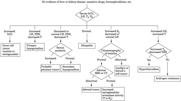
Due To
- Excess GH secretion
- Pituitary adenomas,
hyperplasia, or carcinoma
- Ectopic pituitary tumor
(sphenoid or parapharyngeal sinus)
- Ectopic hormone
production (e.g., tumor of pancreas, lung, ovary, breast)
- Excess secretion of
GH-releasing hormone
- Hypothalamic tumor
(e.g., hamartoma, ganglioneuroma)
- Ectopic hormone
production (e.g., carcinoid of bronchus, GI tract, pancreas; pancreatic
islet cell tumor, small cell carcinoma of lung, adrenal adenoma,
pheochromocytoma)
Other
Causes of Tall Stature in Children
- Klinefelter's syndrome
- Marfan syndrome
(inherited disorder with thin limbs, malformation of eyes and ears,
medionecrosis of aorta, cardiac valve deformities, hypotonia,
kyphoscoliosis)
- Beckwith-Wiedemann
syndrome (hypoglycemia, omphalocele, macrosomia, macroglossia)
- Untreated CAH
- Precocious secretion of
androgens or estrogens
- Obesity
Anorexia
Nervosa
- No diagnostic or typical
laboratory profile; diagnosis by exclusion. Findings may be compensatory
regulatory changes secondary to nutritional deprivation rather than
primary hypothalamic dysfunction.
- ESR is low.
- Vomiting may cause
hypokalemic acidosis.
P.688
- Prerenal azotemia with
increased BUN and serum creatinine
- Decreased serum glucose,
sodium, magnesium
- Renal calculi
- Laboratory findings of
euthyroid sick syndrome
- Basal GH levels may be
increased as in other forms of protein-calorie malnutrition; response to stimulation
tests is usually normal.
- Increased plasma
somatomedin C
- Plasma prolactin level is
normal.
- Plasma LH and FSH may be
low with impaired response to LH-releasing hormone.
- Decreased serum estradiol
- Decreased serum
testosterone
- Adrenal function abnormalities
may be found (e.g., normal or increased plasma corticoids, absence of
diurnal variation of glucocorticoids, hyperresponse to ACTH test,
incomplete suppression by dexamethasone, intact or excessive response to
metyrapone, low 17-KS and 17-KGS in urine; no adrenal insufficiency)
- Atrophic vaginal smear
- Increased serum carotene
(>250 mg/dL) in ~60% of cases and increased cholesterol
- Anemia is unusual;
leukopenia; thrombocytopenia.
- With marked loss of body
weight, serum protein, potassium, and phosphorus may be decreased.
- Vitamin deficiencies are
rare.
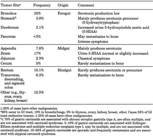
Carcinoid
Syndrome
- See Table
13-25.
- (The
syndrome in malignant carcinoids [argentaffinomas] includes flushing,
diarrhea, bronchospasm, endocardial fibrosis, arthropathy, glucose
intolerance, hypotension.)
- Liver metastases are
present in 95% of cases with syndrome except when lung and ovary are
primary sites, but laboratory tests are not reliable indicators and serum
ALP is frequently normal despite extensive metastases.
- Urinary level of 5-HIAA (a metabolite of serotonin) is increased in 75% of cases (>9
mg/24 hrs in patients without malabsorption or >30 mg/24 hrs with
malabsorption; normal is <6 mg/24 hrs), usually when tumor is far
advanced (with large liver metastases often 300-1000 mg/day), but may not
be increased despite massive metastases. Sensitivity = 73%. Useful in
diagnosis in only 5-7% of patients with a carcinoid tumor but in ~45% of
those with liver metastases. Disease extent and prognosis correlate generally
with urine 5-HIAA excretion; becomes normal after successful surgery. If
urine HIAA is normal, check blood level of serotonin or a precursor,
5-hydroxytryptophan. Urine HIAA may be decreased in renal insufficiency.
Increased
In
- Whipple's disease
- Nontropical sprue
- Small increases may occur
in pregnancy, ovulation, after surgical stress.
- Consumption of various
foods (e.g., pineapples, kiwis, bananas, eggplants, plums, tomatoes,
avocados, plantains, walnuts, pecans, hickory nuts, coffee)
- Use of certain drugs
(e.g., acetanilid, acetaminophen, acetophenetidin, caffeine, glyceryl
guaiacolate, heparin, L-dopa, mephenesin, methocarbamol, phenothiazine
derivatives, Lugol's solution, reserpine, salicylates)
Decreased
In
- Use of certain drugs
(e.g., chlorpromazine, promazine, imipramine, isoniazid, monoamine oxydase
inhibitors, methenamine, methyldopa, phenothiazines, promethazine)
- Serum and urine serotonin may be increased (>0.4 µg/mL) in 20% of
cases but without increased urine 5-HIAA.
- Platelet serotonin and urine serotonin are increased in 64% of cases.
- Increased plasma
chromogranin A predicts adverse prognosis.
P.689
|
|
|
Table 13-25. Carcinoid Tumors of GI Tract
|
- Some tumors can produce
various functionally active substances (e.g., histamine, ACTH,
somatostatin, gastrin, catecholamines, prostaglandins, kinins) causing
different paraneoplastic syndromes. Most are clinically silent because of
small amounts secreted and rapid inactivation.
- VMA and catecholamine
levels in urine are normal.
- Laboratory findings due
to other aspects of carcinoid syndrome (may include pulmonary valvular
stenosis, tricuspid valvular insufficiency, heart failure, liver
metastases, electrolyte disturbances)
- Nonfunctioning tumors can
be diagnosed only by histological examination.
- Some patients may have
decreased serum albumin and pellagra (due to diversion of tryptophan to
synthesis of serotonin).
Diabetes
Insipidus
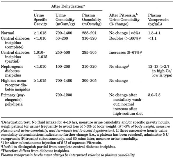
See Table 13-26.
Due To
- Central (pituitary)
- Nephrogenic
- Psychogenic
- High-set osmoreceptor
Diabetes
Insipidus, Central
See Table 13-26.
P.690
|
|
|
Table 13-26. Comparison of Different
Types of Diabetes Insipidus
|
Due To
- Primary
- Idiopathic (now accounts
for <50% of cases)
- Heredity (~1% of cases)
- Secondary
- Supra- and intrasellar
tumors
- Neoplasms (suprasellar
and intrasellar
- Primary (e.g., craniopharyngioma, cyst)
- Metastatic (e.g., carcinoma of breast, lung; leukemias)
- Histiocytosis
(eosinophilic granuloma is most common)
- Hand-Schüller-Christian
disease
- Granulomatous lesions
(e.g., sarcoidosis, TB, syphilis, Wegener's granulomatosis)
- Trauma, with or without
basal skull fracture; neurosurgical procedures
- Vascular lesions (e.g.,
aneurysms, thrombosis, sickle cell disease, Sheehan's syndrome
- Infections (e.g.,
meningitis, encephalitis, Guillain-Barré syndrome, CMV infection)
- Autoimmune disorders
- Others (e.g., hypoxemic
encephalopathy)
- Urine is inappropriately dilute (low specific gravity [usually <1.005] and
osmolality [50-200 mOsm/kg]) in presence of increased serum osmolality
(295 mOsm/kg) and increased or normal serum sodium.
- Large urine volume (4-15
L/24 hrs) is characteristic.
P.691
|
|
|
Table 13-27. Comparison of Hyponatremia
Due to Various Causes
|
- Plasma vasopressin level is decreased
- Dehydration test fails to increase urine specific gravity or osmolality, and serum
osmolality remains elevated. After administration of vasopressin, urine
osmolality increases by 50%.
- Partial central diabetes
insipidus shows intermediate values between complete central and normal.
- See Tables
13-26 and .
Diabetes
Insipidus, Nephrogenic
See Table 13-26.
Due To
- Chronic renal failure
(e.g., GN, pyelonephritis, gout, analgesic nephropathy, polycystic
kidneys, nephrosclerosis)
- Other tubulointerstitial
diseases (e.g., polycystic kidneys, medullary sponge disease, sickle cell
disease or trait, amyloidosis)
- Diuretic phase of acute
tubular necrosis
- After renal transplant or
relief of urinary tract obstruction
- Hypergammaglobulinemia
(e.g., multiple myeloma, amyloidosis, Sjögren's syndrome)
- Drugs (e.g., lithium,
demeclocycline, amphotericin, propoxyphene, methoxyflurane, vincristine)
- Prolonged potassium
depletion and hypokalemia (condition is reversed by restoring potassium
level to normal)
P.692
- Prolonged hypercalciuria,
usually with hypercalcemia (condition is reversed by restoring calcium
level to normal)
- Hereditary renal tubular
unresponsiveness to vasopressin due to X-linked genetic defect; severe
form occurs in males; family history of this condition is frequent.
- Primary
hyperaldosteronism
- Pregnancy
- Laboratory findings are the same as in hypophyseal (central) diabetes
insipidus except that in nephrogenic type
- Plasma vasopressin level
is normal or increased.
- Dehydration test does
not cause urine osmolality to increase above plasma osmolality.
- Dehydration test causes
the plasma vasopressin level to increase.
- Urine osmolality does
not increase with subsequent injection of vasopressin.
Diabetes
Insipidus Due to High-Set Osmoreceptor
- (Rare
entity in which the set point for stimulating release of ADH is ≥
300 mOsm/kg instead of the normal 285 mOsm/kg level)
- See Table
13-26.
- As plasma osmolality
increases, patient becomes thirsty and drinks fluids, thereby diluting the
plasma before it reaches the higher set level to stimulate release of ADH,
initiating cycle of polyuria and polydipsia. If thirst center is also
impaired, patient develops essential hypernatremia.
- Plasma osmolality after dehydration is significantly higher than in normal state.
- Urine osmolality does not increase after administration of vasopressin.
Growth
Hormone (GH) Deficiency
- May be isolated
deficiency with dwarfism or may be associated with TSH deficiency, with
ACTH deficiency, or with TSH and ACTH deficiencies. GH deficiency is
usually due to deficiency of hypothalamic GH-releasing hormone.
- Serum GH basal levels are decreased (<1.0 ng/mL). Use
pooled or average of three samples. Stimulation tests have greater
sensitivity. Increased basal or random serum level excludes this diagnosis
but low levels do not distinguish normal persons from those with GH
deficiency.
- Stimulation (functional)
tests
- Draw serum at 0, 30, 60,
90, and 120 mins.
- Administration of
insulin (regular crystalline, IV, 0.05 to 0.3 U/kg body weight) should
normally produce at least 2× increase in serum GH level and 3× increase
in serum prolactin level at 60-min peak. This is the most reliable
challenge for GH secretion.
- Administration of
levodopa (500 mg orally) should normally produce at least 2× increase in
serum GH level at 60-min peak.
- Administration of
arginine (0.5 gm/kg body weight as 5% solution IV over 30 mins) should
normally produce at least 3× increase in serum GH and at least 2×
increase in serum prolactin level at 30- to 60-min peak.
- Failure to produce these
minimal responses indicates a lesion of pituitary or hypothalamus but
does not differentiate between them.
- A normal response is at
least 10 ng/mL peak value; 5-10 ng/mL is indeterminate, ≤5 ng/mL is
subnormal. (A normal value rules out GH deficiency; in some laboratories
the normal level is ≥ 7 ng/mL.)
- Approximately one-fourth
of patients with normal GH secretory capacity are unable to secrete GH in
response to provocative tests indicated above, at any given time.
Therefore, at least two of these tests should be used to confirm
diagnosis of GH deficiency.
- Nonpituitary factors
that impair GH response include obesity, primary hypothyroidism,
thyrotoxicosis, primary hypogonadism, Kallmann's syndrome, Cushing's
syndrome, use of various drugs (e.g., alpha-adrenergic antagonists,
beta-adrenergic antagonists, serotonin antagonists, dopamine
antagonists). Impaired GH response may even occur in presence of elevated
GH basal level.
P.693
- Normal response may also
occur in patients with partial deficiency.
- GH response is normal or
exaggerated in growth failure due to resistance to GH (Laron dwarfism) or
resistance to somatomedins (African pygmies).
- Glucagon and clonidine
have also been used.
- Decreased fasting blood
sugar (<50 mg/dL) is frequent; responds to GH therapy. Serum phosphorus
and ALP are decreased in prepubertal children but normal in adult-onset
cases.
- Serum prolactin baseline
level is low and does not rise appropriately after TRH administration or
other stimulation. In hypothalamic disease, basal prolactin level is
increased and response may be normal or blunted.
- Laboratory findings due
to involvement of other endocrines
- TSH deficiency (see Sensitive Thyroid-Stimulating Hormone; TRH
stimulation test, Hypothyroidism, and Table 14-3).
- ACTH deficiency (see
tests of adrenal function).
- Gonadotropins are
decreased or absent from urine in postpubertal patients (but increased
levels occur in primary hypogonadism).
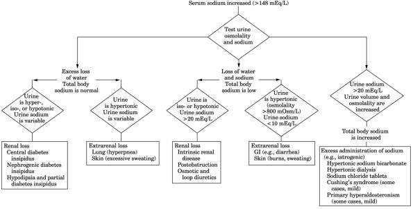
Hypernatremia,
"Essential"
- (Due
to hypothalamic lesions [e.g., infiltration of histiocytes, neoplasm] that
cause impaired osmotic regulation but intact volume regulation of ADH
secretion.)
- See Fig.
13-27.
- Serum sodium shows sustained but fluctuating elevations, corrected by
administration of ADH but not corrected by fluid administration.
- Serum osmolality is increased
- Serum creatinine, BUN,
and creatinine clearance are normal.
- There is spontaneous excretion
of random specimens of urine, which may be very concentrated or very
dilute and opposite to plasma osmolality.
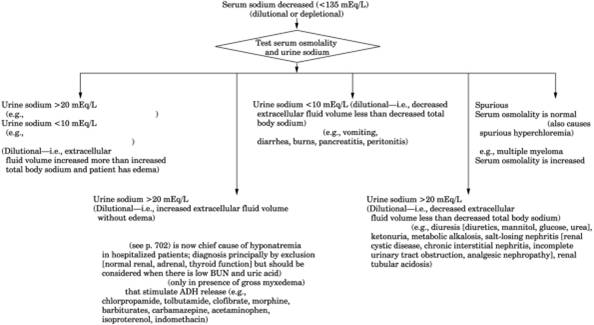
Hyponatremias
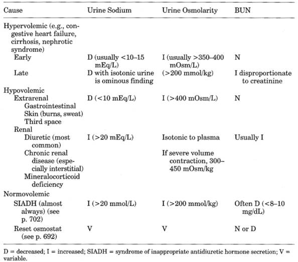
See Table 13-27 and Fig. 13-28.
Due To
- Isotonic (spurious-occurs
with flame photometer but not with ion-selective electrode technology)
- Hyperlipidemia (plasma
looks milky) "falsely" lowers serum sodium; measured serum osmolality
exceeds calculated serum osmolality.
- Calculated serum
osmolality = 2 × Na + (serum glucose/18) + (BUN/2.8)
- Hyperproteinemia (e.g.,
myeloma, macroglobulinemia)
- Hypertonic
- Hyperglycemia (each
increase of blood sugar of 100 mg/dL decreases serum sodium by 1.7 mEq/L)
- Excess mannitol
treatment
- Hypotonic
- Hypervolemic, usually
with clinical edema
- With low urine sodium
(<10 mEq/L) may be due to congestive heart failure, cirrhosis with
ascites, nephrotic syndrome
- With high urine sodium
(>20 mEq/L) may be due to acute tubular necrosis or end-stage chronic
renal failure in which sodium and water intake exceeds excretion. Serum
uric acid and BUN tend to be increased.
- Hypovolemic
- Urine sodium <10
mEq/L. Due to extrarenal loss of sodium (e.g., GI tract, fistulas,
pancreatitis, exercise, sweating, burns).
- Urine sodium >20
mEq/L. Due to renal loss of sodium (e.g., diuretics such as furosemide or
osmotic diuresis due to glucose or urea, diabetic ketoacidosis, renal
tubular acidosis, salt-losing nephritis, adrenal insufficiency,
hyporeninemia, hypoaldosteronism).
- Normovolemic-usually no
edema is present.
P.694
|
|
|
Fig. 13-27. Algorithm for hypernatremia.
(Hypotonic urine-urine osmolality is <800 mOsm/L; isotonic urine-urine
osmolality is between 800 mOsm/L and plasma osmolality; hypertonic
urine-urine osmolality >800 mOsm/L.)
|
P.695
|
|
|
Fig. 13-28. Algorithm for hyponatremia.
(ADH = antidiuretic hormone; SIADH = syndrome of inappropriate antidiuretic
hormone secretion.)
|
P.696
- Large amounts of sodium
appear in urine (>20 mEq/L). May be due to SIADH, hypothyroidism,
hypopituitarism, low-reset osmostat syndrome, physical or emotional
stress, potassium depletion, renal failure, water poisoning, certain
drugs (e.g., ADH analogs, amitriptyline, carbamazepine, chlorpropamide,
cyclophosphamide, diuretics, haloperidol, thioridazine, vincristine).
- Hyponatremic
patients with BUN <10 mg/dL and uric acid <3.0 mg/dL should be
considered to have SIADH or reset osmostat until proved otherwise
- "Pseudohyponatremia"-see
above. Serum osmolality is normal.
Hypopituitarism
Due To
- Pituitary disease
- Neoplasms (e.g.,
craniopharyngioma, chromophobe adenoma, eosinophilic adenoma, meningioma,
metastatic tumor [especially breast, lung]); prolactin-secreting tumor is
the most common pituitary neoplasm.
- Infiltrative diseases
- Granulomatous lesions
(e.g., sarcoidosis, Hand-Schüller-Christian syndrome, histiocytosis X)
- Infection (e.g., TB,
mycoses)
- Hemochromatosis
- Autoimmune inflammation
- Hemorrhage
- Pituitary necrosis secondary
to postpartum hemorrhage (Sheehan's syndrome)
- Hemorrhage into
pituitary tumor
- Infarction (e.g., sickle
cell disease, cavernous sinus thrombosis)
- Miscellaneous
- Head trauma
- Internal carotid artery
aneurysm
- Empty sella syndrome
- Idiopathic
- Isolated hormone
deficiency (e.g., GH, ACTH, TSH, gonadotropin)
- Multiple hormone
deficiency
- Iatrogenic (e.g.,
hypophysectomy, irradiation, section of stalk)
- Familial pituitary
deficiency (deficient hormone production or production of abnormal
hormone)
- Partial GH deficiency
(some forms of "constitutional short stature" with delayed onset of
adolescence)
- Hypothalamic disease
- End-organ resistance to
GH (normal or increased serum GH with low somatomedin level)
- Laron dwarfs
(somatomedin levels are often undetectable and fail to rise when GH is
administered).
- Serum
somatomedin C levels are 5-15% of normal in most hypopituitary dwarfs and
4-12 times normal in all active acromegaly patients.
- Endocrinologic findings: diagnosis is based on low serum level of target organ hormone and
of the corresponding pituitary-stimulating hormone, e.g.,
- Hypogonadism
- Men: low sperm count,
low serum testosterone, inappropriately low serum LH and FSH
- Women: low serum
estradiol, inappropriately low serum LH and FSH
- Hypothyroidism
- Low serum T and FTI, inappropriately low serum thyrotropin
- Hypocorticalism:
- Low serum cortisol and
ACTH
- Low serum GH
unresponsive to provocative tests
- Low serum prolactin
unresponsive to provocative tests. Usually occurs late in course of
hypopituitarism except in Sheehan's syndrome, in which it may be the
earliest manifestation. Rarely or never due to hypothalamic disease.
- See sections on secondary
insufficiency of gonads, thyroid, adrenals. Only one (usually gonadal
first) or all of these may be involved.
P.697
- Dynamic
tests are usually needed to detect partial deficiencies.
- See Diabetes
Insipidus, Central.
Hypothalamus,
Diseases
Due To
- Neoplasms (primary or
metastatic cancer, craniopharyngioma) (most frequent cause)
- Inflammation (e.g., TB,
encephalitis)
- Head trauma (e.g., basal
skull fractures, gunshot wounds)
- Granulomas (e.g.,
histiocytosis X, sarcoidosis)
- Releasing-hormone
deficiency, genetic or idiopathic
- Irradiation for childhood
cancer
Manifestations
- Sexual abnormalities are
the most frequent manifestations of hypothalamic disease.
- Precocious puberty
- Hypogonadism (frequently
as part of Fröhlich's syndrome)
- Diabetes insipidus is a
frequent but not an early manifestation of hypothalamic disease.
- Hypopituitarism-differentiate
primary hypopituitarism from this secondary form of hypopituitarism by
appropriate stimulation tests.
Multiple
Endocrine Neoplasia (Men Syndrome)
MEN
Type I (Wermer's Syndrome)
- (Triad
of parathyroid, pancreatic islet cell, and anterior pituitary tumors)
- ○
Hyperparathyroidism (due to involvement of all four glands) in >88% of
patients; is usual presenting feature; associated renal and bone disease
are infrequent. 15% of cases of HPT have MEN; frequently multicentric. 10%
of parathyroid tumor patients have relatives with MEN.
- ○ Pancreatic
endocrine tumors in ~60% of patients; most are functional; usually
multiple.
- Gastrinomas with Z-E
syndrome occur in ~50% of cases and ~50% are malignant. 50% of cases of
Z-E syndrome have MEN type I.
- Insulinomas (beta cells)
in ~25% of MEN type I patients; usually benign; multiple foci are common.
- Glucagonomas (alpha
cells) syndrome of distinctive rash, diabetes mellitus, anemia, weight
loss.
- Vipomas occur less
often.
- ○ Pituitary
adenomas in 40-50% of cases
- ~25% are prolactinomas.
- ~15% are eosinophilic
adenomas causing acromegaly.
- ~5% are basophilic
adenomas causing Cushing's syndrome.
- ~10% are nonfunctional
adenomas causing hypopituitarism due to space-occupying effect.
- ○ Tumors possible
related to MEN type I
- Adrenal cortical
adenomas or hyperplasia are incidental and nonfunctioning in ~10%,
functioning in ~5% of cases. Adrenal medulla is not involved.
- Thyroid disease in ~20%
of cases including benign and malignant tumors, colloid goiter,
thyrotoxicosis, Hashimoto's disease.
- Uncommon lesions include
carcinoids (~16%), schwannomas, multiple lipomas, gastric polyps,
testicular tumors.
MEN
Type II (or IIa) (Sipple's Syndrome)
- ○ Medullary thyroid
carcinoma in >90% of cases is usually multicentric and preceded by
C-cell hyperplasia (thereby differing from sporadic type). Produces
calcitonin and sometimes ACTH or serotonin. Calcitonin response to IV
pentagastrin stimulation
P.698
has >90% sensitivity and specificity. 25% of these carcinomas occur as part
of MEN type II. May be asymptomatic but lethal.
- ○ Pheochromocytoma
in 10-50% of cases; usually bilateral, often multiple, and may be
extra-adrenal. 10% of pheochromocytomas occur as part of MEN.
- ○
Hyperparathyroidism in ~20% of cases; due to hyperplasia in 84% and
adenoma in 16%; occurs late in disease; may occur without medullary
thyroid carcinoma.
- DNA analysis detected carriers of the gene before biochemical manifestations (100%
sensitivity and specificity).30
MEN
Type III (or IIb)
- (Features
in common with MEN type II but is a separate genetic syndrome)
- ○ Medullary thyroid
carcinoma in 75% of cases.
- ○ Pheochromocytoma
in 33% of cases.
- Hyperparathyroidism is
rare (<5% of cases).
- ○ Other lesions:
- Multiple mucosal
gangliomas in >95% of cases appear early in life.
- Marfan syndrome habitus,
hypertrophy of corneal nerves, ganglioneuromas of GI tract,
characteristic retinal changes and facial appearance are frequent.
- ○ All first-order relatives of MEN patients should have
appropriate serial testing.
Nonendocrine
Neoplasms, Causing Endocrine Syndromes
- (Tumors
secrete proteins, polypeptides, or glycoproteins that have hormonal
activity.)
- Diagnosed by measuring arteriovenous gradient of hormone across tumor bed or between
tumor and nontumor tissue; confirm by in vitro demonstration of hormone
production by tumor cells and by resolution of endocrine syndrome after
successful removal of tumor.
- Cushing's syndrome: increased blood ACTH level (>200 pg/mL), inability
to suppress with high-dose DST (except in bronchial carcinoids), loss of
diurnal variation of cortisol levels (usually >40 µg/dL). Therefore
cannot be distinguished from excessive pituitary secretion of ACTH by use
of DST. Typically malignant disease causing ectopic ACTH production has
acute effects on adrenal glands manifested predominantly by excess mineralocorticoid
production with hypokalemia and hypertension. May sometimes require
selective venous catheterization to measure ACTH levels or in vitro
hybridization assay to demonstrate ACTH-encoding messenger RNA to
establish the diagnosis. Patients with lung cancer may have elevated ACTH
levels without Cushing's syndrome.
Due To
- Bronchogenic oat cell
carcinoma (causes ~50% of cases) and carcinoid
- Thymoma
- Hepatoma
- Carcinoma of ovary
- Also medullary carcinoma
of thyroid, islet cell tumor of pancreas, etc. Hypercalcemia simulating
HPT (see Humoral Hypercalcemia of Malignancy, and Table 13-7)
- Renal carcinoma
- Squamous cell and
large-cell carcinoma of respiratory tract
- Carcinoma of breast
(occurs in 15% of patients with bone metastases)
- Malignant lymphoma,
myeloma, etc.
- Cancer of ovary,
pancreas, etc.
- SIADH
- Especially with oat cell
carcinoma of lung
- Hypoglycemia: serum insulin is low in presence of fasting hypoglycemia. Not
associated with decreased serum phosphorus as in insulin-induced
hypoglycemia.
- Bronchogenic carcinoma
- Carcinoma of adrenal
cortex (6% of patients)
P.699
- Hepatoma (23% of
patients)
- Retroperitoneal
fibrosarcoma (most frequently)
- Thyrotoxicosis: signs and
symptoms are rare, but laboratory findings are present.
- Tumors of GI tract,
hematopoietic tumors, pulmonary tumors, etc.
- Trophoblastic tumors in
women
- Choriocarcinoma of
testis
- Struma ovarii
- Precocious puberty in
boys
- Acromegaly
- Pancreatic tumors
producing GH or GH-releasing factor in presence of normal sella;
increased GH not suppressed by glucose.
- Carcinoid.
- Erythrocytosis (due to
erythropoietin production)
- Carcinoma of kidney,
liver
- Fibromyoma of uterus
- Cerebellar
hemangioblastoma
- See also Carcinoid
Syndrome, Precocious Puberty, Syndrome of Inappropriate Secretion
of Antidiuretic Hormone.
Pineal
Tumors
- Boys-precocious puberty
in 30% of patients
- Girls-delayed pubescence
- Diabetes insipidus occurs
occasionally.
Pituitary
Tumors
- Findings due to increased production of hormones or effect of growing mass.
- Most common tumors are
- Prolactin-secreting
tumors comprise ~30% of all pituitary tumors (see Prolactinomas)
- GH-secreting tumors (see
Acromegaly and Gigantism)
- ACTH-secreting tumors
(see Cushing's Syndrome)
- Nonfunctioning adenomas,
which may produce findings of intracranial mass, especially with visual
changes, and hypopituitarism (sometimes with impaired hypothalamic
function)
- Microadenomas (<10 mm
in size) may be present in 10-20% of the population by autopsy and
radiographic studies ("incidentaloma") but tumors >10 mm in size are
quite rare.
Polydipsia,
Psychogenic
- (Excessive
intake of water causes loss of medullary sodium and urea to renal venous
blood and abnormally reduced tonicity of renal medulla.)
- See Table
13-26.
- ○ Should be
suspected when large volumes of very dilute urine occur with plasma
osmolality that is only slightly decreased or low normal.
- Test dose of vasopressin
often shows failure to concentrate urine, simulating nephrogenic diabetes
insipidus. However, the test is normal when performed after restoration of
normal hypertonicity of renal medulla by a period of high sodium and low
water intake.
- Fluid deprivation test is
least reliable in differentiating this from partial central diabetes
insipidus; e.g., some increase in urine osmolality after dehydration with
an inconclusive (~10%) further increase after vasopressin may be due to
either condition.
Polyglandular
Syndromes, Autoimmune
Type I
- Requires two or more of the following: hypoparathyroidism, Addison's disease,
chronic mucocutaneous candidiasis (all three are present in approximately
one-third
P.700
of patients). Patient may also have associated immune disorders, e.g.,
autoimmune hypothyroidism. Gonadal failure and chronic hepatitis may also
occur.
|
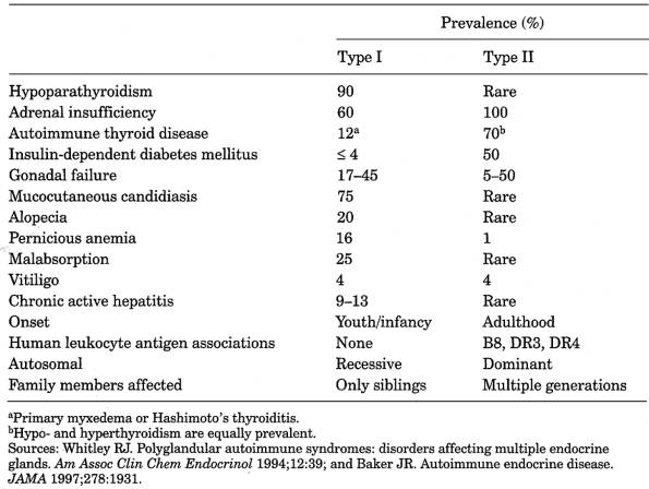
|
|
Table 13-28. Comparison of Polyglandular
Syndromes Types I and II
|
Type
II (Schmidt's Syndrome)
- Autoimmune thyroiditis or insulin-dependent diabetes (15% of all patients with
insulin-dependent diabetes mellitus have type II) with Addison's disease.
Interval between onset of endocrinopathies may be up to 20 yrs. Gonadal
failure may sometimes occur.
- (For comparison of types
I and II, see Table 13-28.)
Type
III
Autoimmune thyroid disease with two other
autoimmune disorders, including insulin-dependent diabetes mellitus, PA, or a
non-endocrine-organ-specific autoimmune disorder (e.g., myasthenia gravis) but
without Addison's disease.
Prolactinomas
Serum
Prolactin Reference Values
- Normal <25 ng/mL in
females; lower in males and children.
- Gradual increase from
birth until adolescence
- 13- to 15-yr-old boys:
2.5× adult levels
- 13- to 15-yr-old girls:
3× adult levels
- Serum samples should be
collected under basal conditions with minimal stress and by pooling three
blood samples collected at 20-min intervals for one assay; all drugs
should be discontinued for at least 2 wks before testing.
Interpretation
- 40-85 ng/mL: seen in
craniopharyngioma, hypothyroidism, effect of drugs
- 50 ng/mL: 25% chance of a
pituitary tumor
P.701
- 100 ng/mL: 50% chance of
a pituitary tumor
- <200 ng/mL with a
macroadenoma, particularly with extrasellar extension, is most likely due
to compression of pituitary stalk rather than prolactinoma.
- 200-300 ng/mL: nearly
100% chance of a pituitary tumor; >200 ng/mL may indicate a
macroadenoma rather than microadenoma and tumor usually is visible on CT
or MRI, but CT or MRI is normal in ≤ 20% of microadenomas.
- High levels may be seen
with simultaneous multiple additive factors that usually cause lesser
increases (e.g., chronic renal failure plus use of methyldopa)
- Immediate postoperative level of <7.0 ng/mL indicates long-term cure but
higher levels are associated with recurrence.
- Repeated serum levels in late morning or early afternoon that are increased
3-5× normal in men or nonlactating women are usually considered diagnostic
of pituitary adenoma or rarely of hypothalamic disease or pituitary stalk
section or hypothyroidism. One elevated level is not adequate for
diagnosis. Level normally increases sharply during sleep; higher in
morning than afternoon.
Increased
Serum Prolactin In
- Amenorrhea/galactorrhea
- 10-25% of women with
galactorrhea and normal menses
- 10-15% of women with
amenorrhea without galactorrhea
- 75% of women with both
galactorrhea and amenorrhea/oligomenorrhea
- Causes 15-30% of cases
of amenorrhea in young women
- Pituitary lesions (e.g.,
prolactinoma, section of pituitary stalk, empty sella syndrome, 20-40% of
patients with acromegaly, up to 80% of patients with chromophobe adenomas)
- Hypothalamic lesions
(e.g., sarcoidosis, eosinophilic granuloma, histiocytosis X, TB, glioma,
craniopharyngioma)
- Other endocrine diseases
- ~20% of cases of hypothyroidism
(second most common cause of hyperprolactinemia). Therefore
serum TSH and T should always be measured
- Addison's disease
- Polycystic ovaries
- Glucocorticoid
excess-normal or moderately elevated prolactin
- Ectopic production of
prolactin (e.g., bronchogenic carcinoma, renal cell carcinoma, ovarian
teratomas, acute myeloid leukemia)
- Children with sexual
precocity-may be increased into pubertal range
- Neurogenic causes (e.g.,
nursing and breast stimulation, spinal cord lesions, chest wall lesions such
as herpes zoster)
- Stress (e.g., surgery,
hypoglycemia, vigorous exercise)
- Pregnancy (increases to
8-20× normal by delivery, returns to normal in 2-4 wks postpartum unless
nursing occurs)
- Lactation
- Chronic renal failure
(20-40% of cases; becomes normal after successful renal transplant but not
hemodialysis)
- Liver failure (due to
decreased prolactin clearance)
- Idiopathic causes (some
probably represent early cases of microadenoma too small to be detected by
CAT scan)
Interferences
- Drugs-most
common cause; usually subsides a few weeks after cessation of using
drug; these elevations are usually <100 ng/mL
- Neuroleptics (e.g.,
phenothiazines, thioxanthenes, butyrophenones)
- Antipsychotic drugs
(e.g., Compazine, chlorpromazine [Thorazine], trifluoperazine hydrochloride
[Stelazine], thioridazine hydrochloride (Mellaril), haloperidol lactate
[Haldol])
- Dopamine antagonists
(e.g., metoclopramide, sulpiride)
- Opiates (morphine,
methadone)
- Reserpine
- Alpha-methyldopa
(Aldomet)
- Estrogens and oral
contraceptives
- TRH
- Amphetamines
- Isoniazid
P.702
Serum
Prolactin May Be Decreased In
- Hypopituitarism
- Postpartum pituitary
necrosis (Sheehan's syndrome)
- Idiopathic
hypogonadotropic hypogonadism
- Use of certain drugs
- Dopamine agonists
- Ergot derivatives
(bromocriptine mesylate, lergotrile mesylate, lisuride hydrogen maleate)
- Levodopa, apomorphine,
clonidine
Interpretation
- Normal value in child with growth
retardation virtually rules out hypopituitarism but a low value is not diagnostic.
Single blood value may be more reliable than multiple measurements of GH
in diagnosis of active acromegaly.
- TRH stimulation of patients with increased prolactin not due to pituitary tumors usually
doubles serum prolactin level to peak of >12 ng/mL in 15-30 mins, but
most patients with prolactinomas do not respond to TRH stimulation
(response <2× baseline level). Enhanced responsiveness in
hypothyroidism and blunted prolactin rise in chronic renal failure.
Unresponsiveness to TRH (<2× baseline level) also occurs in
panhypopituitarism. Measurement of multiple basal prolactin levels has
replaced stimulation tests for diagnosis of prolactinoma.
- Microscopic examination
of breast discharge shows numerous fat globules; if not seen, rule out
intraductal breast carcinoma or infection.
- Normal or decreased serum
FSH, LH, and testosterone may occur in men.
- Women may also present
with hirsutism, infertility. Men may present with decreased libido,
impotence, oligospermia, low serum testosterone levels, and sometimes
galactorrhea.
- Hypothyroidism
- Acute fasting and chronic
protein-calorie deprivation (when GH often rises)
Syndrome
of Inappropriate Secretion of Antidiuretic Hormone (Siadh)
(Syndrome of
continuing release of vasopressin in presence of low plasma osmolality; kidney
responds normally to arginine vasopressin.)
Due To
- CNS disease of all types
(e.g., neoplastic, degenerative, infective, traumatic, vascular,
psychogenic)
- Advanced endocrinopathies
(e.g., myxedema, ACTH deficiency, adrenal insufficiency)
- Neoplasms (most commonly
oat cell carcinoma of lung; adenocarcinoma of lung, carcinoma of pancreas,
carcinoma of duodenum, lymphoma), some of which show ectopic production of
ADH
- Pulmonary diseases (e.g.,
cancer, pulmonary emboli, TB, pneumonia, chronic infections, lung abscess,
aspergillosis)
- Miscellaneous (e.g., acute
intermittent porphyria, postoperative state)
- Idiopathic causes
- Various drugs
- Oral hypoglycemic agents
(chlorpropamide, tolbutamide, phenformin, metformin)
- Antineoplastic agents
(vincristine, cyclophosphamide)
- Diuretics
(chlorothiazide)
- Sedatives, analgesics
(morphine, barbiturates, acetaminophen)
- Psychotropic drugs
(amitriptyline, phenothiazines)
- Miscellaneous
(clofibrate, isoproterenol, nicotine)
- Cause
should be established because some causes are curable with resolution of
SIADH. Cortisol deficiency and hypothyroidism should always be excluded
- Dilutional hyponatremia with appropriately decreased osmolality (usually <280
mOsm/kg) when urine is not at maximum dilution; this is basis for
diagnosis in
P.703
patient with no evidence of cardiac, liver, kidney, adrenal, pituitary, or
thyroid disease, or hypovolemia and not on drug therapy (especially diuretics).
- Increased urine sodium (>20 mmol/L; >30 mmol/day) with inappropriately
high urine osmolality (>500 mOsm/kg) is essential for diagnosis because
it excludes hypovolemia as the cause of hyponatremia (in absence of
abnormal renal function or causative drugs)
- Increased urine osmolality higher than serum osmolality
- Normal serum potassium,
CO , BUN, and creatinine
- Decreased serum chloride
- Decreased AG
- Decreased uric acid (due
to dilution)
- Increase in plasma
vasopressin that is inappropriate for the degree of plasma osmolality is
not helpful in diagnosis because most causes of true hyponatremia are
associated with detectable or increased vasopressin. (See Arginine
Vasopressin.)
- Clinical and biochemical
response to fluid restriction but not to administration of isotonic or
hypertonic saline
Thyroid-Stimulating
Hormone (Tsh)-Secreting Pituitary Adenomas
- (Rare
type of adenoma that causes hyperthyroidism)
- Laboratory findings of hyperthyroidism except serum TSH is increased and does not
increase in response to TRH stimulation or does not decrease in response
to suppressive doses of thyroid hormone.
- Increased molar ratio of alpha subunit of TSH to whole TSH.
- ○ Secretion of
other hormones (e.g., prolactin, GH) occurs in about one-third of these
cases.
REFERENCES
1. Kolesnick RN,
Gershengorn MC. Thyrotropin-releasing hormone and the pituitary. Am J Med
2. Davidson MB, et al.
Relationship between fasting plasma glucose and glycosylated hemoglobin.
Potential for false-positive diagnosis of type 2 diabetes using new diagnostic
criteria. JAMA
3. Data from National
Diabetes Data Group, 1978.
4. McMahon MM, O'Brien PC,
Service FJ. Diagnostic interpretation of the intravenous tolbutamide test for
insulinoma. Mayo Clin Proc
5. Report of the Expert
Committee on the Diagnosis and Classification of Diabetes Mellitus. Diabetes
Care 1997;20:1183.
6. Jensen RT, Fraker DL.
Zollinger-Ellison syndrome. Advances in treatment of gastric hypersecretion and
the gastrinoma. JAMA
7. Chrousos GP, et al. NIH
Conference. Clinical applications of corticotropin-releasing factor. Ann Intern Med
8. Kaye TB, Crapo L. The Cushing
syndrome: an update on diagnostic tests. Ann Intern
Med
9. Doppman JL, Oldfield EH,
Nieman LK. Bilateral sampling of the internal jugular vein to distinguish
between mechanisms of adrenocorticotropic hormone-dependent Cushing syndrome. Ann Intern Med
10. Blumenfeld JD, et al.
Diagnosis and treatment of primary hyperaldosteronism. Ann Intern Med
11. Bornstein SR, et al.
Adrenocortical tumors: recent advances in basic concepts and clinical
management. Ann Intern Med
12. Mann SJ, Pickering TG.
Detection of renovascular hypertension. State of the art. Ann Intern Med
13. Carpenter PC. Cushing's
syndrome: update of diagnosis and management. Mayo
Clin Proc
14. Dunlap NE, Grizzle WE,
Siegel AL. Cushing's syndrome. Screening methods in hospitalized patients. Arch Pathol Lab Med
15. Orth DN. Cushing's
syndrome. N Engl J Med
16. Freda PU. Differential
diagnosis in Cushing syndrome. Use of CRH. Medicine
17. Chrousos GP, Detera-Wadleigh SD,
Karl M. Syndromes of glucocorticoid resistance. Ann
Intern Med
18. Pickering TG. The
role of laboratory testing in the diagnosis of renovascular hypertension. Clin Chem
19. Duncan MW, Compton P,
Lazarus L, Smythe GA. Measurement of norepinephrine and
3,4-dihydroxyphenylglycol in urine and plasma for the diagnosis of
pheochromocytoma. N Engl J Med
20. Lenders JWM, et al.
Plasma metanephrine in the diagnosis of pheochromocytoma. Ann Intern Med
21. Peplinski GR, Norton
JA. The predictive value of diagnostic tests for pheochromocytoma. Surgery
22. Eisenhofer GE, et al.
Plasma normetanephrine and metanephrine for detecting pheochromocytoma in von
Hippel-Lindau disease and multiple endocrine neoplasia Type 2. N Engl J Med
23. Lee MM, et al.
Measurements of serum müllerian inhibiting substance in the evaluation of
children with nonpalpable gonads. N Engl J Med
24. Lippe BM. Ambiguous
genitalia and pseudohermaphrodites. Ped Clin North Am
25. Adams JE. Infertility
in men: diagnosis and treatment. ASCP Check Sample CC 87-9 (CC-187). 1987;27:1.
26. Rothmann SA, Morgan BW.
Laboratory diagnosis in andrology. Cleve Clin J Med 1989;(Nov-Dec):805.
27. Ferrara F, et al.
Automation of human sperm cell analysis by flow cytometry. Clin Chem
28. Kulke MH, Mayer RJ.
Carcinoid tumors. N Engl J Med
29. National Institutes of
Health Conference. Multiple endocrine neoplasia type I: clinical and genetic
topics. Ann Intern Med
30. Lips CJM, et al.
Clinical screening as compared with DNA analysis in families with multiple
endocrine neoplasia type 2A. N Engl J Med
Footnotes
TSH injection causes increase of ≥50%
of RAIU in normal persons.
TSH injection does not cause a normal
increase of ≥50% of RAIU.
Urinary iodine >2000 µg/24 hrs.
