ESTHETIC PROBLEMS OF MISSING TEETH
CHAPTER 21. FIXED REPLACEMENT OF MISSING
TEETH - Steven K. Nelson, DMD, F. Michael Gardner, DDS, MA, Ronald E.
Goldstein, DDS
INTRODUCTION
With the development of many new metal alloy systems, porcelains to fire to
these alloys, all-ceramic crown materials, porcelain margins for metal-ceramic
restorations, porcelain veneers, and much stronger bonding and cementing
systems, both functional and esthetic results may be obtained today that were
unthought of in the past. However, with the evolution of such varied choices in
materials and techniques, the requirements for selecting the correct
combinations for success become ever more difficult.
Once a patient decides to have a lost tooth replaced with a fixed partial
denture, it must be remembered that regardless of how skillfully the
biomechanical requirements have been met, the patient will judge his or her
result primarily on the basis of esthetics, especially if the prosthesis is in
the anterior region. Today's patient is more aware of the interrelationship
between teeth and facial appearance and is entitled to the dentist's best
artistic efforts.
Prior to choosing a fixed partial denture as the restoration of choice for a
given edentulous situation, a logical sequence of diagnosis and treatment
planning must be followed to achieve a successful outcome. A thorough
diagnostic work-up should be performed, which will provide the restoring
dentist with all of the information needed to determine the best treatment plan
for the patient.3,5,20 This diagnostic information is best gathered
through a systematic approach such as the one outlined below.
DIAGNOSIS
Medical and Dental History
The starting point for any patient care begins with thorough medical and dental
histories. Information gathered on the written history sheet and expanded
verbally can provide the restoring dentist with data pertinent to successful
prosthodontic treatment. Noting prior dental experiences and the patient's
attitude toward treatment is of extreme importance in developing a rapport with
the patient. The patient's chief complaint should be ascertained, and if it is
esthetic in nature, specific esthetic desires and needs should be assessed, as
well as any esthetic shortcomings with previous prostheses. Once these facts
have been established and a good working relationship exists between the
dentist and the patient, the correct treatment for that patient can be pursued.
Intraoral Examination
A comprehensive intraoral examination should be performed on all patients. The
charting of preexisting restorations and any new pathoses should be a routine
procedure. All diagnoses should be documented, such as missing teeth,
periodontal status, pulpal pathosis, caries, fractures, wear, unesthetic
restorations, muscle and temporomandibular joint pathosis, and neoplasm. All
data should be recorded, and the patient should be treatment planned as a total
entity, rather than only addressing the specific edentulous area and adjacent
tooth structure. Often, steps for developing comprehensive treatment plans are
overlooked, and the treatment starts before a diagnosis is made. Proper work-up
procedures help achieve a sound prosthodontic outcome.
The intraoral examination can be enhanced by using an intraoral camera (see
Figures 2-7A to C, Chapter 2, Esthetics in Dentistry, Volume 1, 2nd
Edition)16,18 or surgical microscope (Figure 21-1).21 Revelations such as
hidden microcracks, defective restoration margins, and other tooth and tissue
defects can determine a choice of single or multiple retainers when weakened
teeth thought to be in good condition are discovered. Each tooth should be
manually recorded in the chart and/or electronically in the computer for
diagnostic and legal purposes.
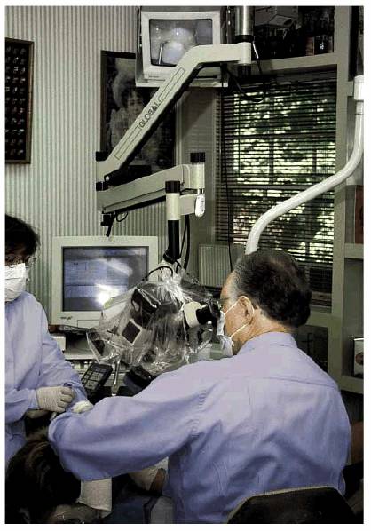
Figure 21-1: The surgical microscope, due to the extremely bright field and high magnification, provides the ultimate in intraoral diagnosis.
Extraoral Examination
The extraoral examination should include assessment of symmetry, muscle
hypertrophy, possible loss of vertical dimension of occlusion, and a smile
analysis that determines the amount of each tooth that will be seen while
smiling, speaking, etc. (see Chapter 2, Esthetics in Dentistry, Volume
1, 2nd Edition).
Radiographs
A full-mouth series 24424q1624y of radiographs or a panoramic radiograph with selected
periapical radiographs of the proposed abutment teeth is necessary in the
evaluation for treatment with a fixed partial denture. The primary purpose of
radiographs is to disclose hidden areas and structures such as the root
morphology, pulpal outline, periodontal ligament space, and the extent of
present and past caries.30 In addition to the intraoral examination,
the radiographs can provide the restoring dentist with information such as
pulpal pathosis, crown-to-root ratio, and quality of remaining bone and aid in
determining the tilt of teeth.
Computerized radiographs37,42 can also be a helpful way to
communicate with the patient. The ability to colorize the findings plus isolate
and enlarge segments of the root or crown in question, while producing a
greatly reduced amount of radiation, provides an avenue to enhance
patient-doctor relationships.
Diagnostic Casts
Diagnostic casts are imperative in diagnosis and treatment planning. The casts
are used as an educational aid for the patient and to provide the dentist with
the preexisting condition of the patient. Casts mounted on a suitable
articulator, at the treatment position, will enable the restoring dentist to
evaluate the condition of the patient's mouth. Clinical crown length, tipped or
rotated teeth, ridge form, and the span of the edentulous area can all be
evaluated, thereby helping the dentist in the decision-making process. The
interarch space and the occlusal plane can be evaluated on the diagnostic
casts, which may lead to the diagnosis of lost interocclusal space or
supraeruption of a segment of the dentition. The treatment, therefore, may
involve crown lengthening, ridge reduction, endodontic therapy, repositioning
of teeth, segmental osteotomy, or extraction. Properly mounted, accurate
diagnostic casts are a very important element of the diagnosis and treatment
planning of a fixed partial denture.
Diagnostic Waxing
A diagnostic waxing of the proposed fixed partial denture can be invaluable in
determining the esthetic criteria for a treatment plan. It allows for the
opportunity to observe the abutment tooth-pontic relationship and the
pontic-ridge relationship. The diagnostic waxing also allows the dentist to
evaluate and work within the exact space of the edentulous area. The edentulous
area itself may be badly resorbed and require surgical correction with grafting
of bone, soft tissue, or both. Often, other treatment issues or necessary
modifications become evident at this point in the planning process.39
Frequently, orthodontic treatment is the best solution for limited space and
rotated, tipped, or malposed teeth. This may be done in place of or prior to
fabrication of a fixed partial denture. In some situations, the diagnostic
waxing will indicate the need for endodontic treatment when tooth preparation
will involve the pulp of slightly malposed or tipped teeth.
Esthetic Considerations
A prime part of diagnosis is ascertaining the requirements relating to
esthetics, especially from the patient's perspective. Communication with the
patient provides a general sense of what his or her expectations are and
therefore provides information that may dictate, for example, the type of
retainer margin, retainer margin placement in relation to the gingiva, or
whether porcelain occlusal surfaces are indicated. The dentist should know what
the patient expects esthetically before treatment begins to avoid esthetic disasters.
For example, although the maxillary anterior region is usually the most
demanding due to its easy visibility, certain patients will place just as much
demand for exact shade duplication in the posterior region.
The arch in which the prosthesis is to be placed, the restoration's position in
that arch, the amount of display of the prosthesis, and the patient's esthetic
awareness all have to be considered when designing the elements of an esthetic
fixed partial denture. These elements include retainer type, material and
amount of coverage of the teeth, margin location and material, ceramic-metal
junction location on metal-ceramic crowns, and pontic design. These esthetic
considerations must be coupled with biologic and functional considerations such
as span length, need for splinting, periodontal support, soft-tissue
management, the use of provisionals, and the need for adjunctive care such as
orthodontics, endodontics, periodontics, and oral and maxillofacial surgery.
The anterior fixed prosthesis often presents the most difficult esthetic
problem. The choice of tooth form, shade, and arrangement used for complete
dentures is not usually available for the fabrication of a fixed prosthesis.
Artistic skill is required to obtain a pleasing result. Correct occlusal
function is difficult, if not impossible, without correct form. They are
inseparable qualities.
Pleasing esthetics can best be achieved when restorations blend inconspicuously
with the patient's remaining natural dentition. An exception to this rule is
when the entire dentition is changed. In addition, existing facial features
should always be evaluated during the diagnostic period before any treatment is
instituted. In other words, all "pieces to the puzzle" should be
evaluated during diagnosis.
Computer-generated analyses and imaging can also be used as an adjunct when
considering esthetic requirements.13,14 One of the greatest
advantages of this technique is the ability to evaluate proposed tooth sizes
and shapes before the final restoration is constructed. This imaging will not
only assist in the planning of the restoration but also in the actual
construction of a provisional fixed partial denture.
Functional Considerations
Functional considerations, by their nature, are most intimately tied to
esthetic values. The type and number of abutments used requires functional
considerations, and the choice can affect the esthetic result. The use of
intracoronal or extracoronal retainers depends on the length of the space to be
restored, the functional stresses that will be placed on the prosthesis, and
the age of the patient. If extracoronal retainers are chosen, the same
considerations apply to the choice of either complete- or partial-coverage
crowns that apply to intracoronal retainers.
Patients with deep vertical overlap or who have severe bruxism or clenching,
especially in protrusive movements, can be at risk for restoration fracture.
The best solution is to improve the occlusal relationship through orthodontic
treatment (Figures 21-2A 21-2B 21-2C 21-2D 21-2E, and 21-2F
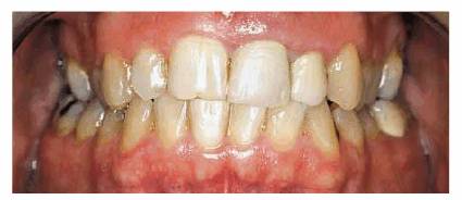
Figure 21-2A: This 50-year-old man had a habit of grinding in a protrusive excursion, continuously chipping his anterior teeth.
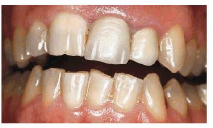
Figure 21-2B: In addition to the chipped anteriors, the left central incisor had advanced periodontal disease, requiring extraction.
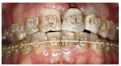
Figure 21-2C: Orthodontic treatment was suggested to improve the occlusion and provide a more favorable protrusive excursion. Unfortunately, the patient lost the left central incisor because of a nonrestorative root fracture.
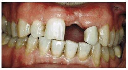
Figure 21-2D: After orthodontic treatment, the occlusion is now in a much more favorable relationship.
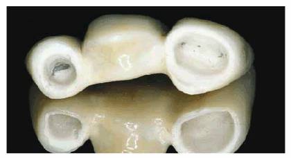
Figure 21-2E: A three-unit all-ceramic
(Inceram, Vident,
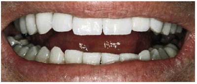
Figure 21-2F: The after smile shows how larger central incisors enhance the esthetics.
Interdisciplinary Consultations
Interdisciplinary consultations and treatment referrals are important to
providing comprehensive care. Multiple treatment modalities and all treatment
options for the patient should be investigated and presented during the
treatment planning phase.17
Once the diagnostic process has been completed, treatment options may be
selected from the following choices:
I. Retainers
A. Partial coverage
1. Cemented
2. Resin bonded
3. Porcelain veneers
B. Complete coverage
1. All metal
2. All ceramic
3. Metal ceramic
a.
Margins
.
Location
.
Material
.
Metal collar margin
.
Disappearing metal margin
.
Porcelain margin
b.
Porcelain-metal junction
C. Other considerations
1. Cantilever fixed partial
denture
2. Implants
3. Splinting
4. Use of telescoping crowns as
abutments
II. Pontics
A. Design
B. Edentulous ridge form
C. Material
RETAINERS
When selecting appropriate retainers for a fixed partial denture, esthetics is
only one of three important factors to be considered. The other two are
biologic considerations and functional or mechanical considerations.
Unfortunately, some of the most esthetically advantageous retainers can be the
poorest from a mechanical standpoint, and other very esthetic retainers can be
the most biologically invasive. Some of these biologic and mechanical
considerations are the size of the abutment tooth; the amount of remaining
tooth structure; the size and type of restorative material in the tooth; the
size, age, and status of the pulp; the clinical crown length; the location of
the tooth in the mouth; the type of occlusal load; the interocclusal space; the
opposing dentition or prostheses; the edentulous span length; and, especially,
the consideration of the insertion path (Figures 21-3A
and B). It
does the patient little good to have a beautifully esthetic restoration that
fails because the biologic and functional issues are not treated correctly.
Figures 21-3A and B: When patients have lost considerable bone support, resulting in extremely divergent roots, linking two fixed partial denture segments together with attachments may be a treatment option.
Fixed partial denture retainers can be separated broadly into two categories,
partial- and complete-coverage retainers. Usually, the most esthetic material
the restoring dentist can choose to match the patient's existing dentition is
natural tooth structure. This display of natural tooth structure in the
esthetic zone is accomplished by using partial-veneer restorations.
Partial-Coverage Retainers
The oldest, and now probably least used, of the partial-coverage retainer
designs is the metal inlay, onlay, or three-quarter crown. These are usually
made of a relatively soft gold alloy and cemented with traditional, mechanically
retentive cements (Figures 21-4A 21-4B 21-4C and D). Due to the buttressing,
retention, and resistance form necessary to make these retainers functionally
successful, it is virtually impossible to avoid some show of metal at the
proximal and incisal or occlusal line angles. Because of this show of metal, this
retainer is unacceptable in the anterior region of the mouth for the
esthetically conscious patient. It can be used very acceptably, however, in
less esthetically critical areas of the mouth. Its best application is for use
on large, relatively unrestored second premolars and first molars in the
maxillary arch.
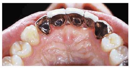
Figure 21-4A: Although the three-quarter crown can be a functionally sound retainer, it is esthetically difficult to avoid showing metal at the proximal, incisal, or occlusal line angles.
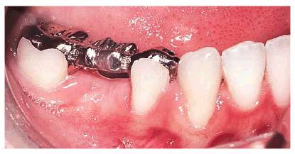
Figure 21-4B: This conservative, economical, three-unit hygienic pontic fixed partial denture is supported with gold onlays and can be esthetically acceptable for some patients.
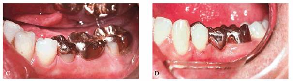
Figure 21-4C and D: These two versions of three-quarter and full-crown abutments supporting all-gold pontics were esthetically acceptable for the patients.
Currently, the most widely used partial-coverage retainer is the resin-bonded
retainer (Figures 21-5A 21-5B 21-5C 21-5D, and 21-5E). In its original form, it was
described as the "prepless bridge."19,32,41 The
preparation design was overly conservative, lacked resistance form, and relied
almost entirely on the resin bond to enamel for retention. The documented
success rates for these early restorations varied widely and left much room for
improvement.
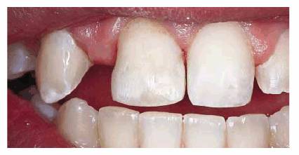
Figure 21-5A: This 17-year-old male is missing his maxillary right lateral incisor.
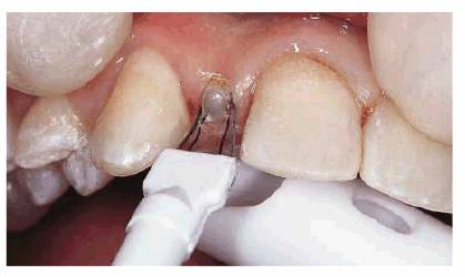
Figure 21-5B: To make a proper pontic site, tissue surgery was accomplished with electrosurgery.
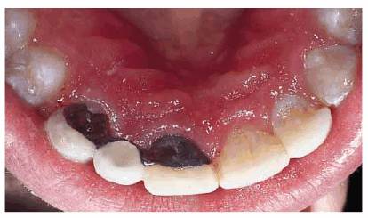
Figure 21-5C: The conventional resin-bonded fixed partial denture is made of thin metal linguals on each of the retainers.
Figure 21-5D: The final result shows a natural-appearing tooth replacement due to a favorably shaped tissue site and the use of the ovate pontic.
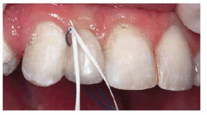
Figure 21-5E: An adequate space must be provided for the use of a floss threader to maintain optimal oral health.
Gradually,
the preparation design for resin-bonded retainers has evolved to look much like
the classic three-quarter crown preparation. It is now advocated that parallel
grooves be used for resistance; that retention be augmented with pins,
potholes, or ledges; and that a definite finish line be created (Figures 21-6A to
C).8,10,28,33
The only concession for the sake of esthetics is the lack of the incisal offset
and proximal metal display seen in the classic three-quarter crown preparation.
This is compensated for by the use of base metal alloys that are relatively
rigid in thin sections and micromechanical retention potential between the
cement and metal and the cement and tooth enamel. The current preparation
design is technically difficult to perform and requires a great deal of
attention to detail. For this reason, properly constructed resin-bonded fixed
partial dentures may fall from favor with the dental profession much as
traditional partial-coverage retainers have.
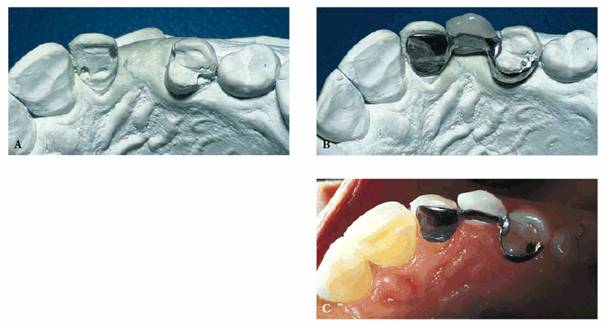
Figure 21-6A to C: The best design for long-term results with a resin-bonded fixed partial denture is to include parallel groves to aid retention and resistance form, in addition to the possible use of potholes, pins, or ledges and a well-defined finish line.
Resin-bonded partial-veneer fixed partial dentures would be the restoration of
choice, particularly in the anterior part of the mouth if the following
conditions are met: the abutment teeth are esthetically acceptable to the
patient in their present size, form, and color; the teeth are free of restorations
or have only minimal restoration that does not involve the crown margins; and
the abutment teeth are of adequate length to afford preparation resistance and
retention and of adequate thickness to prevent metal shadowing from the lingual
surface. For the best results, the resin-bonded partial-veneer retained
prosthesis should only replace one tooth. The teeth should have only normal
mobility. Failure rates rise rapidly with increased numbers of pontics and with
mobile abutment teeth. The pontic space must be of the ideal width since little
widening or narrowing of the edentulous space can be accomplished with
partial-veneer retainers. One of the most frequently seen esthetic problems
with this type of retainer is the difficulty of perfect shade matching. If the
adjacent retainers are metal, light translucency of the abutment teeth is
diminished, resulting in possible shade variance.
A third type of partial-coverage restoration made from traditional crown
materials and cemented or bonded to tooth structure is the porcelain veneer. It
is, no doubt, one of the most esthetic of all partial-coverage restorations but
has limited advocacy as a fixed partial denture retainer. The porcelain veneer
that fits and is bonded to the tooth correctly has adequate strength to survive
most clinical conditions as a single tooth restoration. The problem of
survivability is greatly magnified when porcelain pontics are attached to
porcelain veneers with porcelain connectors. For this type of restoration to
succeed, two requirements are necessary: minimal or no occlusion force and
patient compliance in avoiding biting or occluding on hard foods or objects. In
addition, the patient should be advised to wear an occlusal splint (Figures 21-7A 21-7B 21-7C 21-7D 21-7E 21-7F 21-7G, and 21-7H
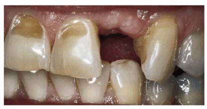
Figure 21-7A: This 52-year-old lady is missing her maxillary left lateral incisor and has severely eroded central incisors.
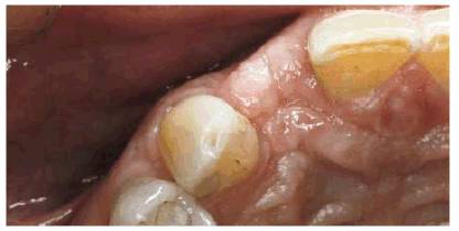
Figure 21-7B: Note the linguoincisal wear on the left central incisor.
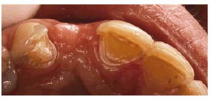
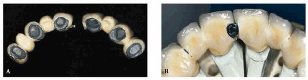
Figure 21-7C: The lingual surfaces are prepared for a resin-bonded bridge. Note the retentive ledges placed.
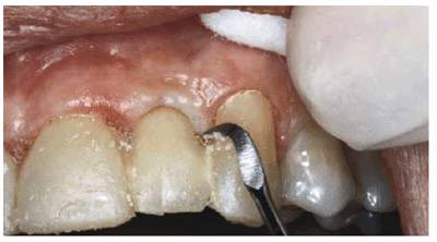
Figure 21-7D: A three-unit all-ceramic
(Inceram, Vident) fixed partial denture is placed, and the resin cement is
trimmed with the Novatek 12 (Hu-Friedy,
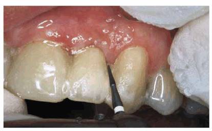
Figure 21-7E: After removing the excess cement,
the interproximal margins are carefully polished with a 30-blade carbide
(ETUF4, Brasseler,
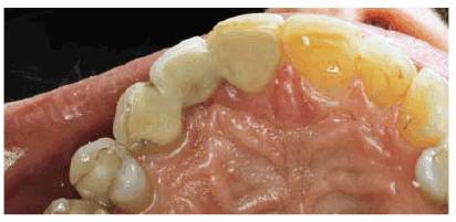
Figure 21-7F: The main advantage for using the all-ceramic retainer is to avoid abutment tooth discoloration.
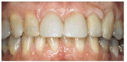
Figure 21-7G: Labial view of the final restoration, which also shows composite resin bonding of the two central incisors.
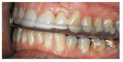
Figure 21-7H: It is essential that the patient be required to wear a well-fitting occlusal night guard.
Complete-Coverage Retainers
Full-veneer retainers are the most popular and the most universally used of all
retainers for fixed partial dentures. They generally fall into three
categories: all-metal, all-ceramic, and metal-ceramic retainers.
All metal crowns are not particularly esthetic and therefore should be used
only in patients who have low esthetic demands. Typically, they are placed in
areas of the mouth that virtually cannot be viewed by observers or the patient.
They are ideal for maxillary and mandibular second and third molar abutments
and for the occasional maxillary first molar in patients with acceptable
esthetic smile lines that do not expose this tooth. It is fortunate that these
areas of the mouth lend themselves to all-metal crowns since it is rare to find
a second or third molar, particularly mandibular, that has sufficient gingivo-occlusal
height to allow preparation reduction sufficient for porcelain occlusal
coverage. The major advantages of all metal retainers are minimal tooth
reduction in comparison with the metal-ceramic or all-ceramic crown
preparation, ease of fabrication, and lack of wear of the opposing dentition.
The chief disadvantage beyond the lack of esthetics is high thermal
conductivity.
The use of all-ceramic restorations as retainers for fixed partial dentures is experimental and is unsupported by any long-term clinical studies. The all-ceramic fixed partial denture should, at best, be used with extreme caution and limited to one-tooth anterior replacements in patients with less than normal occlusal force (see Figures 21-2A 21-2B 21-2C 21-2D 21-2E, and 21-2F). The inferior mechanical properties related to the strength of all-ceramic connectors should be fully explained to the patient, as well as other more traditional alternatives, before selecting this unproven choice. Maximum esthetics rather than longevity must be the overriding consideration in the use of all-ceramic fixed prostheses (Figures 21-8A 21-8B 21-8C 21-8D 21-8E 21-8F 21-8G 21-8H 21-8I 21-8J 21-8K 21-8L and M
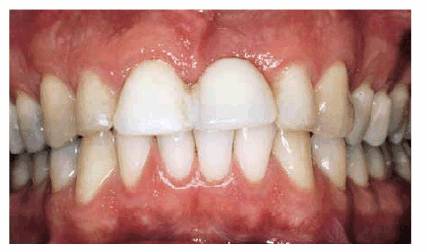
Figure 21-8A: This 33-year-old dentist wanted to improve the appearance of his front teeth.
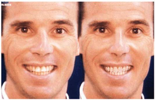
Figure 21-8B: Other than improving the color of
his smile, the main problem was the lack of proportion of the two central
incisors with the rest of the anterior teeth. Esthetic imaging shows the
improvements that would be gained with better-proportioned teeth.
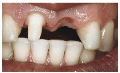
Figure 21-8C: After diagnosis and treatment planning, the next step was to remove the too-wide existing cantilever fixed partial denture.
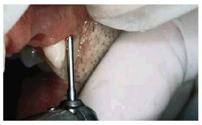
Figure 21-8D: After an all-ceramic three-unit fixed partial denture was selected, a full-shoulder margin on the left lateral incisor is finished with a diamond bur.
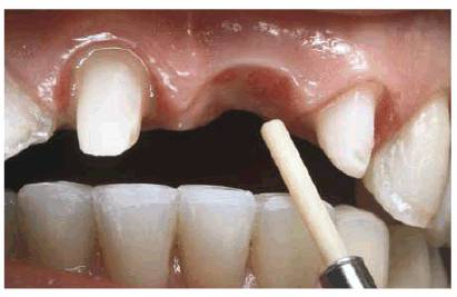
Figure 21-8E: The pontic site is enhanced using a CO2 laser to prepare the tissue for an ovate pontic.
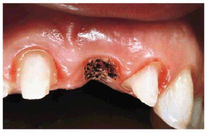
Figure 21-8F: Note the lack of bleeding directly after laser surgery.
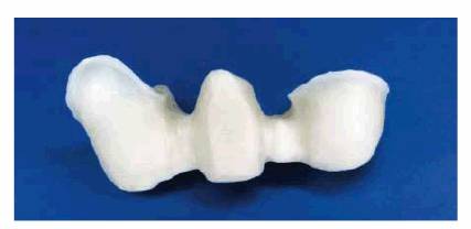
Figure 21-8G: The three-unit all-ceramic core structure (Inceram, Vident) provides the strength for the all-ceramic fixed partial denture.
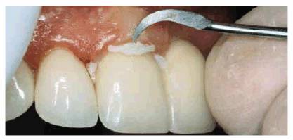
Figure 21-8H: The three-unit all-ceramic fixed partial denture is cemented with a resin cement containing fluoride for both strength and caries protection. The excess cement is trimmed with a double-ended, heavy-duty, cement-removing instrument.
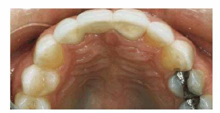
Figure 21-8I: The color and proportion were further enhanced by placing porcelain laminate veneers on the opposing lateral incisor, both canines, and the left first premolar.
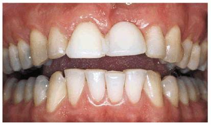
Figure 21-8J: This picture shows the maxillary discolored and disproportionate teeth and the crowded mandibular incisors.
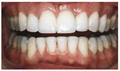
Figure 21-8K: Note the esthetic improvement of the mandibular incisors following esthetic contouring of these teeth. Eventually, it may be necessary to treat the cervical erosion on the maxillary posterior teeth.
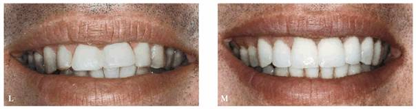
Figure 21-8L and M: The comparison of the before and after smile. Maximum esthetics rather than longevity was the overriding consideration in the choice of an all-ceramic fixed prosthesis. Because this patient is a dentist, he is well aware of occlusal limitations, especially in excursive movements.
By far the most commonly used retainer for fixed partial dentures is the
metal-ceramic crown. During the last 30 to 40 years, this restoration has
proven to be a very satisfactory compromise between functional success and
esthetics. The acceptance of this retainer has fostered many variations and
choices that impact on esthetics, as well as biologic acceptance, functional
success, ease of predictable fabrication, and economics. Some of the variables
that should be considered with metal-ceramic retainers are the location of the
margins in relation to the gingiva, the materials used for margin fabrication,
and the location of the porcelain-metal junction in relation to the occlusal
surface of the retainer.
Margin Location
Studies have shown that with respect to biologic acceptance of an artificial
crown by the gingival apparatus, the situation is generally healthier when the
artificial material remains supragingival.38 Supragingival margins
are also easier to prepare, impress, and evaluate for fit (Figures 21-9A, and 21-9B). Although it has been shown that
soft-tissue health can be maintained in the presence of subgingival margins,31
the general consensus is that margins should only be placed into the gingival
sulcus for reasons of esthetics, wall height for resistance and retention, or
extension beyond existing caries or restorations.25 If subgingival
margins are chosen solely for esthetics, they should be limited to areas in
which the gingival margins are visible to observers of the patient in normal
and extreme facial movement. It was shown that less than 50% of the population
observed in one study revealed the gingival area of any of their mandibular
teeth during movements of facial expression (without manually pulling down
their lower lips).7 Further, a smaller percentage in the same study
did not reveal their maxillary gingival margins during facial muscle use. It
has been the author's observations that many patients do not show their
gingival margin areas of maxillary molars. In general, when the patient
permits, subgingival metal-ceramic margins should be limited to a patient's
visible esthetic zone. This may require some prior education by the restoring
dentist so that the patient will understand that what is visible with mirrors,
cheek retractors, and fingers in the mouth is not necessarily visible in normal
circumstances. However, patients whose primary concern is esthetics will often
only agree to subgingival metal margins (Figures 21-10A 21-10B and C, and 21-10D). With these situations, the
porcelain shoulder butt margin design prepared supragingivally should also be
considered either 180 or 360 degrees (Figures 21-11A, and 21-11B) (see also Esthetics in Dentistry,
Volume 1, 2nd Edition, Figure 15-11).
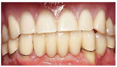
Figure 21-9A: Supragingival margins with exposed metal will generally not be an esthetic problem if the lip line is favorable.
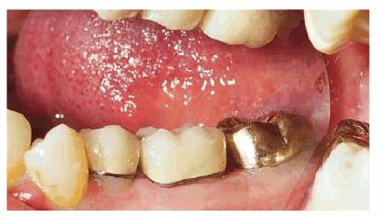
Figure 21-9B: A complete metal crown can be the longest-lasting retainer of choice anytime esthetics permit.
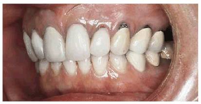
Figure 21-10A: This patient was extremely dissatisfied with the recession of maxillary gingival exposing metal margins.
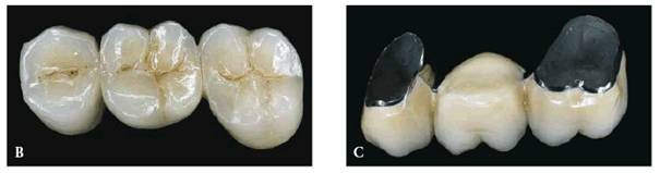
Figure 21-10B and C: A three-unit fixed partial denture replacement was constructed using concealed metal-ceramic margins.
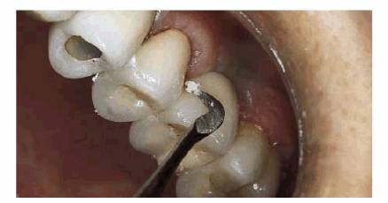
Figure 21-10D: Since caries was present, the fixed partial denture had to be redone and the adjacent Class V area esthetically repaired.
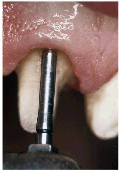
Figure 21-11A: A tissue-protective end-cutting diamond can be used to prepare a 360-degree full-shoulder margin.
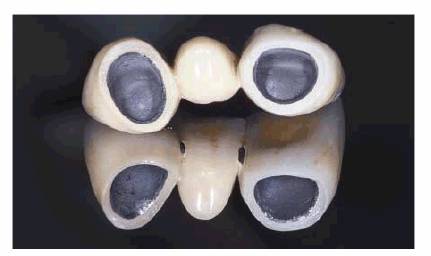
Figure 21-11B: This three-unit metal-ceramic fixed partial denture features a 360-degree all-porcelain margin.
Margin Materials
Certain subgingival margins are not completely natural and esthetic in
appearance. This is usually because the restorative material shows through the
thin gingival sulcular tissue or there is too much artificial crown material
subgingivally either in the form of overcontouring, overextension, or both (Figure 21-12). This unesthetic subgingival
material has led to three different approaches to metal-ceramic crown margins.
The first is the classic metal collar margin, in which a small band of metal
creates the terminus for the crown with no porcelain overlaying it. The metal
collar margin was the technique for all early metal-ceramic crowns and is
currently used for a large proportion of them. Many metal collars have been
placed subgingivally with good esthetic results. This requires a superior
technical approach to margin preparation, retraction, and impression techniques
and soft-tissue management in the interim phase. However, the results are often
less than predictable.
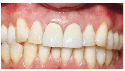
Figure 21-12: Thin, transparent gingiva may not be thick enough to fully mask a subgingival metal margin.
This
unpredictability of consistently hiding the metal collar subgingivally led to
the second approach to metal-ceramic crown margins. This is a compromise
variously named the metal-porcelain margin, the covered metal margin, or the
disappearing metal margin. This is a technique in which the technician creates
the metal collar as thin as possible without opening or shortening the crown
margin and then overlays this thin metal with porcelain, also as thin as
possible, to completely hide the metal margin. In other words, both metal and
porcelain end precisely at the crown margin (Figures 21-13A
and B 21-13C, and 21-13D). This compromise, unfortunately,
has several severe drawbacks. Because the metal is extremely thin at the margin
and is chemically bonded to the porcelain, the marginal metal often distorts
more than the traditional metal collar during the porcelain firing cycles.36
Because of the extreme thinness of the porcelain overlying this metal, it is
almost purely opaque porcelain (the first coat applied in the firing cycle).
This opaque porcelain is virtually unglazable and unpolishable, resulting in a
very rough marginal surface. Since there are two layers of restorative material
attempting to end at the same finite finish line, the porcelain portion must be
external to the normal contour of the tooth prior to preparation, thus creating
a potentially rough, open, overcontoured margin. Even the best attempts to hide
the metal margin are questionable because the overlying porcelain is often too
thin to truly mask the metal color. The margin is still apparent as an
off-color area of porcelain even though it is preferable to a shiny silver
metal collar.
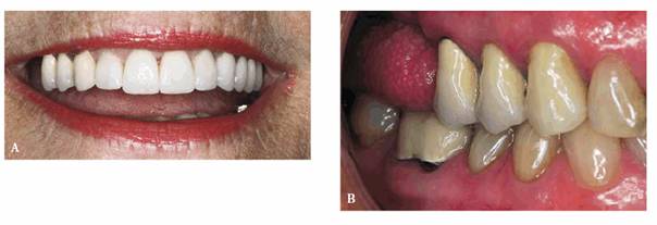
Figure 21-13A and B: This patient was embarrassed to smile fully due to the darkness caused by her missing molars.
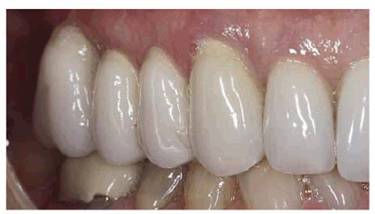
Figure 21-13C: Since she was not a candidate for implants because of advanced bone loss and a large maxillary sinus, this patient elected to have distal extensions for both esthetics and function.
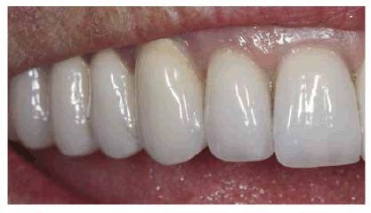
Figure 21-13D: Note how effectively the distal cantilever masks the spaces even with a wide smile.
The third approach to achieving esthetics in the margin
area of metal-ceramic crowns is the all-ceramic margin. This is accomplished by
removing all metal back to the internal line angle of the shoulder of the
preparation as far proximally as is possibly visible and replacing it with a
special higher- fusing shoulder porcelain of the same shade as the body or
gingival porcelain. Some techniques also remove the metal to some distance up
the facial surface of the metal coping.22 It was originally thought
that this technique would not yield sufficiently accurate marginal adaptation
to be clinically acceptable. Several studies have shown that margins of equal
clinical acceptability with metal margins can be created by many different
porcelain application techniques.2,4,44 As technicians become more
comfortable with these various techniques, clinically acceptable margins may be
produced more often. This is due to the virtually infinite ability to repair or
correct porcelain margins, a characteristic not possible with cast metal
margins. Initially, ceramic margins were not expected to be strong enough to
withstand clinical loads. However, research has indicated that once cemented to
the abutment teeth, all-ceramic margins have equal or possibly greater strength
than metal collar margins.12
The authors' recommendation for margin selection for metal-ceramic retainers is
to use metal collar margins in esthetically noncritical areas. These are the
ones previously outlined where supragingival margins are esthetically
acceptable. All-ceramic margins should be used in the patient's esthetic zone
or for patients with higher esthetic demands, and the combination
metal-porcelain margin should be avoided whenever possible.
Porcelain-Metal Junction
The last area to consider in the relative esthetics of the metal-ceramic crown
is the location of the porcelain-metal junction. Obviously, the most esthetic
choice is to cover the entire metal coping with porcelain in all areas of the
mouth where the abutment retainer is visible. As long as the underlying metal
coping is of sufficient thickness and shape to protect and support the
overlying porcelain and the porcelain is of adequate and equal thickness
throughout, this design produces no functional problems. If inadequate occlusal
reduction occurs in the preparations for porcelain overlying metal, this will
usually lead to poor esthetics of the porcelain and often to fracture of the
ceramic under functional load. However, in certain patients and certain
occlusal schemes, these all-porcelain occlusal surfaces can have disastrous
results on the opposing dentition, particularly if the restorations will
occlude with natural tooth structure or metal alloy. Nevertheless, all of these
rules are negated if the patient expects or demands no metal showing even if he
or she has to pull the cheek back to view it.
The most esthetically necessary area for complete-porcelain coverage is the
mandibular premolars and first molar (Figure 21-14). This is because the occlusal and lingual
surfaces of these teeth are readily visible when the mouth is open. In some
patients, even the mandibular second molar is visible. However, except for
public performers, who are observed by audiences or cameras from below, rarely
are the maxillary posterior occlusal surfaces visible, even in wide open
movements. Therefore, from a functional and esthetic perspective, these
retainers can usually have porcelain-metal junctions that end on the lingual
slope of the bucco-occlusal surfaces (Figure 21-15). The exception to this is when the maxillary
fixed prostheses are opposed by all-porcelain occlusal coverage in the
mandibular arch. In this case, complete-porcelain coverage of the maxillary
restorations is advisable to prevent excessive wear by the opposing porcelain.
It does little good to use this buccal-only porcelain coverage in the
previously mentioned area of the mandibular premolars and molars since this
area of these teeth is covered by the cheek when the mouth is opened and the
metal occlusal surface will still be visible (see Figure 21-9B). The delicate balance of porcelain occlusal
coverage for esthetics and prevention of wear of the opposing dentition may be
compensated for in the posterior occlusion if the patient has anterior or
mutually protected occlusion in lateral and protrusive excursive movements. If
the patient does not have or cannot be restored to an anterior disclusive
occlusal scheme, then the problem of porcelain wear of the opposing dentition must
be addressed much as it would in a bruxer or other parafunctional patient with
some sort of protective occlusal splint prescription.
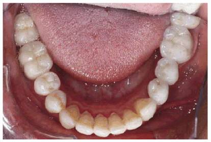
Figure 21-14: The most esthetically necessary use of complete- coverage porcelain is with the mandibular premolar and even the mandibular molars when patients demand them.
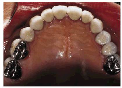
Figure 21-15: This patient was happy with metal occlusal surfaces because he recognized that, under ordinary circumstances, they would not show.
The decision of the location of the porcelain- metal junction for maxillary
anterior crowns for Angle Class I patients is an entirely different matter. The
location of the junction is of little consequence esthetically since the
lingual surface of maxillary anterior teeth is never visible. Some clinicians
feel that having the intercuspal contact in metal on the maxillary anterior
prosthesis will preclude wear. This is not entirely true since all of these
patients will still function to some degree in excursive movements on porcelain
of the lingual surface of these crowns incisively to the porcelain-metal
junction. Since porcelain contact in lateral and protrusive movements with the
opposing mandibular incisors and canines is unavoidable, the best solution is
careful re-creation of the patient's natural incisal guidance in the porcelain
prosthesis. This means avoiding creating a new, steeper incisal guide angle or
longer incisal guide path. If the patient shows tendencies of parafunction,
splint protection should be prescribed. If wear of the opposing mandibular
anterior teeth continues, restoration with porcelain of similar hardness may be
a last resort. For many patients, the porcelain- metal junction location on
mandibular anterior restorations is rarely of importance either esthetically or
functionally. Biologically, owing to the small size of mandibular anterior
teeth, overcontouring of the lingual surface of the restorations is best
avoided by locating the junction as far incisively as esthetics allows.
OTHER CONSIDERATIONS
Anterior Restorations Involving One
Missing Tooth
Cantilever Fixed Partial Denture. The ideal choice for the single
missing tooth is the single-tooth implant. However, there are times when an
implant may not be possible or practical. A conservative alternative would be
the cantilever fixed partial denture involving one or more abutment teeth. The
cantilevered restoration is highly desirable esthetically (especially in areas
adjacent to sound, attractive teeth), and the final result, using a
cantilevered retainer, can be natural looking and quite attractive (Figures 21-16A and B, 21-16C, 21-16D, 21-16E, 21-16F, and 21-16G).

Figure 21-16A and B: This lady, who was congenitally missing her maxillary lateral incisors, was unhappy with the appearance of the replacement fixed cantilever partial dentures.
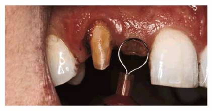
Figure 21-16C: Electrosurgery was used to contour the tissue for the ovate pontic.
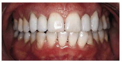
Figure 21-16D: The final replacement has a more harmonious gingival relationship between the pontic and the abutment.
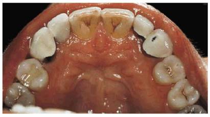
Figure 21-16E: Note that the previous cantilever fixed partial denture had no lingual rest on the lateral incisor.
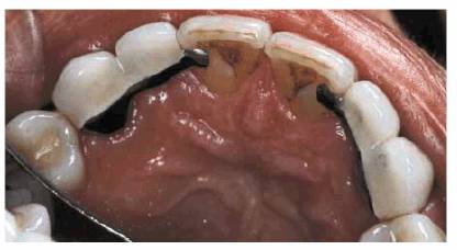
Figure 21-16F: The new bridges were strengthened by adding lingual rests. Note also the severe stain produced by the patient's continued smoking habit.
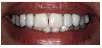
Figure 21-16G: The patient's after smile shows improved shade match, proportion, and fixed partial denture contours.
For esthetic reasons, it may be necessary to
cantilever a posterior abutment. For example, if the patient's smile line shows
the missing molar, then this tooth can be replaced as a posterior cantilever
(see Figures 21-13A and B, 21-13C, and 21-13D).
Implants. It has become the standard of care to also evaluate each
patient possessing an edentulous space for the possibility of replacing that
space with an implant. The patient has a right to know the potential functional
and esthetic success associated with the placement of an implant instead of a
fixed or removable prosthesis.
The question of whether to select an implant prosthesis rather than a
tooth-borne fixed partial denture is generally decided by the dentist and the
patient after a thorough analysis of the advantages and disadvantages, both
esthetically and functionally, of each treatment. A frank discussion must
include an honest analysis of the longevity, costs, and esthetic appearance of
each proposed treatment. Although computer imaging can usually demonstrate the
esthetic appearance of the proposed final result, the esthetic difference may
be too subtle to see on the computer screen. Instead, a diagnostic waxing may
be necessary to reveal the difference, especially if it involves soft-tissue
issues. The form of the lip line will help to determine the treatment choice.
Further, when a tissue recession problem exists, a periodontal procedure may be
necessary regardless of whether an implant or fixed partial denture is chosen.
Splinting. Esthetically, it is much better not to splint the
incisors to achieve individuality between teeth. Either separation or the
appearance of separation helps to make a missing tooth replacement appear
natural. In addition, the lack of splinting will promote easier maintenance and
good oral care. However, the issue of splinting is determined by mobility
patterns of teeth, and esthetic compromises will necessarily have to be made
when functional requirements indicate splinting.
To achieve a natural appearance, restorations should appear bilaterally
symmetric. There should also be harmony in both gingival contour and incisal
levels to prevent a "replacement" look.
Use of Telescoping Crowns as Abutments
Advantages. Telescoping restorations offer certain benefits but are
not without problems. One advantage of telescoping crowns is that the
preparations do not have to conform to a common path of insertion.24
Only the functional surfaces of the copings require waxing to a common path.
Another advantage is the ability to link different segments of fixed partial
dentures while still keeping the span small. This is particularly useful when
using a metal-ceramic prosthesis where a long span may produce excessive
flexure and, subsequently, the possibility of fracture. It allows the smaller
spans to be removed in the event of acrylic resin wear, porcelain fracture, or
need for access to the pulp chambers if endodontic procedures become necessary.
The flexibility of telescoping procedures permits alterations in design that
may not require making the entire restoration. For example, in the event of the
loss of a posterior abutment, the superstructure may be able to be redesigned
without remaking the entire prosthesis. Depending on how the coping linkage is
designed, a great deal of latitude is thus provided.
Building the teeth anterior to the ridge to gain more space should be avoided.
This will alter the occlusal relationship, and if the patient has a high or
medium lip line, unattractive spaces can result. If extractions are
contemplated, the patient's natural teeth should be noted. If they are crowded,
they can be restored with crowded or straight teeth. Since crowding indicates a
space problem, the patient and the dentist need to understand the esthetic
problem and alternative solutions. This situation was treated successfully in a
patient who wanted his natural teeth to be copied in the final restoration (Figures 21-17A, 21-17B, 21-17C, 21-17D, 21-17E, 21-17F, and 21-17G). It is interesting to note that even during the
provisional stage, the patient could not enunciate properly until the crowded
teeth were simulated.
Figure 21-17C shows the use of four copings splinted to serve
as abutments for two possible superstructure combinations. Inserting the
six-unit anterior segment separately achieves splinting (Figure 21-17D). The use of the interlock attachments distally
on the canines aids retention of two future planned implants. If this were not
successful, an alternative plan provides for use of an attachment removable
partial denture retained by the remaining copings and interlocks. Figure 21-17E shows the extra superstructure crowns in place
with flexibility to be used in either capacity.
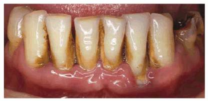
Figure 21-17A: This man had to lose his mandibular incisors due to periodontal disease; he insisted that the replacements copy the look of his natural teeth.
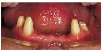
Figure 21-17B: After extraction, even in the provisional stage, the patient could not enunciate correctly until the crowded teeth were simulated.
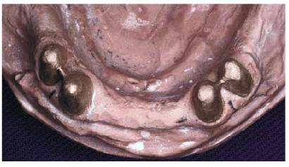
Figure 21-17C: Four copings crosslinked to serve as abutments for two possible superstructure combinations.
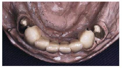
Figure 21-17D: This shows the anterior segment in place.
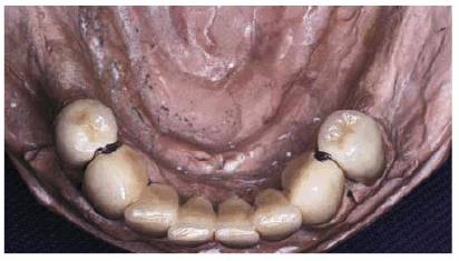
Figure 21-17E: The two premolar crowns can be removed for an attachment removable partial denture in the future.
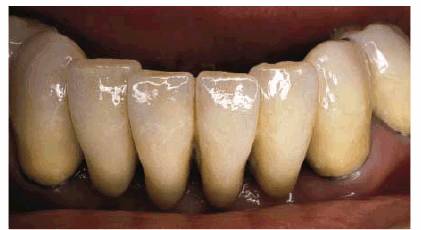
Figure 21-17F: Note how the final metal-ceramic fixed prosthesis accurately simulates his natural dentition.
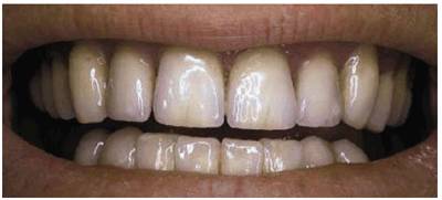
Figure 21-17G: The final smile shows the maxillary denture occluding against the new mandibular prosthesis.
The use of telescopic procedures to splint mobile teeth is seen in Figures 21-18A, 21-18B, and 21-18C. Figure 21-18A shows the copings in place. Note the soldered
joints between the canine-lateral incisor on the left side and the
canine-premolar on the right side. Figure 21-18B shows the posterior metal-ceramic
superstructure in place, and Figure 21-18C shows how the anterior segment completes the
splinting using porcelain occlusal surfaces. The copings are cemented with a
definitive cement and the superstructure with a provisional cement. If the
restoration is to be removed, the superstructure should be designed with
exposed metal at the connector site so that a reverse hammer can be used to tap
off the superstructure without fracturing the porcelain.

Figure 21-18A: Copings are in place with soldered joints between the canine-lateral incisor on the left side and the canine-premolar on the right side.
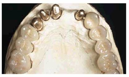
Figure 21-18B: The posterior metal-ceramic superstructure in place.
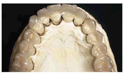
Figure 21-18C: The anterior segment completes the crossarch splinting using porcelain occlusal surfaces.
Disadvantages. The main disadvantage in telescoping is twofold. First, bulk is created
by an extra layer of metal, which can be a problem unless there is adequate
room for preparation. To avoid producing additional stress, the buccolingual
diameter of the superstructure should not be increased. Therefore, using
telescoping crowns in small, flat teeth should be avoided. Another disadvantage
with the coping and telescope procedure is the need for an extra-long gold collar.
This may create an esthetic problem at the gingiva unless there is enough space
to hide or mask the metal. A compromise coping can be constructed for anterior
teeth by not covering the gingival half of the tooth, making crosslinkage
possible without loss of esthetics.26,27
Copings are especially useful in the patient with a low lip line. The patient
should be shown that during normal conversation, laughing, or smiling, the
gingival portion of the tooth will not be seen.
Because of increased bulk, metal occlusal surfaces should be used whenever
possible for coping procedures. A compromise can be made in the anterior
section for esthetic purposes (Figures 21-19A, 21-19B, and 21-19C). Cross-arch splinting is accomplished by using
soldered copings and a three- segment superstructure.
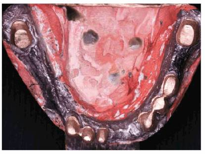
Figure 21-19A: Splinted copings are in place to permit the superstructure to be inserted in three sections.
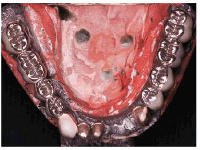
Figure 21-19B: Metal occlusal surfaces were used because this patient had insufficient vertical space for porcelain.
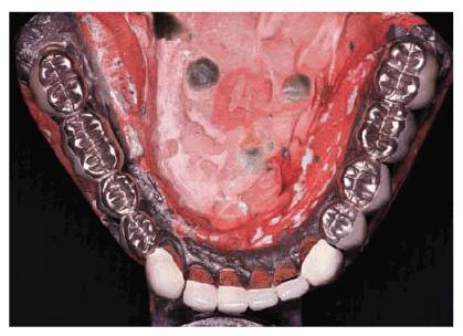
Figure 21-19C: A compromise can usually be obtained anteriorly so that ceramic material can be used.
An alternative in the posterior region, where vertical height is severely
restricted, is the open-telescopic technique. The occlusal surface of the
restoration is wholly or partially a part of the inner coping, and the outer
crown fits around the inner coping. Provision must be made for occlusal seating
by incorporating a shoulder in the coping. Anteriorly, it is possible for the
facing material to be on either the coping or the superstructure (Seymour M, personal
communication, 1974).
The necessity and advantages of the telescoping procedure should be
re-emphasized in a letter to the patient documenting why the procedure is being
used.
Use of Precision Attachments
Where advantages outweigh the disadvantages, copings should be used with
full-arch procedures if space and economic factors permit. When full-arch
copings cannot be used, it is wise to interlock at strategic positions with the
use of four copings. These copings can then be splinted together, and three
segments can be made. If this is not feasible, an interlocking type of
matrix-patrix attachment can be used. It is preferable to use precision
attachments that allow either segment to be removed at will. Other types of
interlocking devices may require that both segments be removed together to
remove the matrix portion. This means more chance for porcelain fracture and is
therefore not as convenient a device for cross-arch splinting.
Semiprecision or precision internal attachments in fixed partial dentures may
improve the quality of the prosthesis significantly. Their two primary uses are
to eliminate problems of parallelism and to interlock smaller segments, which
avoids lengthy spans of porcelain to metal.
PONTICS
Pontic Design
The overall esthetic objective in pontic design is to make the missing tooth
replacement look like a real tooth. A tooth substitute should be in harmony
with the abutment teeth and the remaining dentition. Concealing the fact that
the pontic is an artificial replacement is accomplished by the outline form,
size, alignment, contour, surface texture, and color. In addition, it must
function with the opposing occlusion and provide comfort and support to the
adjacent tissues and continuity to food flow patterns; it must have contours
that are easy to keep clean.
There are several pontic designs available for fixed partial dentures. The
choices include ridge lap, modified ridge lap, conical or bullet, hygienic,
modified hygienic, and the ovate pontic (Figure 21-20). Esthetics, edentulous ridge anatomy, and the
patient's ability to maintain adequate hygiene must be considered during pontic
design selection. Due to the inability of the patient to maintain adequate
hygiene under the ridge lap pontic, the authors do not recommend the use of
this type of pontic design.
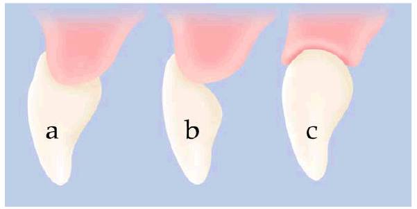
Figure 21-20: Pontic design. (A) Total ridge lap. (B) Modified ridge lap. (C) Ovate.
To optimize esthetics, the modified ridge lap described by Stein40
and the ovate pontic (Figures 21-21A and B, and 21-21C)9,11 are considered the pontics of choice.
These two pontic designs work well because a natural-appearing emergence
profile can be achieved, leading to a more esthetic result. However, certain
requirements are necessary to accomplish a favorable esthetic outcome. The
pontic must have the proper incisogingival or occlusogingival length in
relation to the abutment teeth. Excessively open interproximal embrasures or
"black triangles" must be avoided in the anterior region, and a
proper labiolingual or buccolingual relationship with the abutment teeth should
be obtained, creating a proper emergence profile. To accomplish these three
requirements, proper edentulous ridge tissue form is imperative. Preprosthetic
surgery is often needed to enhance the edentulous area to achieve the desired
esthetic results (Figures 21-22A, 21-22B and C, 21-22D, and 21-22E).
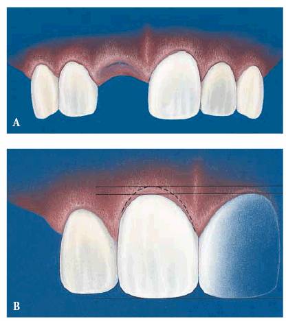
Figure 21-21A and B: The ovate pontic has become one of the most desired forms for maximum esthetics and function.
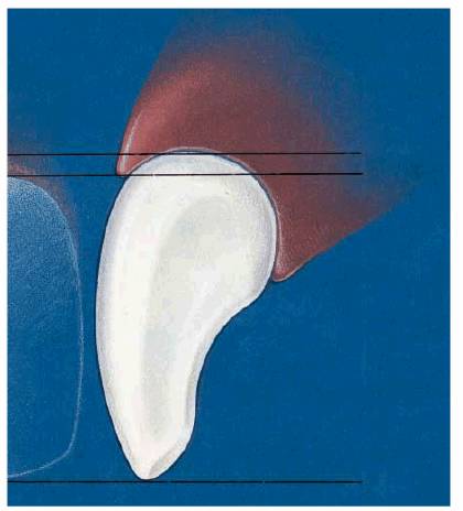
Figure 21-21C: The shape of the pontic, as seen in sagittal section, is conducive to effective cleaning with dental floss.
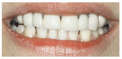
Figure 21-22A: This patient was unhappy with the unnatural-looking "black triangles" caused by loss of interdental tissue.
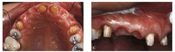
Figure 21-22B and C: Ridge augmentation plus sculpting for ovate pontics was done during the interim phase.
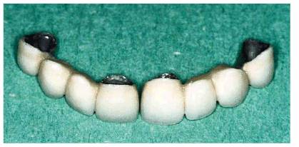
Figure 21-22D: Two four-unit fixed partial dentures were fabricated with ovate pontics.
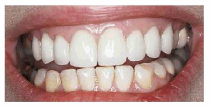
Figure 21-22E: This patient was extremely pleased that her widest smile did not give a hint of missing teeth.
Preparation of Tissue
A diagnostic waxing of the fixed partial denture will aid in assessing the
pontic-ridge relationship to determine if the three design requirements will be
met. If the relationship reveals that an esthetic result would be enhanced
through modification of the edentulous ridge area, then further adjunctive
therapy should be considered to correct the pontic-tissue site.
The edentulous ridge with ideal dimensions both buccolingually and
occlusogingivally can be treated with a modified ridge lap pontic design,
meeting all three esthetic design requirements. Ridge contour for the modified
ridge-lap pontic should be slightly convex in a labiolingual direction and
gently concave mesiodistally.1 For the edentulous ridge that has
excessive hard or soft tissue, surgical reduction can be performed (see Figures 21-5D, and 21-5E). If the soft tissue is thick, scalloping of the tissue
may create a favorable pontic site. If the hard tissue is excessive with a
minimal soft- tissue covering, osseous resection may be necessary.
Ovate pontic designs are generally used in two types of clinical situations:
the healed edentulous ridge and new extraction sites. When a healed edentulous
ridge exists, the recipient site requires a surgical procedure of either hard
tissue, soft tissue, or both to provide proper emergence from the tissue.
However, with a new extraction site, at the time of extraction, the abutment
teeth can be prepared and the fixed partial denture provisional fabricated;
then, the ovate pontic provisional can be placed so that it emerges from the
extraction site. This type of procedure quite often leads to a highly
acceptable esthetic effect. However, be aware that, occasionally, to enhance
esthetics, a surgical procedure may be necessary once the extraction site heals
around the pontic. Ridge anatomy for the ovate pontic requires a wider
labiolingual ridge dimension (see Figures 21-21A and B, and 21-21C).29
Adjunctive Tissue Treatment
Frequently, adjunctive treatment involves a deficient edentulous ridge.
Deficient pontic areas may occur as a result of trauma, developmental defects,
or disease. The edentulous area may be deficient in height, width, or both,
depending on the individual situation. Seibert classified the deficient ridge
based on the dimension of the defect as follows: (1) buccolingual loss of
tissue with normal ridge height (Class I), (2) apicocoronal loss of tissue with
normal ridge width (Class II), or (3) combined loss of ridge contour in both
the buccolingual and apicocoronal dimensions (Class III).35 For the
deficient ridge, adjunctive treatment involves surgical site augmentation,
which can be accomplished using an autogenous or allogenic graft of hard or
soft tissues, an alloplastic graft, or a combination of these grafts depending
on the amount of augmentation needed. The volume of donor tissue needed to
repair the defect and the availability of such tissue will have a bearing on
the source of graft material.15 Larger augmentations quite often
involve multiple surgeries to achieve optimal results. However, for sites that
can be augmented with soft tissue alone, esthetic results can often be obtained
with one surgical grafting procedure. If the deficient site cannot be
augmented, for reasons that may include cost, medical history, or too severe a
defect, another modality such as a removable partial denture should be
considered.
The goal of the pontic site tissue preparation procedure is to provide a ridge
in which the pontic looks natural in its emergence. To achieve this, proper
soft-tissue thickness must be generated. Although the hard tissue gives the
augmented site the necessary support, modifying the thickened-ridge soft tissue
helps to eliminate the "black triangles"; then, a proper emergence
from the ridge area can be generated. Tissue thickness over edentulous ridge
areas can vary depending on the location. In Stein's study of 50 anterior
ridges and 50 posterior ridges, he found that, regardless of the degree of
ridge atrophy, the mean tissue thickness of the posterior regions was 2.05 mm.
The mandibular anterior region was similar to the posterior regions, whereas
the maxillary anterior regions showed a mean tissue thickness of 4.13 mm. This
study and many others have shown that pontic placement against the underlying
ridge can cause a chronic inflammatory reaction. A certain thickness of tissue
needs to be maintained, and encroachment on the tissue leads to an inflammatory
process. If additional tissue thickness is generated over the ridge,
soft-tissue modification can be performed.40 Class I category
defects can be treated with a soft-tissue augmentation procedure buccally to
improve esthetics. This is a highly successful and fairly predictable
procedure. Class II and Class III defects are much less predictable and quite
often require multiple surgeries to increase the likelihood of a successful
result.
The potential pontic site often has a nonrestorable root that needs to be
extracted prior to fabrication of the fixed partial denture. Another
alternative to the ovate pontic fabricated to the extraction site is
orthodontic extrusion of the root, which manipulates "hopeless" teeth
to modify local defect areas.34 Orthodontic extrusion before
extraction can modify the ridge to help in controlling pontic site design by
also erupting the bone as the tooth erupts.
Prosthodontic preparation prior to ridge augmentation can follow this protocol.
Prior to ridge augmentation, the abutments are prepared, and a provisional
acrylic resin fixed partial denture is fabricated. The proper form and function
of the prosthesis are created in the provisional, and the pontic intaglio
surface (the surface that approximates the ridge tissue) is designed to
simulate the position and contour desired in the final prostheses. At the
surgical appointment, the provisional is removed, and the ridge is augmented.
The surgeon uses the intaglio surface of the pontic as a reference point for
the amount of augmentation, making sure to compensate for tissue shrinkage. The
intaglio surface of the pontic is then modified prior to recementation,
ensuring no tissue contact. The surgical site is allowed to heal for 6 to 8
weeks, depending on the location (longer period for anterior esthetic areas).
Once adequate healing has occurred, the provisional fixed partial denture is
removed, and the pontic intaglio surface is modified by forming acrylic resin
to the ideal shape. At this time, the soft tissue is modified either by
electrosurgery, a surgical blade, laser surgery, or rotary instrumentation to a
contour adaptive to the provisional. The highly polished provisional is again
provisionally cemented, and the area is allowed to heal for an additional 6 to
8 weeks prior to making the final impression for the definitive prosthesis.
"Scalloping" the soft-tissue site and adapting the fabricated
provisional to the scalloped site affords the clinician the opportunity to
shape the tissue, creating an esthetic prosthesis. The tissue scalloping allows
the pontic to closely mimic the emergence of the abutment teeth. The
pontic-ridge relationship will look natural, and the three requirements for an
esthetic pontic/edentulous ridge will be met.
If attempts at surgery are unsuccessful or even only moderately successful,
resulting in small black triangles, then esthetic masking must take place in
the fabrication of the prosthesis. This can take the form of either fixed or
removable tissue inserts. The fixed tissue insert can be fabricated from
tissue- colored ceramic or composite resin material. Greater longevity if
ceramics are used to replace the interdental tissue should be expected (Figures 21-23A, 21-23B, 21-23C, 21-23D, 21-23E, 21-23F, 21-24A and B, and 21-24C).
As an alternative, some patients use a removable tissue insert fabricated from
acrylic resin. Certain patients prefer these, particularly for photographic or
social occasions (Figures 21-25A, 21-25B, 21-25C, 21-25D, and 21-25E).
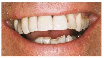
Figure 21-23A: This patient wanted to improve his fixed partial denture without implants or tissue surgery.
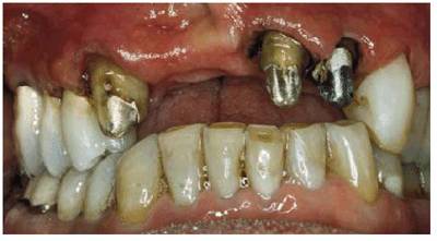
Figure 21-23B: The old fixed partial denture was removed and the abutments reprepared.
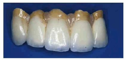
Figure 21-23C: The new five-unit fixed partial denture included fixed pink porcelain to resemble gingival papilla to mask the interdental space.
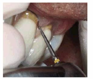
Figure 21-23D: A diamond bur is used to carefully create sufficient space for a floss threader.
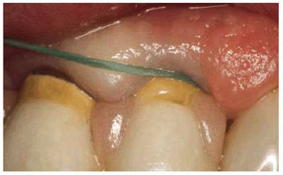
Figure 21-23E: The space allows the floss to effectively clean below the pontic.
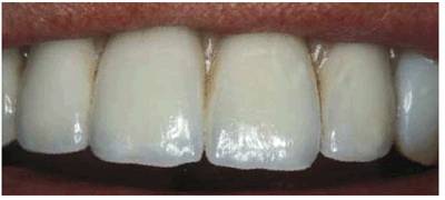
Figure 21-23F: A maximum smile reveals an
esthetic result.
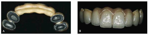
Figure 21-24A and B: This eight-unit fixed partial denture featured four anterior pontics. Note how a pink porcelain fixed-tissue insert was constructed to blend in with this patient's tissue.
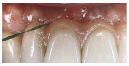
Figure 21-24C: The dental floss can effectively clean above this well-fitting tissue insert.
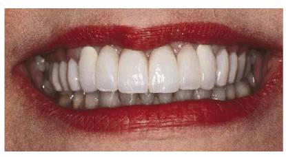
Figure 21-25A: This high fashion model wanted to mask the length of her anterior fixed partial denture so that she could do photographic modeling.
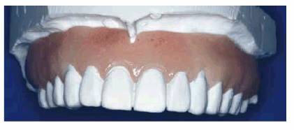
Figure 21-25B: An impression is made of the seated maxillary fixed partial denture. It is then poured and the laboratory waxes, invests, and cures the insert in pink acrylic resin. A slightly flexible removable tissue insert is fabricated that will then lock into her premolars and molars.
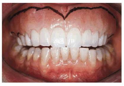
Figure 21-25C: The removable tissue insert is tried in and marked approximately where the flange will be trimmed.
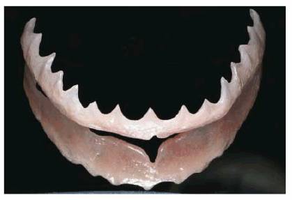
Figure 21-25D: The trimmed and polished, slightly flexible removable insert is photographed on a mirror to view both aspects.
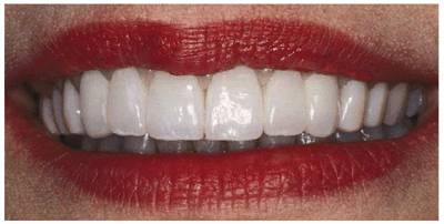
Figure 21-25E: The removable tissue insert camouflages the uneven and excessively long teeth, resulting in an attractive smile.
Pontic Materials
The type of material used to fabricate the pontic also depends on the esthetic
result required. Pontic material types can be all metal, metal ceramic, all
ceramic, or metal with acrylic resin. Porcelain covering all visible areas is
the selection of choice for an esthetic situation. As mentioned earlier,
all-ceramic fixed partial dentures should be avoided due to the inherent lack
of strength. Metal with acrylic resin is occasionally used today in the
posterior regions when retainer design dictates Type III gold, but more often
than not the esthetic pontic is fabricated as a metal-ceramic prosthesis. The
length of span of a fixed partial denture can influence material choice. Many
failures associated with the fixed partial denture can be related to the choice
of materials.43 For longer-span fixed partial dentures, the more
rigid (higher modulus of elasticity) predominantly base metal alloy such as
Rexillium III (Jeneric/Pentron, Wallingford, CT) may be the alloy of choice to
minimize flexure.
Proper pontic-tissue contours and surface finish are the key to healthy tissue
response. Pontic design has been found to be the foremost factor in obtaining
inflammatory-free pontic-ridge relationships. According to Stein, the ideal
pontic design is the modified ridge lap with a pinpoint contact on the facial
slope of the residual ridge. Surface smoothness and a fine finish are
prerequisites; there is no observable distinguishing advantage with porcelain, acrylic
resin, or gold. However, Stein also found that modification of the pontic
outline form without attention to the surface smoothness did not prevent
gingival inflammation.40 Other studies have found that from a
hygienic perspective, glazed porcelain and highly polished gold are preferable
choices at this time for tissue contact.6,23
CONCLUSION
Although the field of fixed prosthodontics has been greatly enhanced by the
emerging field of implant dentistry, patients will continue to desire
nonsurgical fixed prosthetics. The future will, no doubt, be influenced by
further improvements in the science of dental materials.
REFERENCES
1. Abrams L. Augmentation of the deformed residual edentulous ridge for fixed
prosthetics. Compend Cont Educ Dent 1980;3:205-14.
2. Belser UC, MacEntee MI, Richter WA. Fit of three porcelain-fused-to-metal
margin designs in vivo: a scanning electron microscope study. J Prosthet Dent 1985; 53:24-9.
3. Bowley JF, Stockstill JW, Attanasio R. A preliminary diagnostic and
treatment protocol. Dent Clin North Am 1992;36:551-68.
4. Boyle JJ Jr, Naylor WP, Blackman RB. Marginal accuracy of metal ceramic
restorations with porcelain facial margins. J Prosthet Dent 1993;69:19-27.
5. Brehm TW. Diagnosis and treatment planning for fixed prosthodontics. J Prosthet Dent 1973;30:876-81.
6. Cavazos E Jr. Tissue response to fixed partial denture pontics. J Prosthet Dent 1968;20:143-53.
7. Crispin B, Watson J. Margin placement of esthetic veneer crowns. Part 1:
anterior tooth visibility. J Prosthet Dent 1981;45:278-82.
8. De Kanter RJ, Creugers NH, Verzijden CW, Van't Hof MA. A five-year
multi-practice clinical study on posterior resin-bonded bridges. J Dent Res 1998;77: 609-14.
9. Dewey KW, Zugsmith R. An experimental study of tissue reactions about porcelain
roots. J Dent Res 1933;13:459-72.
10. El Salam Shakal MA, Pfeiffer P, Hilgers RD. Effect of tooth preparation on
bond strengths of resin-bonded prostheses: a pilot study. J Prosthet Dent 1997;77:243-9.
11. Garber DA, Rosenberg ES. The edentulous ridge in fixed prosthodontics. Compend Cont Educ Dent 1981;2:212-23.
12. Gardner FM, Tillman-McCombs KW, Gaston ML, Runyan DA. In-vitro failure load
of metal-collar margins compared with porcelain facial margins of metal-ceramic
crowns. J Prosthet Dent 1997;78:1-4.
13. Goldstein CE, Goldstein RE, Garber DA. Computer imaging: an aid to
treatment planning. J Calif Dent Assoc 1991;19:47-51.
14. Goldstein RE, Miller MC. High technology in esthetic dentistry. Curr Opin
Cosmet Dent 1993;1:5-11.
15. Johnson GK, Leary JM. Pontic design and localized ridge augmentation in
fixed partial denture design. Dent Clin North Am 1992;36:591-605.
16. Johnson LA. A systemic evaluation of intraoral cameras. J Calif Dent Assoc 1994;22:34-42, 44-7.
17. Johnson PF, Taybos GM, Grisius RJ. Prosthodontics; diagnostic, treatment
planning, and prognostic considerations. Dent Clin North Am 1986;30:503-18.
18. Levin RP. Building your practice with an intraoral video camera. Compendium 1990;11:52, 54, 56.
19. Livaditis GJ. Cast metal resin-bonded retainers for posterior teeth. J Am
Dent Assoc 1980;110:926-9.
20. McCracken WL. Differential diagnosis: fixed or removable partial dentures?
J Am Dent Assoc 1961;63: 767-75.
21. Musikant BL, Cohen BI, Deutsch AS. The surgical microscope, not just for
the specialist. N Y State Dent J 1996;62:33-5.
22. O'Boyle KH, Norling BK, Cagna DR, Phoenix RD. An investigation of new metal
framework design for metal ceramic restorations. J Prosthet Dent 1997; 78:295-301.
23. Podshadley AG. Gingival response to pontics. J Prosthet Dent 1968;19:51-7.
24. Preiskel H. Telescopic prosthesis. Israel J Dent 1969;18:12.
25. Preston JD. Rational approach to tooth preparation for ceramo-metal
restorations. Dent Clin North Am 1977;21:683-98.
26. Prichard JP. Advanced periodontal diseases. 2nd edn. Philadelphia: WB
Saunders, 1972.
27. Prichard JF, Feder M. A modern adaptation of the telescopic principle in
periodontal prosthesis. J Periodont 1962;33:360.
28. Priest G. An 11-year reevaluation of resin-bonded fixed partial dentures. Int J Periodont Restor Dent 1995;15:238-47.
29. Reel DC. Establishing esthetic contours of the partially edentulous ridge. Quintessence Int 1988;19: 301-10.
30. Reynolds MJ. Abutment selection for fixed prosthetics. J Prosthet Dent 1968;19:483-8.
31. Richter WA, Ueno H. Relationship of crown margin placement to gingival
inflammation. J Prosthet Dent 1973;30:156-61.
32. Rochette AL. Attachment of a splint to enamel of lower anterior teeth. J Prosthet Dent 1973; 30:418-23.
33. Saad AA, Claffey N, Byrne D, Hussey D. Effects of groove placement on
retention/resistance of maxillary anterior resin-bonded retainers. J Prosthet Dent 1995; 74:133-9.
34. Salama H, Salama M. The role of orthodontic extrusive remodeling in the
enhancement of soft and hard tissue profiles prior to implant placement: a
systematic approach to the management of extraction site defects. Int J
Periodont Restor Dent 1993;13:313-33.
35. Seibert JS. Reconstruction of deformed, partially edentulous ridges, using
full thickness onlay grafts. Part I. Technique and wound healing. Compend Cont Educ Dent 1983;4:437-53.
36. Shillingburg HT, Hobo S, Fisher DW. Preparation design and margin
distortion in porcelain-fused-to-metal restorations. J Prosthet Dent 1973;29:276-84.
37. Shrout MK, Russell CM, Potter BJ, et al. Digital enhancement of
radiographs: can it improve caries diagnosis? J Am Dent Assoc 1996;127:469-73.
38. Silness J. Periodontal conditions in patients treated with dental bridges.
2. The influence of full and partial crowns on plaque accumulation, development
of gingivitis and pocket formation. J Periodont Res 1970;5: 219-24.
39. Silverman SI. Differential diagnosis. Fixed or removable prosthesis? Dent Clin North Am 1987;31:347-62.
40. Stein RS. Pontic-residual ridge relationship: a research report. J Prosthet Dent 1966;16:251-85.
41. Thompson VP, Del Castillo E, Livaditis GJ. Resin-bonded retainers. Part 1:
resin bond to electrolytically etched non-precious alloys. J Prosthet Dent 1983;50: 771-9.
42. van der Stelt PF. Improved diagnosis with digital radiography. Curr Opin Dent 1992;2:1-6.
43. Walton JN, Gardner FM, Agar JR. A survey of crown and fixed partial denture
failures: length of service and reasons for replacement. J Prosthet Dent 1986; 56:416-21.
44. Wanserski DJ, Sobczak KP, Monaco JG, McGivney GP. An analysis of margin
adaptation of all-porcelain facial margin ceramometal crowns. J Prosthet Dent 1986;56:289-97.
ADDITIONAL RESOURCES
Garber DA, Adar P, Goldstein RE, Salama H. The quest for the all-ceramic
restoration. Quint Dent Tech 2000;23:27-37.
Goldstein RE. Esthetics in dentistry. Philadelphia: JB Lippincott, 1976.
Goldstein RE. Diagnostic dilemma: to bond, laminate, or crown? Int J Periodont
Restor Dent 1987;87(5): 9-30.
Goldstein RE. Esthetic principles for ceramo-metal restorations. Dent Clin
North Am 1988;21:803-22.
Goldstein RE. Change your smile. 3rd edn. Carol Stream, IL: Quintessence, 1997.
Goldstein RE, Adar P. Special effects and internal characterization. J Dent
Technol 1989;17:11.
Goldstein RE, Feinman RA, Garber DA. Esthetic considerations in the selection
and use of restorative materials. Dent Clin North Am 1983;27:723-31.
Goldstein RE, Garber DA, Goldstein CE, et al. The changing esthetic dental
practice. J Am Dent Assoc 1994;125:1447-57.
Goldstein RE, Garber DA, Schwartz CG, Goldstein CE. Patient maintenance of
esthetic restorations. J Am Dent Assoc 1992;123:61-6.
Gregory-Head B, Curtis DA. Erosion caused by gastroesophageal reflux:
diagnostic considerations. J Prosthodont 1997;6:278-85.
Grippo JO. Abfractions: a new classification of hard tissue lesions of teeth. J Esthet Dent 1991;3:14-9.
Grippo JO. Noncarious cervical lesions: the decision to ignore or restore. J Esthet Dent 1992;4(Suppl):55-64.
Hacker CH, Wagner WC, Razzoog ME. An in vitro investigation of the wear of
enamel on porcelain and gold in saliva. J Prosthet Dent 1996;75:14-7.
Harris EF, Butler ML. Patterns of incisor root resorption before and after
orthodontic correction in cases with anterior open bites. Am J Orthod Dentofac Orthop 1992;101:112-9.
Hazelton LR, Faine MP. Diagnosis and dental management of eating disorder
patients. Int J Prosthodont 1996;9:65-73.
Hertzberg J, Nakisbendi L, Needleman HL, Pober B. Williams syndrome-oral
presentation of 45 cases. Pediatr Dent 1994;16:262-7.
Heymann HO, Sturdevant JR, Bayne S, et al. Examining tooth flexure effects on
cervical restorations: a two year clinical study. J Am Dent Assoc 1991;122:41-7.
Hicks RA, Conti P. Nocturnal bruxism and self reports of stress-related
symptoms. Percept Mot Skills 1991; 72:1182.
Hicks RA, Lucero-Gorman K, Bautista J, Hicks GJ. Ethnicity and bruxism. Percept Mot Skills 1999;88:240-1.
Horsted-Bindslev P, Knudsen J, Baelum V. 3-year clinical evaluation of modified
Gluma adhesive systems in cervical abrasion/erosion lesions. Am J Dent 1996;9:22-6.
Hsu LK. Epidemiology of the eating disorders. Psychiatr Clin North Am 1996;19:681-700.
Hudson JD, Goldstein GR, Georgescu M. Enamel wear caused by three different
restorative materials. J Prosthet Dent 1995;74:647-54.
Hugoson A, Ekfeldt A, Koch G, Hallonsten AL. Incisal and occlusal tooth wear in
children and adolescents in a Swedish population. Acta Odontol Scand 1996;54: 263-70.
Ikeda T, Nishigawa K, Kondo K, et al. Criteria for the detection of
sleep-associated bruxism in humans. J Orofac Pain 1996;10:270-82.
Imfeld T. Dental erosion. Definition, classification and links. Eur J Oral Sci 1996;104:151-4.
Imfeld T. Prevention of progression of dental erosion by professional and
individual prophylactic measures. Eur J Oral Sci 1996;104:215-20.
Ingleby J, Mackie IC. Case report: an unusual cause of toothwear. Dent Update 1995;22:434-5.
Jagger DC, Harrison A. An in vitro investigation into the wear effects of
selected restorative materials on enamel. J Oral Rehabil 1995;22:275-81.
Jagger DC, Harrison A. An in vitro investigation into the wear effects of
selected restorative materials on dentine. J Oral Rehabil 1995;22:349-54.
Jarvinen VK, Rytomaa II, Heinonen OP. Risk factors in dental erosion. J Dent Res 1991;70:942-7.
Johansson A. A cross-cultural study of occlusal tooth wear. Swed Dent J Suppl 1992;86:1-59.
Josell SD. Habits affecting dental and maxillofacial growth and development. Dent Clin North Am 1995;39:851-60.
Josephson CA. Restoration of mandibular incisors with advanced wear. J Dent Assoc S Afr 1992;47:419-20.
Kaidonis JA, Richards LC, Townsend GC, Tansley GD. Wear of human enamel: a
quantitative in vitro assessment. J Dent Res 1998;77:1983-90.
Kampe T, Hannerz H, Strom P. Ten-year follow-up study of signs and symptoms of
craniomandibular disorders in adults with intact and restored dentitions. J Oral Rehabil 1996;23:416-23.
Kelleher M, Bishop K. The aetiology and clinical appearance of tooth wear. Eur J Prosthodont Restor Dent 1997;5:157-60.
Khan F, Young WG, Daley TJ. Dental erosion and bruxism. A tooth wear analysis
from south east Queensland. Aust Dent J 1998;43:117-27.
Kidd EA, Smith BG. Toothwear histories: a sensitive issue. Dent Update 1993;20:174-8.
Kiliaridis S, Johansson A, Haraldson T, et al. Craniofacial morphology,
occlusal traits, and bite force in persons with advanced occlusal tooth wear. Am J Orthodont Dentofac Orthop 1995;107:286-92.
Kleinberg I. Bruxism: aetiology, clinical signs and symptoms. Aust Prosthodont J 1994;8:9-17.
Knight DJ, Leroux BG, Zhu C, et al. A longitudinal study of tooth wear in
orthodontically treated patients. Am J Orthod Dentofacial Orthop 1997;112:194-202.
Kokich VG. Esthetics and vertical tooth position: orthodontic possibilities. Compend Cont Educ Dent 1997; 18:1225-31.
Lambrechts P, van Meerbeek B, Perdigao J, et al. Restorative therapy for
erosive lesions. Eur J Oral Sci 1996;104:229-40.
Lavigne GL, Rompre PH, Montplaisir JY. Sleep bruxism: validity of clinical
research diagnostic criteria in a controlled polysomnographic study. J Dent Res 1996; 75:546-52.
Lee CL, Eakle WS. Possible role of tensile stress in the etiology of cervical
erosive lesions of teeth. J Prosthet Dent 1984;52:374-80.
Lee CL, Eakle WS. Stress-induced cervical lesions: review of advances in the
past 10 years. J Prosthet Dent 1996;75:487-94.
Leinfelder KF, Yarnell G. Occlusion and restorative materials. Dent Clin North Am 1995;39:355-61.
Leung AK, Robson WL. Thumb sucking. Am Fam Physician 1991;44:1724-8.
Levine RS. Briefing paper: oral aspects of dummy and digit sucking. Br Dent J 1999;186:108.
Lussi A. Dental erosion clinical diagnosis and case history taking. Eur J Oral Sci 1996;104:191-8.
Lussi A, Portmann P, Burhop B. Erosion on abraded dental hard tissues by acid
lozenges: an in situ study. Clin Oral Investig 1997;1:191-4.
Mair LH, Stolarski TA, Vowles RW, Lloyd CH. Wear: mechanisms, manifestations
and measurement. Report of a workshop. J Dent 1996;24:141-8.
Marchesan IQ, Krakauer LR. The importance of respiratory activity in
myofunctional therapy. Int J Orofac Myol 1996;22:23-7.
Maron FS. Enamel erosion resulting from hydrochloride acid tablets. J Am Dent Assoc 1996;127:781-4.
Matis BA, Cochran M, Carlson T. Longevity of glass-ionomer restorative
materials: results of a 10-year evaluation. Quintessence Int 1996;27:373-82.
McCoy G. The etiology of gingival erosion. J Oral Implantol 1982;10:361-2.
McCoy G. On the longevity of teeth. J Oral Implantol 1983;11:248-67.
McIntyre JM. Erosion. Aust Prosthodont J 1992;6:17-25.
Mehler PS, Gray MC, Schulte M. Medical complications of anorexia nervosa. J Womens Health 1997;6:533-41.
Menapace SE, Rinchuse DJ, Zullo T, et al. The dentofacial morphology of bruxers
versus non-bruxers. Angle Orthod 1994;64:43-52.
Mercado MD. The prevalence and aetiology of craniomandibular disorders among
completely edentulous patients. Aust Prosthodont J 1993;7:27-9.
Mercado MD, Faulkner KD. The prevalence of craniomandibular disorders in
completely edentulous denture-wearing subjects. J Oral Rehabil 1991;18:231-42.
Metaxas A. Oral habits and malocclusion. A case report. Ont Dent 1996;73:27.
Millward A, Shaw L, Smith AJ. Dental erosion in four-year-old children from
differing socioeconomic backgrounds. ASDC J Dent Child 1994;61:263-366.
Millward A, Shaw L, Smith AJ, et al. The distribution and severity of tooth
wear and the relationship between erosion and dietary constituents in a group
of children. Int J Paediatr Dent 1994;4:151-7.
Milosevic A. Tooth wear: an aetiological and diagnostic problem. Eur J Prosthodont Restor Dent 1993;1:173-8.
Milosevic A, Brodie DA, Slade PD. Dental erosion, oral hygiene, and nutrition
in eating disorders. Int J Eat Disord 1997;21:195-9.
Milosevic A, Dawson LJ. Salivary factors in vomiting bulimics with and without
pathological tooth wear. Caries Res 1996;30:361-6.
Milosevic A, Lennon MA, Fear SC. Risk factors associated with tooth wear in
teenagers: a case control study. Community Dent Health 1997;14:143-7.
Morley J. The esthetics of anterior tooth aging. Curr Opin Cosmet Dent 1997;4:35-9.
Moses AJ. Thumb sucking or thumb propping? CDS Rev 1987;80:40-2.
Moss RA, Lombardo TW, Villarosa GA, et al. Oral habits and TMJ dysfunction in
facial pain and non-pain subjects. J Oral Rehabil 1995;22:79-81.
Murray CG, Sanson GD. Thegosis-a critical review. Aust Dent J 1998;43:192-8.
Nel JC, Bester SP, Snyman WD. Bruxism threshold: an explanation for successful
treatment of multifactorial aetiology of bruxism. Aust Prosthodont J 1995;9:33-7.
Nel JC, Marais JT, van Vuuren PA. Various methods of achieving restoration of
tooth structure loss due to bruxism. J Esthet Dent 1996;8:183-8.
Nemcovsky CE, Artzi Z. Erosion-abrasion lesions revisited. Compend Cont Educ Dent 1996;17:416-8.
Neo J, Chew CL. Direct tooth-colored materials for noncarious lesions: a 3-year
clinical report. Quintessence Int 1995;27:183-8.
Neo J, Chew CL, Yap A, Sidhu S. Clinical evaluation of tooth-colored materials
in cervical lesions. Am J Dent 1996;9:15-8.
Nunn JH. Prevalence of dental erosion and the implications for oral health. Eur J Oral Sci 1996;104:156-61.
Nunn J, Shaw L, Smith A. Tooth wear-dental erosion. Br Dent J 1996;180:349-52.
Nystrom M, Kononen M, Alaluusua S, et al. Development of horizontal tooth wear
in maxillary anterior teeth from five to 18 years of age. J Dent Res 1990;69:1765-70.
Okeson JP. Occlusion and functional disorders of the masticatory system. Dent Clin North Am 1995;39:285-300.
Osborne-Smith KL, Burke FJ, Farlane TM, Wilson NH. Effect of restored and
unrestored non-carious cervical lesions on the fracture resistance of
previously restored maxillary premolar teeth. J Dent 1998;26:427-33.
O'Sullivan EA, Curzon ME, Roberts GJ, et al. Gastroesophageal reflux in
children and its relationship to erosion of primary and permanent teeth. Eur J Oral Sci 1998;106:765-9.
Owens BM, Gallien GS. Noncarious dental "abfraction" lesions in an
aging population. Compend Cont Educ Dent 1995;16:552-62.
Paterson AJ, Watson IB. Case report: prolonged match chewing: an unusual case
of tooth wear. Eur J Prosthodont Restor Dent 1995;3:131-4.
Pavone BW. Bruxism and its effect on the natural teeth. J Prosthet Dent 1985;53:692-6.
Pierce CJ, Chrisman K, Bennett ME, Close JM. Stress, anticipatory stress, and
psychologic measures related to sleep bruxism. J Orofac Pain 1995;9:51-6.
Pintado MR, Anderson GC, DeLong R, Douglas WH. Variation in tooth wear in young
adults over a two-year period. J Prosthet Dent 1997;77:313-20.
Powell LV, Johnson GH, Gordon GE. Factors associated with clinical success of
cervical abrasion/erosion restorations. Oper Dent 1995;20:7-13.
Principato JJ. Upper airway obstruction and craniomandibular morphology. Otolaryngol Head Neck Surg 1991;104:881-90.
Ramp MH, Suzuki S, Cox CF, et al. Evaluation of wear: enamel opposing three
ceramic materials and a gold alloy. J Prosthet Dent 1997;77:523-30.
Ribeiro RA, Romano AR, Birman EG, Mayer MP. Oral manifestations of Rett
syndrome: a study of 17 cases. Pediatr Dent 1997;19:349-52.
Ritchard A, Welsh AH, Donnelly C. The association between occlusion and
attrition. Aust Orthod J 1992;12:138-42.
Rivera-Morales WC, McCall WD Jr. Reliability of a portable electromyographic
unit to measure bruxism. J Prosthet Dent 1995;73:184-9.
Robb ND, Cruwys E, Smith BG. Regurgitation erosion as a possible cause of tooth
wear in ancient British populations. Arch Oral Biol 1991;36:595-602.
Rogers GM, Poore MH, Ferko BL, et al. In vitro effects of an acidic by-product
feed on bovine teeth. Am J Vet Res 1997;58:498-503.
Schmidt U, Treasure J. Eating disorders and the dental practitioner. Eur J Prosthodont Restor Dent 1997;5: 161-7.
Schneider PE. Oral habits-harmful and helpful. Update Pediatr Dent 1991;4:1-4, 6-8.
Schwartz JH, Brauer J, Gordon-Larsen P. Brief communication: Tigaran (Point
Hope, Alaska) tooth drilling. Am J Phys Anthropol 1995;97:77-82.
Seligman DA, Pullinger AG. The degree to which dental attrition in modern
society is a function of age and of canine contact. J Orofac Pain 1995;9:266-75.
Seow WK. Clinical diagnosis of enamel defects: pitfalls and practical
guidelines. Int Dent J 1997;47:173-82.
Sherfudin H, Abdullah A, Shaik H, Johansson A. Some aspects of dental health in
young adult Indian vegetarians. A pilot study. Acta Odontol Scand 1996; 54:44-8.
Silness J, Berge M, Johannessen G. A 2-year follow-up study of incisal tooth
wear in dental students. Acta Odontol Scand 1995;53:331-3.
Silness J, Berge M, Johannessen G. Longitudinal study of incisal tooth wear in
children and adolescents. Eur J Oral Sci 1995;103:90-4.
Silness J, Berge M, Johannessen G. Re-examination of incisal tooth wear in
children and adolescents. J Oral Rehabil 1997;24:405-9.
Smith BG, Bartlett DW, Robb ND. The prevalence, etiology and management of
tooth wear in the United Kingdom. J Prosthet Dent 1997;78:367-72.
Smith BG, Robb ND. The prevalence of toothwear in 1007 dental patients. J Oral Rehabil 1996;23:232-9.
Sognnaes RF, Wolcott RB, Xhonga FA. Dental erosion I. Erosion-like patterns
occurring in association with other dental conditions. J Am Dent Assoc 1972;84:571-6.
Speer JA. Bulimia: full stomach, empty lives. Dent Assist 1991;60:28-30.
Spranger H. Investigation into the genesis of angular lesions at the cervical
region of teeth. Quintessence Int 1995;26:183-8.
Steiner H, Lock J. Anorexia nervosa and bulimia nervosa in children and
adolescents: a review of the past 10 years. J Am Acad Child Adolesc Psychiatry 1998;37:352-9.
Stewart B. Restoration of the severely worn dentition using a systematized
approach for a predictable prognosis. Int J Periodont Restor Dent 1998;18:46-57.
Suzuki S, Suzuki SH, Cox CF. Evaluating the antagonistic wear of restorative
materials when placed against human enamel. J Am Dent Assoc 1996;127:74-80.
Taylor G, Taylor S, Abrams R, Mueller W. Dental erosion associated with
asymptomatic gastroesophageal reflux. ASDC J Dent Child 1992;59:182-5.
Teaford MF, Lytle JD. Brief communication: diet-induced changes in the rates of
human tooth microwear: a case study involving stone-ground maize. Am J Phys Anthropol 1996;100:143-7.
Teo C, Young WG, Daley TJ, Sauer H. Prior fluoridation in childhood affects
dental caries and tooth wear in a south east Queensland population. Aust Dent J 1997;42:92-102.
Thompson BA, Blount BW, Krumholz TS. Treatment approaches to bruxism. Am Fam Physician 1994;49: 1617-22.
Timms DJ, Trenouth MJ. A quantified comparison of craniofacial form with nasal
respiratory function. Am J Orthod Dentofac Orthop 1988;94:216-21.
Touyz LZ. The acidity (pH) and buffering capacity of Canadian fruit juice and
dental implications. J Can Dent Assoc 1994;60:448-54.
Turp JC, Gobetti JP. The cracked tooth syndrome: an elusive diagnosis. J Am Dent Assoc 1996;127:1502-7.
Tyas MJ. The Class V lesion-aetilogy and restoration. Aust Dent J 1995;40:167-70.
Ung N, Koenig J, Shapiro PA, et al. A quantitative assessment of respiratory
patterns and their effects on dentofacial development. Am J Orthod Dentofac Orthop 1990;98:523-32.
Villa G, Giacobini G. Subvertical grooves of interproximal facets in Neandertal
posterior teeth. Am J Phys Anthropol 1995;96:51-62.
Waterman ET, Koltai PJ, Downey JC, Cacace AT. Swallowing disorders in a
population of children with cerebral palsy. Int J Pediatr Otorhinolaryngol 1992;24:63-71.
West NX, Maxwell A, Hughes JA, et al. A method to measure clinical erosion: the
effect of orange juice consumption on erosion of enamel. J Dent 1998;26:329-35.
Westergaard J, Moe D, Pallesen U, Holmen L. Exaggerated abrasion/erosion of
human dental enamel surfaces: a case report. Scand J Dent Res 1993;101:265-9.
Woodside DG, Linder-Aronson S, Lundstrom A, McWilliam J. Mandibular and
maxillary growth after changed mode of breathing. Am J Orthod Dentofac Orthop 1991;100:1-18.
Yaacob HB, Park AW. Dental abrasion pattern in a selected group of Malaysians. J Nihon Univ Sch Dent 1990;32:175-80.
Yamaguchi H, Tanaka Y, Sueishi K, et al. Changes in oral functions and muscular
behavior due to surgical orthodontic treatment. Bull Tokyo Dent Coll 1994;35:41-9.
Young DV, Rinchuse DJ, Pierce CJ, Zullo T. The craniofacial morphology of
bruxers versus nonbruxers. Angle Orthod 1999;69:14-8.
|
