ESTHETICS: THE COMPLETE DENTURE - Walter F. Turbyfill Jr., DMD
INTRODUCTION
We see all about us the emphasis placed on beauty and health. Dental esthetics
and the beauty of the smile are of prime importance in today's society. The
edentulous patient is no exception, yet creating a natural-appearing smile for
this patient is very difficult to obtain. The edentulous patient will no longer
accept the prosaic straight line over the ridge denture esthetics of the past (Figure 28-1A). Dentists, not patients, must be
educated that it does not have to be this way. The dentist has an awesome
responsibility to the edentulous patient to produce a prosthetic appliance that
appears so natural that it defies detection as a prosthetic replacement (Figure 28-1B
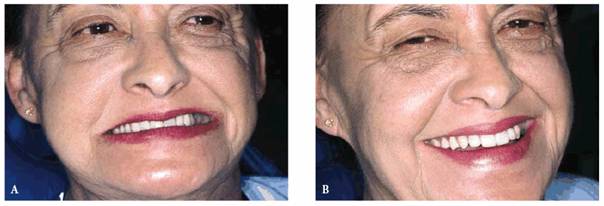
Figure 28-1A and B: (A) Unesthetic denture. (B) Esthetic denture. Maxillary anterior teeth are in the proper position; therefore, the entire denture is esthetic. The proper positioning of the anterior teeth guide all of the teeth in the denture.
This chapter is primarily concerned with denture esthetics; however, comfort
and function must be addressed. The failure of a complete denture treatment can
be traced to three areas: comfort, function, and esthetics. A denture can be
functional and comfortable. If, however, it is ugly in the eyes of the patient,
it is a total failure. On the other hand, a denture can be esthetically
superior, and if it is not functional and comfortable, it is still a failure.
Complete denture prosthetics has been taught in schools the very same way since
the turn of the century.4 Materials are far superior (ie, impression
materials, teeth, acrylic, base tints, and precision processing equipment);
however, the basic approach to satisfying the patient's needs has remained the
same. Impressions are made, and bases and wax rims are constructed. The wax
rims are adjusted in the mouth for tooth display, high lip line, and midline,
and a jaw relation is determined. The teeth are set on the articulator by the
technician or the dentist many times, with few guidelines. Then the wax-up is
presented for patient approval. This is frustrating and may result in several
resets to achieve patient acceptance. The denture is processed and delivered.
In many cases, it is now when the patient begins to speak, eat, and observe the
esthetics over a period of several days that the real problems arise.
Unfortunately, many patients and dentists are far too familiar with the
heart-breaking results using this unpredictable approach.
In the practice of fixed restorative dentistry, comfort, function, and patient
acceptance of the esthetics are ensured prior to final prosthesis construction
by first providing a provisional prosthesis (Figures 28-2, and ). In the modern practice of
complete denture prosthetics, the edentulous patient is first provided with a
provisional denture.2,10,25,27,28 This provisional denture will
allow the dentist to refine all of the functional esthetic aspects of the
denture to his or her and the patient's satisfaction (Figures 28-4, and ). After complete acceptance by the
dentist and the patient, the provisional denture is used much like a blueprint
to construct the final continuance denture. This approach leads to patient
happiness without the frustrating surprises of the past. This technique makes
the practice of denture prosthetics very predictable. While the patient wears
the treatment denture, an added benefit is the creation of functional
impressions (Figures 28-6, and ). It is the author's experience
that after final delivery of the denture, few, if any, postinsertion
adjustments are necessary.
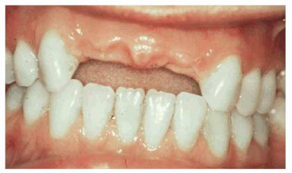
Figure 28-2: Maxillary anterior edentulous area to be treated with a fixed partial denture.
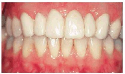
Figure 28-3: Provisional prosthesis placed to gain patient acceptance and to test esthetic and functional values.
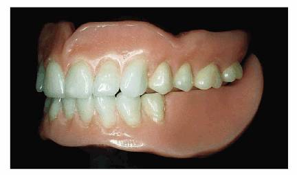
Figure 28-4: Maxillary and mandibular provisional training denture.
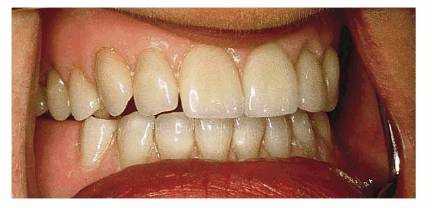
Figure 28-5: Provisional training denture placed to prove all aspects of denture function, esthetics, and comfort and to gain patient acceptance.
Figure 28-6: Mandibular functional impression is created as the patient wears the provisional denture.
Figure 28-7: Maxillary functional impression is created as the patient wears the provisional denture.
OCCLUSION: THE COMPLETE DENTURE
No discussion of complete dentures can be complete without addressing the
occlusion. Of all of the causes of denture failure, the lack of a balanced
occlusion in centric relation accounts for 90% of all denture failures. The
most difficult challenge in denture prosthetics is occlusion. All of the
denture teeth must occlude evenly as the mandible opens and closes on the arc
of closure. Personal experience tells the dentist that patients with natural
teeth many times function for a lifetime in a maximum intercuspal position that
is not coincidental with centric relation. This is not true with the edentulous
patient. These patients have lost most of the occlusal awareness, and the
occlusion must be built to the repeatable position of centric relation.
This is often a difficult task because the precise jaw relation must be
registered on two movable bases. In the treatment denture in Figure 28-5, the mandibular posterior teeth are
replaced with a noninterfering bite block. This bite or chewing block acts as a
superior repositioning splint to help the dentist obtain the optimum position
of centric relation.
Registering and confirming the occlusal relation position is done with various
waxers and central-bearing recording devices.
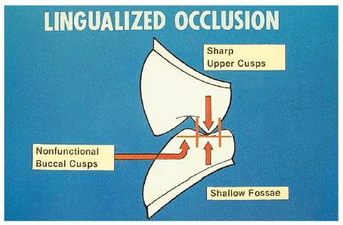
The occlusal scheme is referred to 22122n1316w as a lingualized occlusion that is
characterized with a single maxillary lingual cusp that functions into a
mandibular fossa. This occlusion seems to be very efficient and stable for the
denture patient (Figures 28-8, and
Figure 28-9 shows the posterior esthetics of
lingualized occlusion with the natural maxillary facial cusp.
Figure
28-8: Schematic of lingual contact occlusion. Maxillary lingual cusp will
function in a mandibular central fossa with no contact of the mandibular buccal
cusp and the occlusal incline of the maxillary buccal cusp. (Reproduced with
permission from Turbyfill WF. Regaining pleasure and success with complete
denture services. Int J Prosthet 1989;2:472-82.)
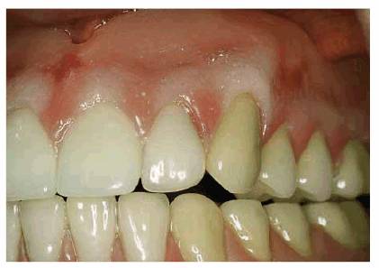
Figure 28-9: Lingualized occlusion in the completed denture.
THE ART OF CREATING ESTHETIC DENTURES:
THE ESTHETIC HARMONIES
There are four esthetic harmonies that must be considered to produce a denture
that will satisfy the patient's esthetic demands. These esthetic harmonies are
(1) tooth size and form, (2) tooth color, (3) tooth position, and (4)
background. The background is the denture base, which should be formed and
colored to look like human gingiva and tinted to blend with the patient's
overall complexion.
Of the four harmonies, the most important are tooth position and size. If the
teeth are placed into the position that the natural teeth once occupied and in
a size that is in harmony with the face, most of the esthetic requirements will
have been achieved. In the consideration of tooth position and arrangement, it
must be understood that everything that is done in this area has an influence on
the esthetics. These considerations include the proper midline, incisal plane,
posterior occlusal plane, horizontal and vertical positions of the maxillary
anterior teeth, and horizontal and vertical positions of the mandibular
anterior teeth.
Tooth Selection
Size and Form. Tooth size and form are considered simultaneously.
The selection of the maxillary incisors is the starting point in creating
esthetic dentures. There are many suggested ways to select teeth, including (1)
pre-extraction records, (2) patient photographs, (3) patient desires, and (4)
facial measurements.
The four methods of tooth selection are used routinely in complete denture
prosthetics. There have been several theories set forth. It must be understood
that none of these ways are accurate in all cases.16-18,33,36
However the selection is made, it is a guide or a starting place.
In 1887, the temperamental theory was proposed.13 It was one of the
earliest to propose that a person's personality might influence the morphology
of the teeth. In 1914, Williams37 rejected the temperamental theory
as a fallacy, proposing what is known as the geometric theory, and concluded
that the shape of the face and the shape of the central incisor are related.
This approach is still being used by many dentists. In 1939, House and Loop12
expanded on Williams's works to include not only pure typal forms (square,
tapered, and ovoid) but also combinations of typal forms and the discovery of
the relationship of the width of the face and the width of the central incisor.
In a study of 555 subjects, House and
In 1955, Frush and Fisher8 brought forth the sex, personality, and
age (SPA) theory of tooth selection. By 1959, five additional articles followed
describing the methods of applying the SPA factors. They concluded that tooth
size is related to the width of the nose. With the use of the Alameter
(Productivity Training Corporation,
At the annual session of the American Academy of Esthetic Dentistry in 1981 in
San Francisco, an interesting study was conducted by Abrams.1 One
hundred slides of human teeth were chosen, and the audience, consisting of
several hundred dentists, was asked to choose whether each slide was a male or
a female (lips were blocked out so that only the teeth were visible). After the
results were tabulated, it was determined that (1) gender cannot be determined
by tooth morphology or arrangement and (2) the older the patient, the more the
audience thought that the patient was a male purely because wear denotes
vigorousness to most dentists. Nevertheless, using this approach can produce
many quite esthetic and pleasing results.30 Although none of these
methods is absolutely accurate,16-18,33 it must be reiterated that
there must be a starting point.
Clinical Tooth Selection. From a clinical standpoint, measurement
of the face is the first step. House and
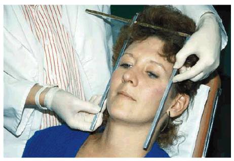
Figure 28-10: The width of the face is measured from 1 inch behind the outer canthus of the eye. (Reproduced with permission from Turbyfill WF. The union of natural contour, color, and shape. Signature 1995; Fall:14-17.)
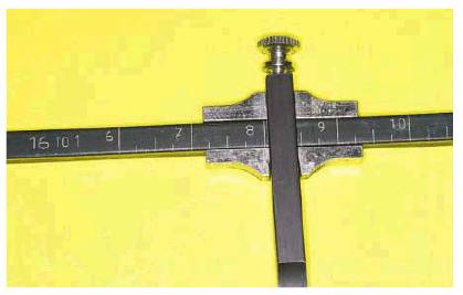
Figure 28-11: The measuring device reads the facial width in millimeters at a ratio of 1 to 16.
Tooth
Form and Mold. Most
instruction on tooth selection suggests that tooth form be matched to the
patient's facial form, that is, square, tapered, ovoid, or, in some
manufacturers' teeth, combination form types. These combinations include
square/tapered, square/ovoid, tapered/ ovoid, etc. Some tooth manufacturers
make only the basic square, tapered, and ovoid but do not make the combination
forms. Other manufacturers make only different size teeth and no teeth
designated for the different facial forms. These manufacturers do, however,
make molds that exhibit different amounts of incisal wear.
The author rarely considers tooth mold in terms of square, tapered, or ovoid.
Once the proper tooth size is selected, the mold is selected to fit the
patient's maturity. Younger patients get more rounded molds with unworn tips on
the cuspids. More mature patients will get teeth that show more incisal wear
and cuspids where the incisal tip is worn flatter. The author puts tapered
teeth in ovoid faces or square teeth in tapered faces and does not like ovoid
teeth in most cases (see Tip 2 below).
After the upper anterior teeth are selected, the lower anterior teeth are
selected as recommended by the manufacturer. For example, the 44E
(DENTSPLY/Trubyte,
Tips on Tooth Selection.
1. Facial width of one-sixteenth is determined to give the width of the
central incisors. The laterals and cuspids in any mold are sized to be in
harmony with this measurement. The facial length is never used because the
length of the teeth is more determined by lip height and the size of the
residual ridge. This maxillary central size is always in harmony with the size
of the patient's face. Next, the shape or mold of the teeth must be selected.
The author uses another method that is called "heart and
imagination." This is hard to explain. Credit is to be given to Fillastre
(Fillastre, Lakeland, FL, personal communication, 1980). After the appropriate
size is selected, the dentist will picture the patient in his mind's eye with
the mold chart in front of him and picture what mold would look good for the
patient.
2. Facial types-square, tapered, or ovoid-have little to do with mold
selection. The amount of incisal wear is a more appropriate guideline. The
older the patient, the flatter the incisal edges. The younger patient will
require more rounded edges that show less wear. Older patients tend to have
flatter, more worn cuspids. There is basically no difference in molds for male
or female.
3. Patients should be asked to bring pictures of themselves before they lost
their natural teeth. This can show anterior tooth arrangement and situations
such as diastemas. A trick to using a portrait-size picture is to measure tooth
width on the photograph and also measure the interpupillary width on the
photograph. Then the interpupillary width on the patient is measured. By using
a simple mathematical proportion, the actual tooth size is determined.
4. Patients should bring pictures of people from magazines who have teeth that
they think are attractive. This is done to point out that teeth are not set
over the residual ridge27 with small teeth hidden back in the mouth.
Pretty teeth are prominent and support the lip. Most pretty people show all of
their teeth when they smile, and many show some or all of their gingiva. This
exercise allows the teeth to be placed more in the position that the natural
teeth once occupied.
5. The molds are mixed. For example, the central and cuspids from one mold and
the laterals from another are used. Also, the use of laterals from two
different molds can create a nice effect. The patient must be educated to the
fact that bilateral symmetry does not occur in nature and that this is not
esthetic.
6. Manufacturers' suggestions for a mandibular anterior mold to use with a
specific maxillary anterior mold are dictated by a combined upper and lower
width that will allow the first bicuspids, maxillary and mandibular, to blend
in harmony with the maxillary and mandibular cuspids and with the cuspids in a
Class I relationship. To be esthetic, a denture must be in harmony with nature.
It is good to remember that in establishing natural denture esthetics, the
teeth are to be set in harmony with original jaw relations, whether it is Class
I, II, or III. In these cases, the mandibular anterior mold may have to be
varied in size to produce a nice harmonic transition from the cuspids to the
first bicuspids during set-up.
7. In selecting posterior teeth, the author's preference is to use an anatomic
maxillary posterior tooth (33-degree cuspid tooth) that occludes in a
lingualized fashion into the central fossae of the lower.5,27 The
esthetics is far superior to flat plane and other lesser degree teeth. The
beautiful maxillary buccal cusps look natural (see Figure 28-9). The purpose of the maxillary
buccal cusp is esthetics, food manipulation, and overjet to prevent jaw biting.
8. Facial profiles can be important in denture tooth selection. For example, an
individual with a flat profile might look better with a flat mold tooth, and
the patient with a curved profile might look better with a more curved or
rounded facial contour.
9. A choice is made between porcelain or acrylic teeth. Porcelain teeth keep
their luster. They will not wear excessively, causing loss of vertical
dimension, which leads to serious esthetic problems. There are times when,
because of interarch space, acrylic teeth must be used. In these cases, metal
occlusal surfaces should be used to prevent further loss of vertical dimension.
The use of acrylic teeth does not reduce the pressure on the underlying bone.
Rapid bone loss is caused by malocclusion. With plastic teeth, the malocclusion
is soon worn in; with porcelain teeth, the malocclusion is there forever.
10. The newer composite resin teeth have certain advantages. Ro Youdalis
pointed out that the hardness of these teeth will make many metal occlusal
surfaces unnecessary because of their wear resistance (personal communication,
1985).
Tooth Color. The hues found in most natural teeth fall into the
ranges of yellow, brown, and orange. The lightness and darkness of teeth are
controlled by the value (degree of gray or white). Chroma (the saturation of
hue) increases with advanced age, whereas the value decreases.
Tooth color and gingival tissue seem to be related to tissue tones and the
general overall complexion of the individual. The light-complexion, blue-eyed
blond will usually display very light teeth, with little or no yellow.
Contrasting this is the dark- complexion brunette and redhead whose teeth
generally show more yellow, brown, and orange.
As these individuals grow older, the yellow-brown of the brunette is
intensified by a deeper color and lower value. As the blue-eyed blond advances
in age, the greatest change is lower value. The teeth become more gray but
still exhibit very little yellow-brown.
There are always exceptions. It is not unusual at all to see a dark-complexion
brunette with very white teeth with no yellow. There are exceptions to all of
these rules.
Tips on Tooth Color.
1. Consultation with the patient is the best way to establish rapport in the
matter of tooth color.11 No attempt should be made to convince a
patient to accept any other shade than what he or she wants.35 All
patients want nice white teeth, and the older denture patient is no exception.
(A personal note on this subject is in order. In the early days, the author
tried to select teeth as his mentor did to be in harmony with the patient's age
and complexion. It seemed that he was always at odds with patients who wanted
white teeth.) Seminars are conducted during which dentures are provided for
patients. Since all of the shades cannot be available during the seminars, only
light shades are stocked. There is never a discussion about shades at the seminars.
The fact that every patient gets young bright teeth is a key to success.
2. Every female patient is asked when looking at their old denture,
"Wouldn't you like a brighter, more youthful tooth on your new
denture?" The response is interesting: it is always yes.
3. Many patients love to see the artist in their dentist emerge. The dentist
could try using central incisors of one shade and then change the shades of the
laterals. The patient with the love of naturalness will respond.
4. Lower anteriors one shade darker than the upper incisors should be used.
Tooth Arrangement
Once a proper tooth has been selected for the patient, the maxillary anterior
teeth will be positioned on the base. The most important consideration in
creating an esthetic denture is the position of the maxillary anterior teeth.
This position will directly influence the position of every other tooth in the
denture.
Frank Lloyd Wright said, "Form and function are one." So it is with
dentures: the closer the artificial teeth are placed to the position once
occupied by the natural teeth, the better the function will be. Another
advantage of natural tooth placement is that they will fall within the neutral
zone, which is the neutral point of muscle balance between the lips and cheeks and
the tongue.
E. Pound (personal communication, 1975) said that, in the early days, when
motion pictures became talking pictures, many actors lost their jobs because of
poor dentistry and poor phonetics. He said that the sound people at the studios
knew as little about speech as he did. Pound said that he strove to make
dentures exquisitely esthetic and that the better they looked, the better they
spoke. This is how his lifetime study of phonetics began.
In the practice of complete denture prosthetics, if the dentist can position
the artificial teeth in the position the natural teeth occupied, the better the
esthetics and function will be.
Key to
The single most important thing that must be done to create an esthetic denture
is proper placement of the maxillary anterior teeth. The key is the placement
of the maxillary central incisors. If these two teeth are correct, they will
directly influence the position of every other tooth in the denture. Correctly
placed, the maxillary six incisors should be as close as possible to the exact
position once occupied by the natural teeth. The notion that teeth should be
set over the ridge to gain a mechanical advantage is simply outdated and
untrue. If the teeth are set to anatomic harmony, then they will be in the
neutral zone and vice versa.
Setting the Maxillary Anterior Teeth in
Anatomic Harmony
Over the years, the author has measured casts of natural maxillary anterior
teeth to find a common position. A common average position of the maxillary
anterior teeth to constant landmarks has been found by measuring hundreds of
casts of natural healthy teeth. These measurements are (1) the distance of the
incisal labial one-third of the maxillary central incisors from the center of
the incisive papilla (Figure 28-12) and (2) the distance down the
incisal edge from the general height of the maxillary anterior labial vestibule
(Figure 28-13
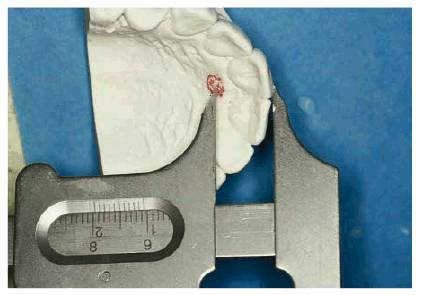
Figure 28-12: Average horizontal position of the maxillary central incisors is determined by measuring the distance from the center of the incisive papilla and the labial incisal one-third of the central incisors.
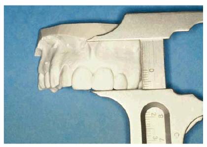
Figure 28-13: Average vertical position of the maxillary central incisors is measured from the general height of the anterior labial vestibule down to the incisal edge of the central incisors.
This position will not be appropriate for every patient. It will put the teeth
in a reasonable position all of the time. Variations from this position require
a judgment on the dentist's part. The author has observed that when positioned
in this manner, about 4 in 10 patients are close to ideal. The other 60% will
need slight changes.
In Figure 28-14A, the patient has very unesthetic
dentures. The ridges are healthy and well formed (Figure 28-14B). A stabilized base is constructed
onto the cast of the maxillary edentulous ridge. A wax rim is placed onto the
stabilized base. The wax rim is built 10 mm out from the center of the incisal
papilla and 20 mm down from the general height of the labial vestibule. In
addition, the wax rim is built level to the interpupillary line and parallel to
Camper's line. Camper's line runs from the middle of the tragis of the ear to
the base of the ala of the nose. The wax rim is tried into the edentulous
mouth. The level of the incisal plane and Camper's line is verified. The facial
midline is marked (Figures 28-14C 28-14D, and 28-14E).
The maxillary central incisors are set onto the wax rim (Figure 28-14F) and confirmed in the mouth (Figures 28-14G, and 28-14H).14,21,23 Once the
proper position of the central incisors is verified, then the remainder of the
maxillary incisors is set (Figure 28-14I). At this point, the basic tooth
position has been determined. Detailed esthetics, such as lapping of the
lateral incisors and tipping of the cuspids, is done in the laboratory.
Once the maxillary anterior basic tooth position has been verified, as a
demonstration, a silicone matrix has been made onto the set-up. In this case,
the central incisors ended up 9 mm anterior to the center of the incisive
papilla. The teeth follow the curvature of the edentulous ridge (Figure 28-14J). If the ridge should be more
square or more "V" shaped, this would have been reflected in the
arrangement. The cuspid begins to be tucked closer to the ridge as the corner
is turned from the anterior ridge to the posterior area of the ridge.
The much improved esthetics of the finished denture is shown. Figure 28-14K shows the superior esthetics that
has been achieved using this approach.
Once the maxillary teeth are set to anatomic harmony and the dentist is
satisfied with this position, the mandibular anterior teeth are set so that
they exhibit one half to 1 mm of clearance as the sibilant sounds are being
enunciated (Figure 28-14L). The vertical dimension of
occlusion is recorded by arcing the mandible in the arc of closure in centric
relation and closing the vertical down until the anterior stop comes into
contact (Figure 28-14M
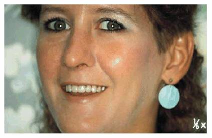
Figure 28-14A: A 32-year-old edentulous patient with teeth set over the ridge with a straight line set-up resulting in the typical denture look.
Figure 28-14B: The edentulous ridges appear adequate and healthy.
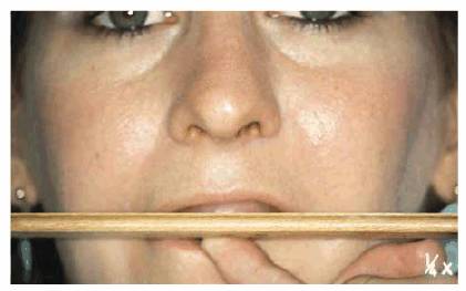
Figure 28-14C: The maxillary bite rim is built
10 mm anterior from the center of the incisive papilla horizontally and
vertically 20 mm down from the general height of the labial vestibule. It is
built level so that it is parallel to the interpupillary line. (Reproduced with
permission from Turbyfill WF.
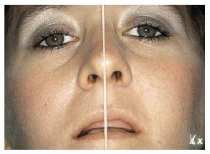
Figure 28-14D: The midline is marked.
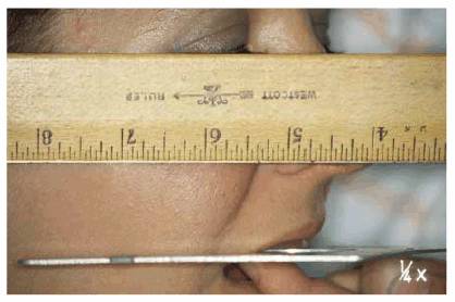
Figure 28-14E: The maxillary bite rim is built parallel to Camper's line.
Figure 28-14F: The maxillary central incisors
are set to the previously determined midline and the horizontal and vertical
determinants. (Reproduced with permission from Turbyfill WF.
Figure 28-14G: The central incisor position is verified in the patient's mouth as to tooth display and midline.
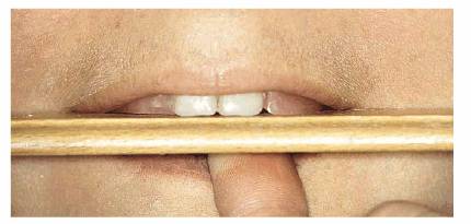
Figure 28-14H: The level of the central incisors is verified. (Reproduced with permission from Turbyfill WF. Union of natural contour, color, and shape. Signature 1995; Fall:14-17.)
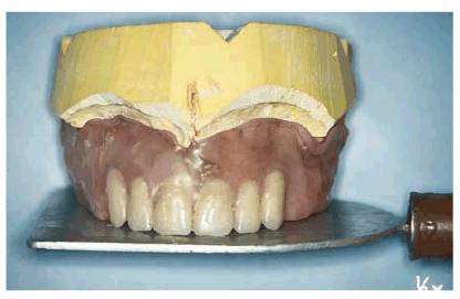
Figure 28-14I: The maxillary anterior tooth position is completed.
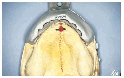
Figure 28-14J: The natural position of the maxillary teeth to the edentulous ridge.
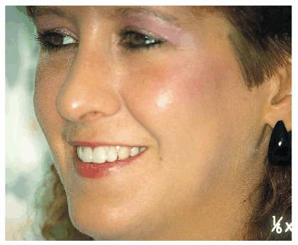
Figure 28-14K: The superior esthetic results achieved by placing the maxillary anterior teeth to anatomic harmony.
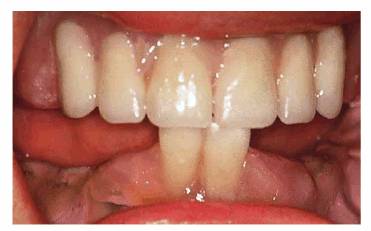
Figure 28-14L: The mandibular anterior teeth are positioned to exhibit a 1- to 1/2-mm clearance with the maxillary anterior teeth as the patient enunciates "S" sounds.
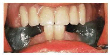
Figure 28-14M: The mandibular position of centric relation is determined by a simple wax recording. This is considered a treatment position, and final centric relation determination is achieved by using a central bearing point and Gathic arch tracing.
A denture may appear to be reasonably esthetic at first glance, as is the
maxillary denture shown in Figures 28-15A 28-15B, and 28-15C. The denture was remade because of
poor function and comfort. The teeth are now set to a position more in keeping
with anatomic harmony. This position is 20 mm down from the height of the
maxillary vestibule and 10 mm out from the center of the incisive papilla. Figures 28-15D 28-15E, and 28-15F show the subtle but exquisite
improvement in esthetics and maxillary lip support.
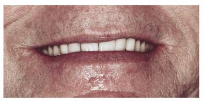
Figure 28-15A: A denture with a poor maxillary anterior tooth position.
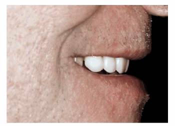
Figure 28-15B: Note the depressed position of the incisors.
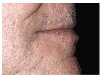
Figure 28-15C: Note the look of the maxillary lip support.
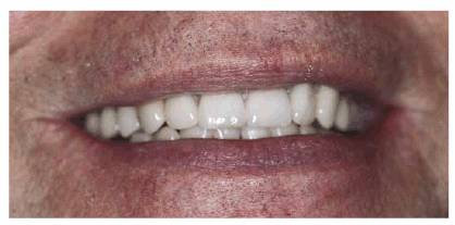
Figure 28-15D: An esthetic denture with the maxillary central incisors set to the 10 × 20 rule.
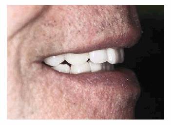
Figure 28-15E: Note the more natural tooth position.
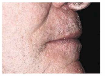
Figure 28-15F: Note the improved lip support.
Placement of the Mandibular Anterior Teeth
The mandibular anterior teeth are set using phonetics. Dawson6 noted
that the vertical dimension of occlusion that has been lost can be regained by
noting the closest speaking level and then establishing the vertical dimension
of occlusion slightly more closed from that closest speaking position. Pound24
referred to this as the vertical dimension of speech, and since the teeth are
not to touch while a person is speaking, then the vertical dimension of
occlusion should be slightly more closed than the "S" position.
Further, the "S" position is the most forward, most closed position
the mandible ever assumes during speech.
Two mandibular incisors are set to the "S" position. Pound defined
the "S" position as the most intimate relationship of the teeth
during speech.24 There are intimate relationships that occur between
the incisal edges of the mandibular teeth and the incisal edges and lingual
surfaces of the maxillary anterior teeth. This allows the dentist to verify the
accuracy of the maxillary tooth arrangement and place the mandibular incisors
in an anatomically natural position that produces an articulate speech pattern.
After this position is verified, the anterior stop has been re-established (see
Figure 28-14L
Anterior Denture Occlusion
No anterior teeth should be in contact when the posterior teeth are in maximum
occlusion. This anterior pressure will cause destruction of the bone of the
premaxilla. It also causes instability of the dentures. Once the anterior teeth
are set to exhibit a phonetic clearance when the "S" sounds are
enunciated, the centric relation registration is then taken with the anterior
stop in contact. In Class I and II jaw relationships, the mandible leaves the
centric relation posture position and moves forward to the phonetic
"S" position. Therefore, the articulator pins are opened slightly on
Class I and II occlusions so that the anterior teeth will not contact in the
centric relation. Since the mandible always moves forward in phonetics, the
condylar guidance will keep the posterior teeth from contacting in speech.
In Class III occlusion, there is no forward movement of the mandible during
speech. Slight anterior contact in the Class III jaw relationship is
inevitable. The occlusal contact should be heavier in the posterior than in the
anterior.
Vertical Dimension
The author uses phonetics and the closest speaking position to develop the
incisal edge position of the mandibular anterior teeth, and the vertical
dimension of occlusion is determined from this. It must be understood that this
method is not always accurate. Some patients exhibit an adapted position that
is obviously overclosed. In cases like this, the vertical is opened on the
trial bases until the facial profile looks more normal. In other words, the
patient should not look older below the nose than above the nose. Since all
patients are treated with a provisional treatment denture, this
"arbitrary" vertical dimension is tested before denture finalization.
Even though phonetics is not accurate in every case, it is still the preferred
way because the different movements of the mandible for the different classes
of occlusion help to position the incisal edges in a more natural position.
There are many ways to establish the vertical dimension of occlusion on
dentures, such as phonetics, relaxation of the mandible to establish the
resting level of the mandibular free way space, having the patient wet the lips
and breathe out, and measuring dots on the nose and chin and other facial
dimensions. Volumes have been written about vertical dimension. The subject of
vertical dimension is a very emotional one, and some heated arguments can erupt
over it.
The most important thing that the dentist must remember about vertical
dimension is that if the vertical is opened too far so as to cause the
posterior teeth to hit as the patient speaks, a failure will always result.
Another important observation is that the dentist should give each denture
patient the greatest vertical dimension possible. The patient will look, chew,
and feel better.
Improper vertical dimension can have a profound effect on esthetics. In Figure 28-16A, the patient looks prognathic and
old below the nose with a vertical dimension that is overclosed. Figure 28-16B shows excellent esthetics when the
vertical dimension is properly restored. Figure 28-16C demonstrates the poor esthetics
created by improper vertical and horizontal positioning of the maxillary
anterior teeth and the denture look. Figure 28-16D shows the improved esthetics with
proper maxillary anterior tooth positioning.
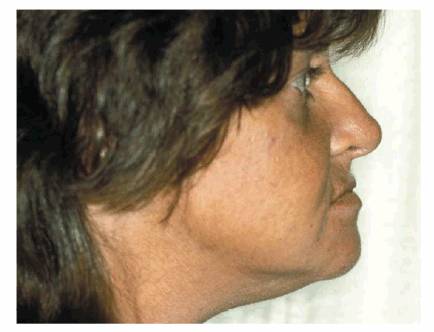
Figure 28-16A: Overclosure of the vertical dimension of the occlusion. Note the prognathic appearance and decreased lower facial length.
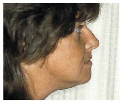
Figure 28-16B: Proper vertical dimension of occlusion. Note how much younger the patient appears.
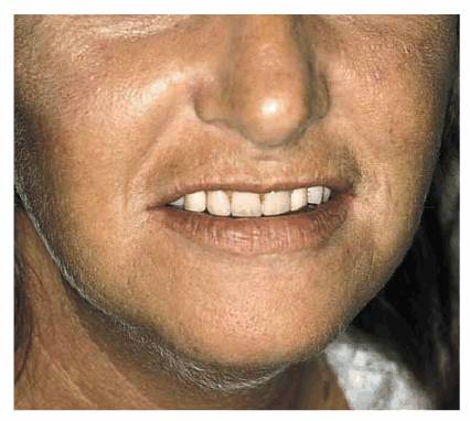
Figure 28-16C: Everything looks bad: the denture look.
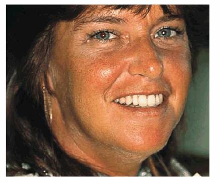
Figure 28-16D: The natural look: correct maxillary anterior tooth placement and correct vertical dimension of occlusion.
Consideration of the vertical dimension of the occlusion for the edentulous
patient as it differs from the patient with natural teeth needs to be
addressed. Patients with natural teeth are very adaptive to changes in the
vertical dimension of the occlusion because of the exquisite proprioception of
natural teeth. Many times, a slight opening of the vertical dimension is needed
to facilitate restorative procedures. Edentulous patients do not adapt nearly as
well. Generally, when the closest speaking position is determined, any opening
from that position should be done while the patient is wearing the provisional
training denture. An excessive vertical dimension of the occlusion in complete
dentures results in a restriction of normal muscle activity, and the posterior
and anterior teeth will hit during speech. When opened experimentally using the
training denture, if the teeth continue to hit during speech for 1 week, then
adaptation is not possible, and the teeth will hit during speech forever.
Posterior Occlusion As It Relates to
Denture Esthetics
One area often overlooked or misunderstood is the effect of the posterior tooth
position on esthetics. An extremely poor esthetic denture can result by
establishing the posterior plane of occlusion too high or too low. This is
demonstrated by the patient who smiles, and the maxillary posterior teeth can
be seen hanging down below the plane of the maxillary incisors. Camper's line
will position the posterior occlusal plane on a line beamed from the maxillary
anterior incisal edge posterior to the middle of the retromolar pad (Figure 28-17
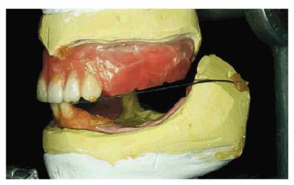
Figure 28-17: Posterior occlusal plane lines up from the maxillary incisal edges to a point halfway up the retromolar pad (Camper's line).
The buccolingual placement of the posterior teeth can also affect esthetics. If
the maxillary teeth are placed too far to the buccal aspect, then the buccal
corridor between the maxillary posterior teeth and the corner of the mouth is
lost. If the maxillary teeth are placed too far lingually or palatally, then
they appear not to exist. Either extreme produces unacceptable esthetics. The
guideline for the maxillary posterior tooth position is found in the mandibular
arch. The lingual central line is from the mesial contact of the cuspid to the
lingual aspect of the retromolar pad. The lingual surface of the mandibular
posterior teeth should fall on this line. The mandibular posterior teeth are
positioned closer to the tongue than this line (Figure 28-18
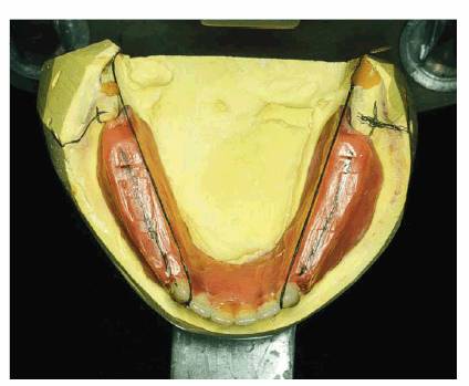
Figure 28-18: Buccolingual placement of the posterior occlusal plane. The lingual control line runs from the mesial contact of the cuspid to the lingual aspect of the retromolar pad.
Tips for Tooth Arrangement
1. The position of the maxillary central incisors is the key to denture
esthetics. Once they are placed and accepted, all other tooth positions are a
product of these two teeth.
2. Placement of the anterior teeth must be done at chairside in the
presence of the patient. The author never lets the patient see the results of
this initial placement because the setting is a very straight-line prosaic
set-up and represents basic tooth position. Detailed esthetics is done at the
laboratory bench.
3. The appointment for this initial setting is from 1 to 11/2
hours. The rapport that is built with the patient at this time is unbelievable.
The patient will always say, "I've never had a dentist spend so much time
with me."
4. The "S" position is the most intimate relationship of any teeth
during speech.22,26 As the "S" sounds are formed, the
anterior teeth will exhibit a space of 1 to 11/2 mm. The
"S" sounds are produced by forcing air between the incisal edges of
the maxillary incisors and the mandibular incisors. The "S" sounds
can also be produced by forcing air between the incisal edges of the mandibular
incisors and the lingual surfaces of the maxillary incisors, as will be found
in many Class II occlusal relationships. It should be remembered that the teeth
do not touch when the "S" sounds are being enunciated.
5. The vertical dimension of occlusion is very easy to determine since it is
always less than the vertical dimension of speech.19,20,32
Therefore, when the anterior teeth are set to the "S" position, the
mandible is retruded and closed down 2 mm to tooth contact or, if no teeth
touch, merely closes in a centric relation 2 mm less than the vertical
dimension of speech.
6. The "F" and "V" sounds are produced when the incisal
edges of all six maxillary anterior teeth make a fleeting seal at the vermilion
border or the wet dry line of the lower lip. If the maxillary teeth are placed
in anatomic harmony, the "F" and "V" position will always
be correct. This is an extremely valuable check on the accuracy of the
maxillary anterior tooth placement.
7. The anterior teeth, both maxillary and mandibular, should appear as if they
are coming from the bone at a slightly different angle. Sharry31
wrote, "There is one prominent guide for providing an excellent
arrangement of anterior teeth; they must be separate." Bilateral symmetry
is not found in nature. The laterals can be mesially lapped or winged out
distally. Laterals can be set to be shorter than the centrals or cuspids;
however, the older the patient, the more even the incisal edges should be. If
photographs are available and show diastemas, they can be placed subject to
patient approval. It should be remembered that the younger the patient, the
more open the incisal embrasures.
8. Always keep the incisal plane level and slightly curved to follow the smile
line of the lower lip. There is nothing more unesthetic than a slanted occlusal
or incisal plane.
9. In a few patients, flaring of the cuspid can be esthetic, but, in most
cases, it is best to set the cuspid so that only the mesial surface can be
seen.
10. It should be remembered that the first introduction to dental esthetics to
dental students and dental technology students was a Columbia Dentiform.
11. Pictures of "pretty" people should be used to show how beautiful
smiles are made in nature. Dentists should point out the asymmetry and how
prominent the teeth appear in a full smile.
12. There are some interesting studies being done concerning the arrangement of
teeth for the edentulous patient by the use of cephalometric radiographs.29
Orthodontists use the method of fixed bony landmarks to determine the ideal
placement of natural teeth. It seems that this would be a valuable aid for
tooth placement in complete denture prosthetics, particularly in the advanced
resorbed dental arch.
13. The author uses "heart and imagination" in selecting and
arranging teeth. In the laboratory, there should be a large selection of teeth,
and as the dentist looks at the basic tooth position set at chairside, he or she
must picture the patient in his or her mind's eye and set and select teeth to
what he or she feels will be pleasing.
BACKGROUND
The denture base is important in esthetics.34,38 Its normal contour
aids in support of the soft tissues of the lips and face. If the patient has a
short upper lip and would normally show gingival tissues in a broad smile, an
unesthetic denture base can destroy an otherwise esthetically successful
denture.
An anatomically accurate denture base is important to function since "form
and function are one." The food tables that we find facially at the tooth
neck in normal healthy tissue help the buccinator muscles keep food out of the
vestibule and up onto the occlusal surfaces. The form of the lingual surfaces
is important in that it departs a feeling of naturalness to the tongue. It is
also paramount that the neutral zone of the tongue not be violated by an
overcontoured mandibular lingual flange.
The denture base that copies nature is also self-cleaning. The interdental
papilla are full and rounded, and there are no "festoons." These only
create food traps and prevent a sweeping of the tongue from its cleaning
action.
Tinting of the denture base is important in several respects (Figure 28-19). A natural-looking base is
desirable on the facial aspect and on the palatal as well. Nothing gives away a
denture faster than an individual laughing with head held back as the slick
mono color of the palate is viewed by others (Figure 28-20
Figure 28-19: Denture base carved and tinted to appear natural. (Reproduced with permission from Turbyfill WF. Regaining pleasure and success with complete denture services. Int J Prosthet 1989;2:474-82.)
Figure 28-20: Correct anatomy of the palate with singulum carved on anterior teeth and lingual surfaces carved on the posterior teeth. The palate is also tinted.
Tips on Creating a Natural-Looking
Denture Base
1. Casts of human tissue should be studied, noting stippling, gingival collars,
and the interdental papilla. There should be no slick, flat, and shiny surfaces
in human gingival tissues.
2. Whatever is wanted in the finished base should be carved in wax. No carving
with rotary acrylic finishers can be done after it is processed (Figures 28-21 , and ).
3. The denture should be invested with the same degree of care as was used in
investing an inlay (Figure 28-24).
4. A denture base tinting acrylic (Kay-See Dental Manufacturing, Kansas City,
MO) should be used (Figure 28-25).
5. Tints should be placed in eye dropper bottles with the glass droppers turned
upside down to control the placement of the tints.
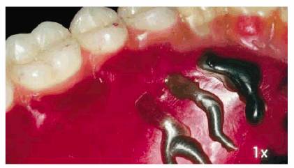
6. The tints are sifted into the boiled out flasks and then are wet with
monomer as the technician goes around the arch three or four teeth at a time (Figures 28-26 , and ).
7. Cases are tinted in four basic shades: (1) light-complexion blue-eyed
blonds, (2) medium-complexion brunettes, (3) dark-complexion brunettes, and (4)
non-Caucasians.
Figures 28-29A
and B 28-29C and D, and 28-29E and F are examples of esthetic dentures.
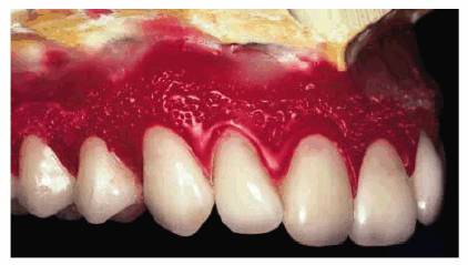
Figure 28-21: Anatomic wax-up.
Figure
28-22: Anatomic wax-up.
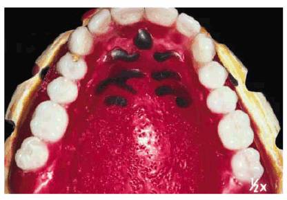
Figure 28-23: Anatomic wax-up. Note that the palate is lightly stippled so as not to appear shiny.
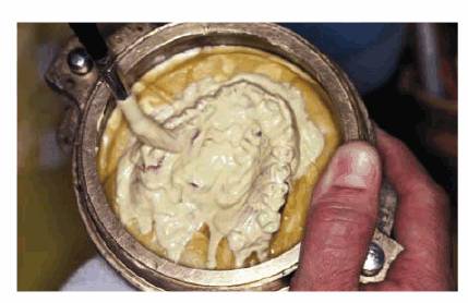
Figure 28-24: Investment is poured using a sable brush to capture the full anatomic wax-up.
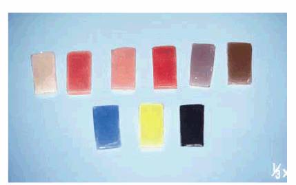
Figure 28-25: Shades of tints available for the wax-up. There is no carving of the base with rotary instruments.
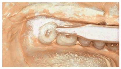
Figure 28-26: Tints are sifted around teeth.
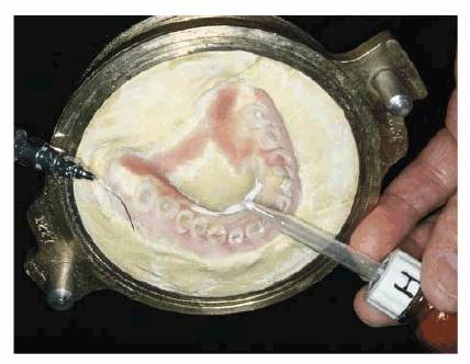
Figure 28-27: The palate is tinted.
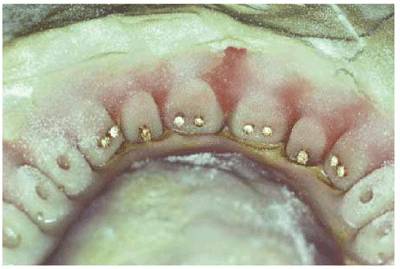
Figure 28-28: The maxillary case tinted. Dentures are then processed with acrylic of a color to complement the overall complexion of the patient.
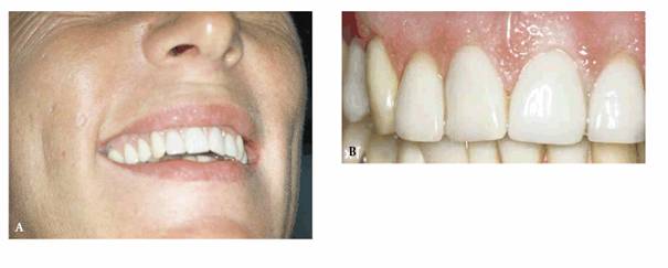
Figure 28-29A and B: (A) Full smile view of esthetic dentures. (B) Close-up of esthetic dentures. (Reproduced with permission from Turbyfill WFJ. The provisional denture: key to denture success. Aurum Ceramic Classic News 1995;8(2):1-4.)
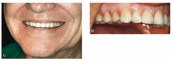
Figure 28-29C and D: (C) Full smile view of esthetic dentures. (D) Close-up of esthetic dentures.
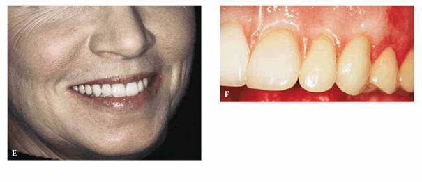
Figure 28-29E and F: (E) Full smile view of esthetic dentures. (F) Close-up of esthetic dentures.
REFERENCES
1. Abrams L. Male or female-can you tell by the teeth? Report of sexual
dimorphism study.
2. Appelbaum MB. The practical dynamics of the interim denture concept: a
comparison with the conventional immediate denture technique. J Am Dent
Assoc 1983;106:826-30.
3. Berg E, Johnson TB, Ingebretsen R. Patient motives and fulfillment of
motives in renewal of complete dentures. Acta Odontol
Scand 1984;42:235-40.
4. Boucher CO. Complete denture prosthodontics-the state of the art. J Prosthet
Dent 1975;34:372-83.
5. Clough HE, Knodle JM, Leeper SH, et al. A comparison of lingualized occlusion
and monoplane occlusion in complete dentures. J Prosthet
Dent 1983;50: 176-9.
6. Dawson P. Evaluation, diagnosis, and treatment of occlusal problems.
7. Fisher RD. Personalized restorations vs. plates. J Prosthet
Dent 1973;30:513-4.
8. Frush JP, Fisher RD. Introduction to dentogentic restorations. J Prosthet
Dent 1955;5:586.
9. Goldstein RE. Study of need for esthetics in dentistry. J Prosthet Dentist
1969;6:589-98
10. Hansen CA. Diagnostically restoring a reduced occlusal vertical dimension
without permanently altering the existing dentures. J Prosthet
Dent 1985;54: 671-3.
11. Hirsch B, Levin B, Tiber N. Effects of patient involvement and esthetics
preference on denture acceptance. J Prosthet
Dent 1972;28:127-32.
12. House MM,
13. Ivy RS. Dental and facial types. In: Litch WF, ed. American system of
dentistry. Vol. 2.
14. Krajicek D. Guides for natural facial appearance as related to complete
denture construction. J Prosthet
Dent 1969;21:654-62.
15. LaVere AM, Marcroft KR, Smith RC, Sarka RJ. Denture tooth selection: an
analysis of the natural maxillary central incisor compared to the length and
width of the face. Part 1. J Prosthet
Dent 1992;67:661-3.
16. Lieb ND, Silverman SI, Garfinkel L. An analysis of soft tissue contours of
the lips in relation to the maxillary cuspids. J Prosthet
Dent 1967;18:292-303.
17. Marunick MT, Chamberlain BB, Robinson CA. Denture aesthetics: an evaluation
of laymen's preferences. J Oral Rehabil
1983;10:399-406.
18. Mavroskoufis F, Ritchie GM. The face-form as a guide for the selection of
maxillary central incisors. J Prosthet Dent 1980;43:501-5.
19. Murray CG. Re-establishing natural tooth position in the edentulous
environment. Aust Dent J 1978;23: 415-21.
20. Murrell GA. Phonetics, function, and anterior occlusion. J Prosthet Dent 1974;32:23-31.
21. Pound E. Apply harmony in selecting and arranging teeth. Dent Clin North Am
1962;Mar:241-58.
22. Pound E. Controlling anomalies of vertical dimension and speech. J Prosthet Dent 1976;36:124-35.
23. Pound E. Fine arts in the fallacy of the ridge. J Prosthet Dent 1954;4(1).
24. Pound E. Mandibular movements of speech and their seven related values. J Prosthet Dent 1966;16:835.
25. Pound E. Preparatory dentures: a protective philosophy. J Prosthet Dent
1965;15:5-18.
26. Pound E. Utilizing speech to simplify personalized denture service. J Prosthet Dent 1970;24:586-600.
27. Pound E, Murrell GA. An introduction to denture simplification. J Prosthet Dent 1971;26:570-80.
28. Pound E, Murrell GA. An introduction to denture simplification. Phase II. J Prosthet Dent 1973;29: 598-607.
29. Rayson JH, Rahn AO, Wesley RC, et al. Placement of teeth in a complete
denture: a cephalometric study. J Am Dent Assoc 1970;81:420-4.
30. Ruffino AR. Personality projection in complete dentures: traits
transmissible to the viewer through variations in maxillary anterior tooth
arrangement. J Prosthet Dent 1984;50:661-2, 664.
31. Sharry J. Essential concepts in denture esthetics. In: Esthetics in
dentistry. Philadelphia: JB Lippincott, 1976.
32. Sherman H. Phonetic capability as a function of vertical dimension in
complete denture wearers-a preliminary report. J Prosthet Dent 1970;23:621-32.
33. Smith BJ. The value of the nose width as an esthetic guide in
prosthodontics. J Prosthet Dent 1975;34: 562-73.
34. Starcke EN Jr. The contours of polished surfaces of complete dentures: a
review of the literature. J Am Dent Assoc 1970;81:155-60.
35. Tau S, Lowenthal U. Some personality determinants of denture preference. J Prosthet Dent 1980;44:10-2.
36. Turner LC. The profile tracer: method for obtaining accurate pre-extraction
records. J San Antonio Dent Soc 1970;25:13.
37. Williams JL. The temperamental selection of artificial teeth. A fallacy.
Dent Dig 1914;20:63.
38. Zimmerman DE, Cotmore JM. Denture esthetics (I). Denture base contour. Quintessence Int 1982;13: 543-9.
|
