ORAL HABITS - Ronald E. Goldstein, DDS, James W. Curtis
Jr., DMD, Beverley A. Farley, DMD
INTRODUCTION
Habits can, and all too frequently do, cause esthetic and/or functional
problems in the mouth. For this reason, destructive habits need to be diagnosed
and corrected as early as possible. Many patients are unaware that they have
habits involving their mouths, particularly unconscious behaviors such as
bruxism. Most have no idea that simple behaviors such as
"occasionally" holding their glasses in their mouth or chewing ice
can cause permanent problems. Adequate diagnosis of damaging habits requires a
thorough evaluation of each patient's stomatognathic state. This must include
examination of the form and function of the teeth and the status of the
temporomandibular joints and related musculature.
Oral habits should be foremost in the examination and diagnosis of pediatric patients.
Later in life, the permanent teeth and mouth should be carefully examined for
changes related to oral habits that often occur in response to stress.
Hygienists can play a key role in initially detecting wear patterns in teeth
that could be arrested. Most people are surprised, but pleased, that their
destructive habits can be stopped or the damage from them controlled. Dental
procedures and corrective behavioral techniques may be helpful in breaking such
oral habits. However, unless these habits are totally discontinued, treatment
will inevitably serve as only a stopgap measure.
DIGIT SUCKING
Digit sucking is a habit that usually begins and ends in childhood (Figure 20-1). Failure to stop this behavior can
result in adult arch deformities that make correction more difficult (Figures 20-2A 20-2B, and 20-2C
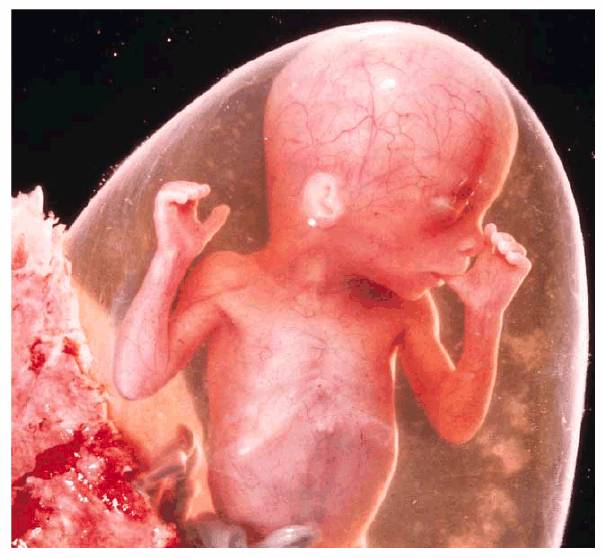
Figure 20-1: This unusual photograph demonstr 646h72g ates the early age (18-week-old fetus) at which thumb sucking may be manifested. Whereas many habits may be acquired, some seem to be genetically inbred as evidenced in this magnificent photograph. (Reproduced with permission from Nilsson L. A child is born. Stockholm: Albert Bonniers, 1976:125.)
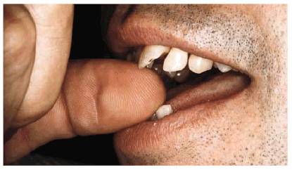
Figure 20-2A: This 33-year-old education director told of sucking his thumb as a child, which graduated into a finger-biting habit. Note the position of the thumb during the biting habit.
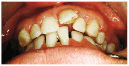
Figure 20-2B: Both maxillary and mandibular left central incisors are in labioversion as a result of the finger-biting habit.
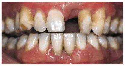
Figure 20-2C: Treatment in this type of habit sometimes consists of orthodontics and/or prosthodontics, depending on whether bone loss is present. Since there was considerable bone loss in this patient, treatment consisted of extraction of maxillary and mandibular left central incisors, plus additional periodontal therapy. Maxillary and mandibular resin bonded fixed partial dentures followed.
It is estimated that roughly 4 of 10 children between the ages of birth and 16
years of age engage in digit sucking at some time during their lives. This
habit may also involve several digits or fist sucking as well.
For example, Larsson has presented the results of longitudinal studies of
children using lateral cephalometric radiographs and observation of the
occlusion.45-47 In the younger children, thumb sucking increased the
incidence of open bite with proclined and protruded maxillary incisors, a
lengthened maxillary dental arch, and anteriorly displaced maxillary base. He
found that finger sucking frequently caused a unilateral abnormal molar
relationship on the sucking side of the mouth when the child consistently
sucked a thumb. Finger sucking was also an important etiologic factor in the
development of a posterior crossbite in the primary dentition. However, if the
children stopped thumb sucking, these malocclusions were somewhat corrected by
increased growth of the alveolar process and the eruption of the incisors.
Similarly, others have found a tendency to an open bite and an elongated
maxillary arch length among children with strong sucking habits between ages 7
and 16 years.61,81,86 The dental effects of the thumb sucking were
primarily in the anterior region of the mouth, with 80% of the children shoving
the tongue over the lower incisors during swallowing. Eliminating the sucking
habit tended to produce spontaneous closure of the open bite and cessation of
the tongue thrust.
Bowden found yet other disturbances in persistent thumb suckers: significant
increases in the proportion of the protrusive maxillary dental base
relationships, tongue thrust activities, tongue-to-lip resting positions, and
open bite tendencies.10 Haryett and colleagues noted crowding of the
mandibular incisors and facial asymmetries resulting from tooth interferences
in the molar area because of maxillary contraction from sucking.31
Infante's study of preschool children found posterior lingual crossbite and
protrusive position of the maxillary molars relative to the mandibular molars
to be more prevalent and pronounced among those children who were thumb
suckers.35 Popovich and Thompson concluded that, as the habit
persisted, the probability increased that a child would develop a Class II
malocclusion.63 In each of these studies, the problems appeared to
diminish in prevalence and severity as digit sucking declined, usually
occurring naturally as the child grew older.
Massler believes that some of these displacements can be self-corrected by the
molding action of normal labial and lingual musculature once thumb sucking is
discontinued. For example, the continued and forceful placement of the thumb
against the long axis of the erupting tooth may temporarily displace the
erupting anterior teeth.56 Massler suggested that more marked
protrusions probably have a genetic basis. Although this protrusive tendency
can be maximized by thumb-sucking behaviors, it can also manifest itself in
children who have never been habitual digit suckers.
In addition to the orofacial effects caused by digit sucking, other injuries
may arise as a result of this habit. Rayan and Turner described hand
complications that may develop from prolonged digit sucking.65
Because thumb sucking is such an obvious oral habit, and perhaps because it
occurs at a time when parental attention is most focused on the child, the
general public has taken part in a sometimes acrimonious open debate over the
possible permanent effect of the habit. The debate extends to when, or even if,
the parent and/or dentist should intervene. In the 1930s and 1940s,
pediatricians, pediatric dentists, and parents were frequently united in their
battle against thumb sucking to prevent malocclusion. Infants were sometimes
wrapped in elbow cuffs or had the sleeves of their nightgowns tied to prevent
fingers from reaching their mouth. However, as Massler described, the result
was "that we now are treating a generation of tongue suckers with anterior
open bites and lip suckers with the so-called mentalis habit."56
These habits, he pointed out, persist much longer than thumb sucking and are
considerably more difficult to discontinue.
The accepted wisdom of our own age is that most children give up sucking by the
age of 3 to 7 years. Until a child passes this age, it is just as well, and
much simpler, to avoid intervention. There is much agreement that digit-sucking
habits are unlikely to produce permanent damage to the orofacial structures if
the habits are abandoned by 4 to 5 years of age. Beyond this period, the
likelihood of harmful effects is increased. At that time, the help of a
behavioral therapist or psychiatrist may be warranted. Techniques available for
eliminating the habit include (1) prevention of the habit, (2) positive
reinforcement, and (3) aversive conditioning methods.
Management of the habit should involve enlisting the parent and child in a
cooperative effort to stop the digit sucking.23,25,57 Treatment may
require the insertion of a fixed intraoral appliance to stop the sucking
activity. For example, a palatal crib appliance blocks the habitual placement
of the thumb, alleviates the suction stimuli, and works to restrain the tongue
from thrusting against the incisors.
HABITS IN ADULT LIFE
None of us outgrows the need for oral gratification. Few adults lack some type
of learned oral habit to meet these needs. Levitas explains that an action
repeated constantly becomes a habit.49 Usually, the original
stimulus or cause quickly becomes lost in the unconscious. Because the need for
oral gratification never quite disappears, even as adults, the most common of
the unconscious habits center in and around the mouth.
It is the dentist's responsibility to detect habits that are destructive.
Unlike children, adult patients seldom make your task easier by displaying the
action. Most often, you cannot look for the habit itself but rather only the
product of the habit. Unfortunately, by the time the problems are visible, the
chances are that the habit has been present for a long time and is fairly
ingrained. This is even more reason for vigilance in detection.
HOW TO DETECT EVIDENCE OF DESTRUCTIVE
HABIT PATTERNS
The following are signs that may help discover destructive occlusal habit
patterns:
1. Loss of enamel contour, especially on the incisal edges of the anterior
teeth.
2. A change in the smile line over the years. This can be observed by asking
the patient for earlier photographs beginning at age 13 or 14 years and
studying the progressive facial changes. An 8× loop should magnify photographs
enough to see these changes.
3. Changes in vertical dimension showing facial collapse.
4. Wear facets that are destroying the natural esthetic contour of the teeth.
In particular, any change in the canine's silhouette form should be noted.
5. Newly apparent spaces in the mouth or the enlargement of previously existing
spaces.
6. Newly flared, erupted, or submerged teeth.
7. Ridges, lumps, or masses in the tissue of the tongue, lips, or inside the
mouth.
Evaluate the patient's stomatognathic state, including examination of the teeth
in form and function and the temporomandibular joints and related musculature.
BRUXISM
The most damaging, most frequently seen, and most frequently missed of all of
the destructive oral habits is bruxism, which can destroy the form and
integrity of the incisal edges of the anterior teeth (Figure 20-3A)
Esthetic treatment of the ravages of bruxism first involves habit correction or
control. Second, if possible, restore the lost tooth form with bonding,
laminating, or crowning combined with cosmetic contouring (Figure 20-3B) of the opposing teeth.5,7,8,64
This usually consists of beveling the opposing teeth and replacement of the
worn tooth structure. Sinuocclusal pathways must remain the same; try not to
contour areas that are involved in excursive movement. If it is not possible to
restore missing tooth structure, it may be possible to restore esthetics
through cosmetic contouring. Also, if the anterior teeth have worn evenly,
reshape the laterals to create more interincisal distance. This technique can
be effective in achieving an illusion of greater incisal length, thus providing
enhanced esthetics.
Bruxism may be a learned behavior that is a reaction to stress associated with
various dental or medical conditions, such as malocclusions, missing or rough
teeth, infections, malnutrition, and allergies.16,19,20,33,34,37
These conditions may contribute to the extent to which bruxism is manifested (Figures 20-4A
and B).
Studies by Hicks and colleagues showing an increase in bruxism among college
students implicated stress as a major etiologic factor.32,33
Cigarette smoking has been shown to exacerbate nocturnal bruxism.48,54
Numerous reports have shown bruxism to be related to sleep disorders and sleep
apnea.3,53,62,90
Bruxism can sometimes begin after orthodontic treatment for crowded teeth.
After incisors are realigned, the patient can develop a habit of clinching and
grinding in the anterior region that can eventually destroy incisal anatomy.
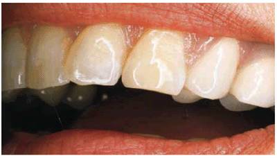
Figure 20-3A: This 30-year-old teacher had worn her left canine flat due to bruxism.
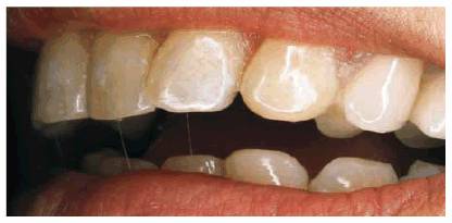
Figure 20-3B: After treatment for the condition, which consisted of appliance therapy, the anteriors were cosmetically contoured rather than adding to the tooth surface. In cases like this, it is important for the patient to wear an appliance afterward to make certain that additional bruxism will no longer destroy enamel.
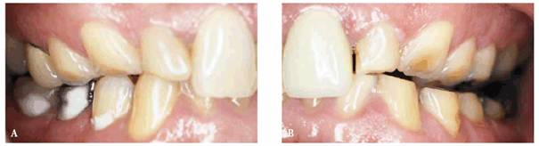
Figure 20-4A and B: This 50-year-old man was completely unaware of his nocturnal bruxism. In fact, during waking hours, it was difficult for him to get his teeth to fit together in the excentric position.
BRUXISM WITH TEMPOROMANDIBULAR JOINT PAIN
Esthetic destruction of the patient's teeth can be sufficient to enable us to
recognize the disease process of temporomandibular joint pain long before the
patient actually begins treatment.1,41,51,60,73,77,83-85
The following case illustrates this position. A young woman had been treated
without success by several physicians for headaches, dizzy spells, and neck,
back, and shoulder pain (Figures 20-5A 20-5B, and 20-5C,and 20-5D and E). Her problems with pain, as well
as with the destruction of her teeth, appeared to be related to her bruxism.
When asked to open her mouth, she deviated sharply to one side. The intraoral
muscles (pterygoid and masseter) and ligaments were in acute spasm and tender
to palpitation. Treatment began with insertion of a maxillary bruxing
appliance. (With bruxism patients, it is important to obtain study casts to
determine if there are wear facets, where they are located, and why they
occurred.) The bruxing appliance was constructed to help stop the incisal wear.
The patient's teeth were then reshaped to improve the smile line. Most pain and
headaches stopped within a period of 3 to 6 weeks after the insertion of this
appliance, together with muscle therapy to the affected areas.
The patient must realize the importance of continuing to wear the bruxing
appliance to maintain the esthetic correction and to avoid reintroducing
temporomandibular joint pain and dysfunction.91
Figure 20-5A: Bruxism was the chief cause of wear for this 31 year old.
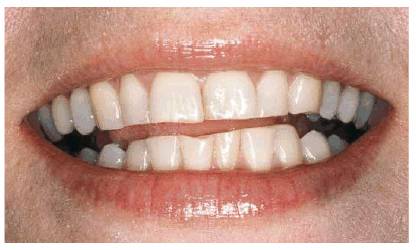
Figure 20-5B: Note how she would unconsciously put the tongue behind the front teeth to hide the space that shows a jagged outline. In addition to poor esthetics, the patient also suffered constant headaches and neck and back discomfort because of further temporomandibular joint dysfunction.
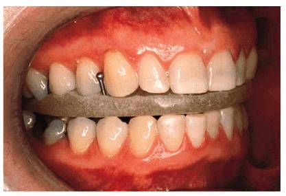
Figure 20-5C: A removable appliance was made to correct the temporomandibular joint dysfunction and prevent teeth from further wearing away. Following 3 months of temporomandibular joint treatment to cure the symptoms and relax the muscles, the patient wore the appliance only at night.
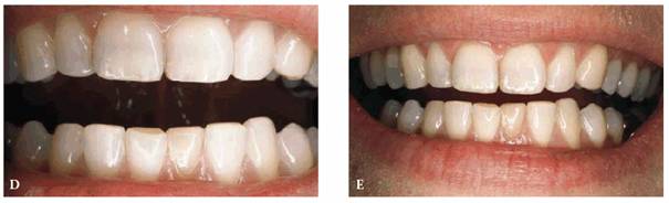
Figure 20-5D and E: After several months of appliance therapy, the square, masculine-looking upper and lower teeth were cosmetically contoured to produce a more feminine and prettier smile.
CHEWING HABITS
The use of smokeless tobacco is another habit that causes excessive wear on the
dentition, in addition to the potential of causing oral cancer.11,55,71
The lingual cusps of maxillary teeth and buccal cusps of mandibular teeth are
the most affected, often worn to the gingival margin. Staining of exposed
dentin is also readily apparent.
Dark brown/black stains on the teeth, marked abrasion of anterior teeth, and
pathologic changes of the oral mucosa are seen in many Eastern countries, such
as India, Malaysia, and Thailand, in those who chew betel nuts for medicinal
and/or psychological purposes67,92,93 (Figure 20-6). Patients who refuse or cannot
stop the habit should be on monthly "cosmetic" cleanings. This type
of prophylaxis is most readily accomplished using a high-powered, mildly
abrasive spray. These patients should be warned that the sharp edges of the
betel nut may cut the gingiva, leading to ulceration. In addition, the betel
nut contains carcinogens that can lead to the development of oral cancer in
habitual users. Coca leaf chewing was shown to cause similar effects on the
dentition in ancient cultures.44
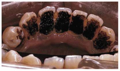
Figure 20-6: This 35-year-old male had the habit of placing betel nuts under his tongue, which helped to produce black stain as shown.
TONGUE HABITS
The tongue is one of the strongest muscles in the human body. The most frequent
signs of tongue thrust are protrusion of the tongue against or between the
anterior teeth and excessive circumoral muscle activity during swallowing.
Although pushing the tongue against teeth, particularly between spaces in the
teeth, does not invariably cause harm, it is certainly a potential cause of
damage. Either maxillary or mandibular teeth can become involved (Figure 20-7). Indentations in the tongue have
been reported to provide an indication of clenching.75 Gellin,
however, feels that anterior tongue positioning does improve with time, and the
continual growth and development of the lower face allow for diminishing
anterior tongue positioning.23 Various studies showed the
relationship between tongue thrust and malocclusion.2,58,59
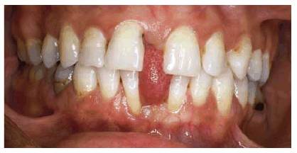
Figure 20-7: This 47-year-old teacher developed a habit of forcing her tongue between her maxillary and mandibular incisors. Note the large space created as a result of years of tongue pressure.
There are
clinicians who have observed a large number of patients with malocclusions who
demonstrate a protrusive tongue tip pattern against or between the anterior
teeth while speaking or swallowing. This group suggests that tongue thrusting
is one of the primary etiologic factors in open bite and incisor protrusion.
There is also much controversy concerning the use of removable appliances in
the treatment of tongue twisting.
The beautiful young woman in Figures 20-8A, and 20-8B would like to advance her modeling
career, but a space between her teeth appears as a black hole in photographs.
Consequently, she poses with her tongue pressed behind the space in her teeth
in an attempt to hide the darkness. This trick helps with the photographic
illusion, but if she presses her tongue too much in a labial direction, the
space can increase over the years. The habit of putting the tongue between the
teeth to disguise a space will almost certainly cause the space to increase
with time (Figure 20-8C). Treatment of spaces between the
teeth is usually best handled through orthodontic care and possibly
myofunctional therapy to promote a more positive resting and swallowing tongue
position. However, some patients may not mind, and may even prefer their teeth
with a space. Although no space should be corrected without approval from the
patient, even these patients need to be referred to an orthodontist for
monitoring and control of any further widening that might have functional
implications.
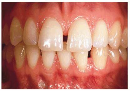
Figure 20-8A: This 23-year-old model had a diastema between the maxillary central incisors.
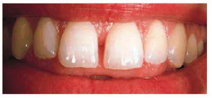
Figure 20-8B: When smiling, she would place her tongue behind the two front teeth to hide the space, which would otherwise show up dark. Note how the tongue creates a pink filler similar to gingival tissue. Many models unconsciously develop similar habits, which can create additional space because of the tongue pressure if done over a long period of time. It is much wiser to either close these spaces orthodontically or compromise with restorative means.
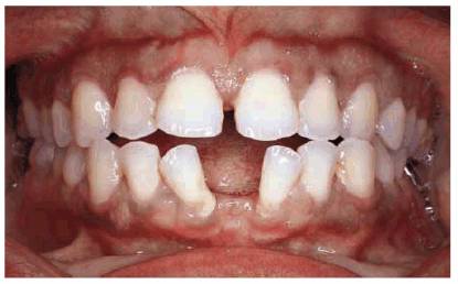
Figure 20-8C: The habit of placing the tongue between the teeth to disguise a space will almost certainly cause the space to increase with time.
For those patients who desire closure of the diastema, referral to an
orthodontist can determine if repositioning of the teeth is appropriate. In
discussing the referral with the patient, make certain that it is understood
that orthodontics does not need to be a matter of metal brackets. One of the
most common solutions to gaps is the construction of a retainer that the
patient wears at night. After the teeth have stabilized, the retainer can be
worn a few nights each week to maintain tooth position and prevent reopening the
space.
An alternative or compromise treatment consists of bonding composite resin to
close the spaces. Crowns and porcelain veneers can also serve this function,
but bonding has the advantage of reversibility. The patient could later elect
to have orthodontic treatment, especially if the spaces continue to widen.
Sometimes, orthodontics may not be the patient's choice, and the following case
illustrates an alternative, restorative means of treatment. The young man shown
in Figures 20-9A 20-9B and C 20-9D and E 20-9F to H was extremely self-conscious about
a space between his central incisors caused by a tongue thrust. He was referred
for orthodontics, both to correct the space and to correct his destructive
habit. However, the appliance needed to correct the spacing between this young
man's teeth gave him what he considered a freakish appearance, and he asked for
an alternative treatment. Treatment was then planned using composite resins,
which, although not permanent, produce immediate results.
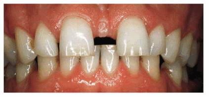
Figure 20-9A: This 32-year-old was self-conscious about a space between his front teeth that was originally caused by tongue thrusting.
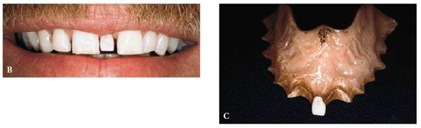
Figure 20-9B and C: He felt that people were noticing his smile, and since he did public speaking, he wanted to improve his appearance. This replacement had been in the mouth for 16 years.
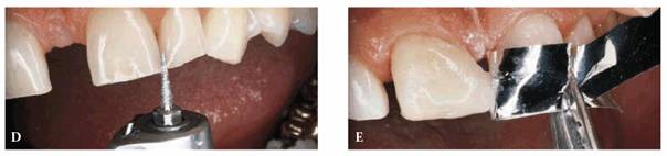
Figure 20-9D and E: The patient was referred for orthodontic consultation but elected to have composite resin bonding as a compromise treatment.
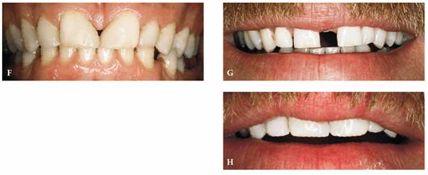
Figure 20-9F to H: Although closing the space created a disproportionate overbuilding of the two central incisors, through judicious carving of the finished bonded restorations, a more proportionate and not unattractive arrangement can be achieved. This consisted of opening the incisal embrasures, as well as creating a greater interincisal distance.
LIP OR CHEEK BITING
The signs of lip or cheek biting are usually telltale marks from the teeth (Figures 20-10A, and 20-10B). Glass and Maize have described
the appearance of oral tissues that have been chewed or bitten over a period of
time.24 This results in the appearance of hard fibrous knots or
masses known as morsicatio buccarum et labiorum. Sometimes, the patient uses
the teeth to suck or knead the altered tissue. If the habit continues over an
extended period of time, it can also cause tooth abnormalities by enlarging any
small diastema or interdental space. The more a patient chews or sucks, the
more pressure is created between the teeth and the wider the space.
Other lip habits such as lip wetting or lip sucking and a swallowing pattern
that includes a hyperactive mentalis muscle can cause damage to developing
orofacial structures in children. Lip wetting is frequently unnoticed by the
average dental practitioner. Clinically, the entire lip looks soft and moist
and does not have a sharply demarcated vermilion border.
Cheek biting is one of the most frequently seen destructive oral habits and can
reflect a circular pattern (Figure 20-10C). Sometimes, loss of part of a
tooth or an entire tooth can initiate cheek biting. The presence of resulting
fibrous tissue may cause the patient to pull the knot of tissue between the
teeth and begin to suck. Diagnosis of cheek biting can be made by examining the
inside border of the cheek for a flickered, sometimes white fibrous ridge
midway between the arches.
Treatment of lip biters and cheek biters consists of several steps. The first
usually involves reshaping the teeth to round, smooth, and polish any sharp
edges. It is also important in working with such patients to find out if there
has been any recent crowning, lengthening, or shortening of the teeth or other
changes that might have induced this habit. If so, they may need to be
modified.
The second part of treatment usually involves creating an appliance to prevent
the patient from biting the lip or cheek (Figures 20-10D, and 20-10E).89 This should be as
thick as feasible and rounded on the labial surface. As a temporary measure, a removable
acrylic interdental spacer or a vacuform matrix can be used to prevent the
patient from biting or sucking (Figures 20-11A 20-11B, and 20-11C
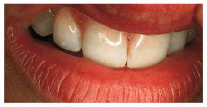
Figure 20-10A: This 29-year-old woman felt that her upper teeth were "growing down more" and irritating her lower lip. She was told to let us know exactly when she closed her lip and felt that it was fitting tightly into the teeth.
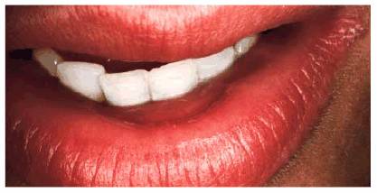
Figure 20-10B: During her next appointment, she stated that she realized that she is both sucking and biting down on her lip at the same time.
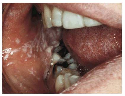
Figure 20-10C: This 50-year-old woman had a habit of biting her cheek. Note the pattern of white fibrous tissue.
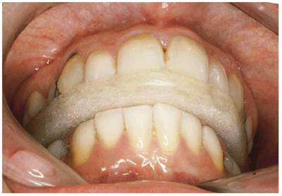
Figure 20-10D: Since her external pterygoid muscles were in spasm, it was also felt that she could be grinding her teeth, so an upper temporomandibular joint appliance was constructed, rounding the labial incisal angle in particular.
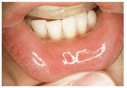
Figure 20-10E: After 5 months of wearing the appliance, at first all of the time and then at night only, the patient's tissue returned to normal. In addition, her muscle spasms disappeared, and all other symptoms subsided.
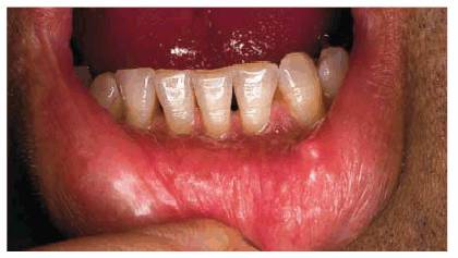
Figure 20-11A: This 50-year-old man had been sucking the lower left lip into an open interdental space between the mandibular left central and lateral. Note the lesion created on the lower left inner border of the lip due to the patient's sucking habit.
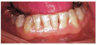
Figure 20-11B: A vacuform matrix was made to close up this area to see if the patient could break the sucking habit. Unfortunately, the only time the patient could eliminate the habit of sucking was when the appliance was being worn.
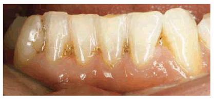
Figure 20-11C: It was decided to bond the mandibular incisors to eliminate space and thereby help to eliminate his sucking habit. Had the patient been able to alleviate the habit with the matrix, it would have been possible to eliminate the need for closure of this space.
One of the added advantages of such devices, which are worn around the clock
during the early phases of treatment, is that they make the patient more aware
of the intensity and frequency of such habits and the circumstances under which
they most often manifest themselves. Many patients are not aware that they bite
or suck their lips or cheeks, especially in relation to stress. Once the
patient stops biting the lip, the use of the appliance can be reduced to
evenings only. Most patients will require between 3 and 6 months to correct the
problem (Figures 20-12A 20-12B 20-12C, and 20-12D).
Finally, any space between the teeth can be corrected via orthodontics or the
application of composite resin bonding or porcelain veneers to close the
diastema (see Figure 20-11C). The use of full crowns would be a
third choice to close the space.
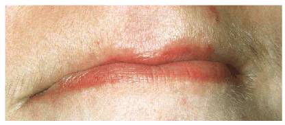
Figure 20-12A: This young lady developed a habit of sucking her upper lip.
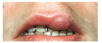
Figure 20-12B: A bulbous lesion was the result of the constant suction action that prompted the patient to seek treatment only for the lip but not the obvious caries. (Reproduced with permission from Goldstein RE. Change your smile. 3rd edn. Carol Stream, IL: Quintessence, 1997:287.)
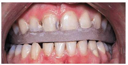
Figure 20-12C: A removable maxillary appliance was made for the patient to wear full time until she completely broke the habit. Note also advanced caries, which needed to be treated.
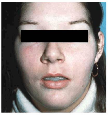
Figure 20-12D: It required only several weeks for the patient to gain back her normal-appearing lip. Then the patient could focus her attention on her other dental treatment.
MOUTH BREATHING
Common in childhood, mouth breathing is the habit of using the mouth instead of
the nose for respiration regardless of whether the nose is obstructed. However,
there are often specific reasons for mouth breathing, such as allergies,
enlarged nasopharyngeal lymphoid tissues, and asthma.6,14,50,87 The
prevailing hypothesis is that prolonged mouth breathing during certain critical
growth periods in childhood causes a sequence of events that results in dental
and skeletal changes. Excessive eruption of the molars is almost always a
constant feature of chronic mouth breathing. This molar eruption causes
clockwise rotation of the mandible during growth, with a resultant increase in lower
facial height. The increased lower facial height is often associated with
retrognathia and anterior open bites. Low tongue posture is seen with mouth
breathing and impedes the lateral expansion and anterior development of the
maxilla.15,18,26-28,30,39,40,42,78,80,88 The dentofacial effects
that develop in children persist into adulthood, and the mouth-breathing and
tongue-thrusting behavior may continue. Barber has found that mouth breathing
can lead to dryness and irritation of the throat, mouth, and lips, as well as
chronic marginal gingivitis.4 Also, it is strongly associated with
both lip-biting and lip-wetting habits. It may be necessary to treat the
mouth-breathing habit prior to or in conjunction with the treatment of the lip
habit.
EATING DISORDERS AND POOR DIETARY
HABITS
Anorexia nervosa and bulimia are psychosomatic eating disorders that have
associated oral symptoms. The exact prevalence of these eating disorders is
unknown. However, they are most frequently seen in young women, ranging from
adolescence into early adulthood. In some studies,21,38,68,76 it has
been estimated that eating disorders affect up to 20% of the women on college
campuses. As our culture continues to emphasize outward appearance, it is
likely that this problem will not resolve soon. Dentists are often the first
health care professionals to recognize the signs of eating disorders,
particularly bulimia. It may well be that the obsession to improve outward
appearances that drives some individuals to develop eating disorders may also
fuel their desire for esthetic dental services. Thus, practices that have a
strong emphasis on esthetic care should be diligent in assessing their
patients, especially young women, for signs of eating disorders.
The bulimic patient ingests large amounts of food, followed by voluntary or
involuntary purging. The purging may occur with the use of high doses of
laxatives or induced vomiting. In those bulimics who purge by vomiting, the pH
of the gastric acid is low enough to initiate dissolution of the enamel.
Further compounding the problem is the frequent vigorous tooth brushing that is
used to rid the mouth of the taste and telltale odor of the vomitus. Brushing
the teeth immediately after they have been exposed to gastric acids will
accelerate the loss of enamel. Numerous studies show that a high percentage of
patients seen with bulimia exhibit lingual erosion of the maxillary anterior
teeth caused by regurgitation of gastric acids (Figures 20-13A 20-13B 20-13C, and 20-13D).9,12,70,72,74 If the
disorder persists, the erosion will eventually affect the occlusal surfaces of
the molars and premolars. If the bulimia is not controlled, the entire
dentition may be destroyed, necessitating complete dental rehabilitation with
full-coverage restorations. Unfortunately, if the habit persists after the
rehabilitation, these patients are likely to develop recurrent caries around
the margins of the crowns.
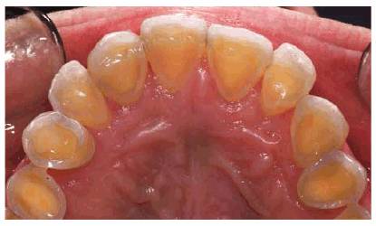
Figure 20-13A: Preoperative upper anterior palatal view of a bulimic patient shows extreme erosion of the lingual and occlusal surfaces. (Courtesy of Dr. Vincent Celenza.)
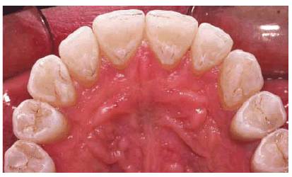
Figure 20-13B: Final restorations in place. (Courtesy of Dr. Vincent Celenza.)
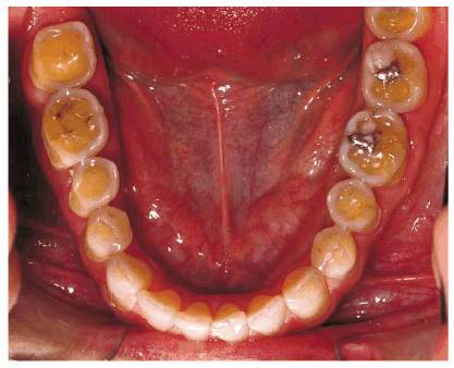
Figure 20-13C: Preoperative occlusal view of the lower arch showing extreme occlusal acid erosion. (Courtesy of Dr. Vincent Celenza.)
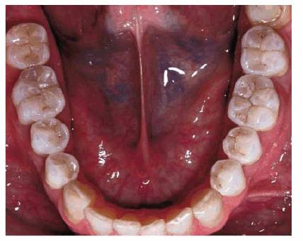
Figure 20-13D: Full arch view, 21/2 years after placement. (Courtesy of Dr. Vincent Celenza.)
Patients with anorexia nervosa pose an entirely different set of problems.
Although bulimia and anorexia are disorders that involve a severely altered
self-perception, individuals with anorexia may tend to fully lose self-esteem
and fall into a state of total oral neglect. In its severest form, patients
with anorexia may present with rampant caries and notably dry mucosa (Figures 20-14A
and B, and 20-14C). They are very prone to the
effects of metabolic imbalance and should be treated cautiously in the dental
office.
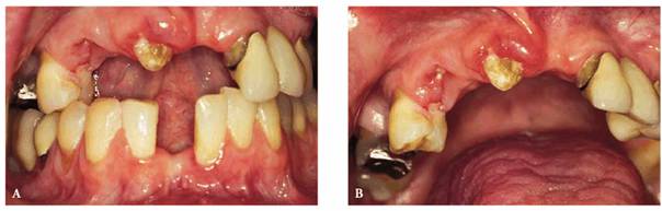
Figure 20-14A and B: This 28-year-old female had been anorexic since age 17. She had unsuccessfully participated in numerous counseling programs, and her self-image continued to be extremely poor. Although she weighed only 92 pounds, she perceived herself to be grossly overweight. She was so focused on her body's appearance that she totally neglected her oral health. Note the loss of teeth and decay.
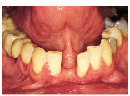
Figure 20-14C: Although not completely visible, this photograph illustrates drying and atrophy of the oral mucosa as seen on the lateral and ventral surfaces of the tongue.
In addition to the intraoral ravages of bulimia and anorexia, outwardly visible
signs of these disorders can be seen. Figure 20-15 shows a 35-year-old female who
suffered from both bulimia and anorexia. Notice the swelling of the parotid
gland, clearly seen at the angle of the mandible. This hypertrophy of the gland
is commonly seen in bulimics. It is caused by repeated vomiting and is present
bilaterally. Also, on close examination of the photograph, a fine, downy facial
hair can be detected. This facial hair is called lanugo and may be found in
anorexics.
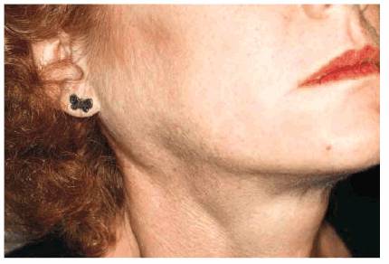
Figure 20-15: Bulimia and anorexia can occur in the same patient. This 35-year-old female initially manifested her eating disorder as bulimia when she was about 15 years old. Over the years, she has continued her purging and while in college began to exhibit behavior characteristic of anorexia. She is now firmly entrenched in both bulimia and anorexia. Her parotid hypertrophy is a manifestation of her bulimia. The lanugo (fine facial hair) is a sign sometimes seen in anorexics.
The dental manifestations of eating disorders can be treated immediately, but
treatment must be limited to emergency, preventive, and/or temporary measures
until the disorder is brought under control. Preventive measures to reduce the
damaging effects of gastric acids can be immediately employed. The first
measure is to have the individual refrain from brushing the teeth after
vomiting. Second, oral rinses to reduce the pH in the mouth can be extremely
helpful. Water can be used to rid the mouth of the acids that are present. If
available, sodium bicarbonate (baking soda) rinses can neutralize the acidity
in the mouth following an episode of vomiting. Various topical fluoride
preparations will aid in minimizing the acidic destruction of the enamel and
dentin. Because of the psychosomatic nature of anorexia and bulimia, it is
imperative for the dentist to approach these patients in a factual,
nonconfrontational, concerned manner that will encourage them to seek proper
medical attention.
People who habitually eat or suck on lemons or drink large amounts of
lemon-flavored water may exhibit acid erosion (Figures 20-16A, and 20-16B). This erosion is seen on the
labial surfaces of the anterior teeth if they suck the fruit or the lingual
surfaces if they chew it. Those who actually eat lemons may present with
lingual erosion that mimics that of bulimia. Excessive consumption of fruits
and drinks with high acid content can cause decalcification of enamel and dissolution
of dental tissues (refer to Chapter 17
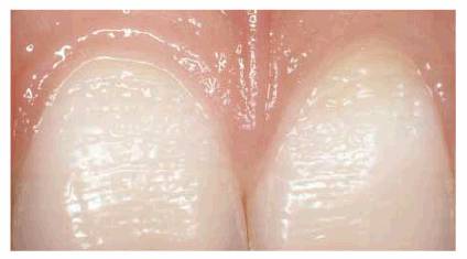
Figure 20-16A: People who have a habit of sucking lemons seldom are aware of the potential for damage to their enamel. This patient was diagnosed early in her habit, so a minimum of damage was done.
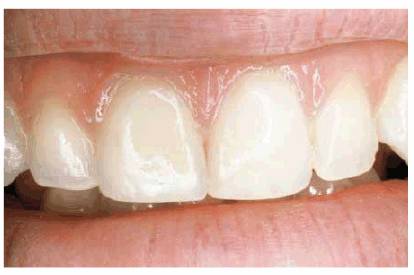
Figure 20-16B: Composite resin bonding was the treatment of choice.
In cases of dental erosion, the teeth first exhibit a diminished luster. As
continual erosion leads to smoothing of enamel pits, the eroded areas
eventually appear smooth and polished. Advancing erosion results in exposed
dentin that wears down rapidly and often exhibits extensive sensitivity.
Early detection of erosive lesions and identification of patients at high risk
for developing erosion are most important. If you detect erosion, it is
essential to ask if the patient has changed his or her eating habits or diet
recently.
Caries is frequently seen in patients who have a habit of sucking hard
citrus-flavored candies with high sugar content (Figure 20-16C). Thus, rebuilding the lost tooth
structure offers only palliative therapy. Dietary changes must be made in these
patients who insist on continuing with this habit pattern. Dental management of
patients with these disorders should be conservative. Bonded composite resin or
glass ionomer materials may reduce sensitivity and prevent the erosion from
progressing. Any extensive dental treatment, such as crown and bridge, should
be postponed until the disorder/habit itself is controlled or stabilized.
Otherwise, dental treatment may not be effective.
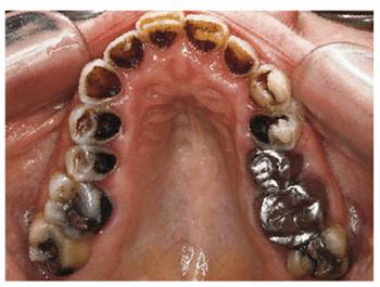
Figure 20-16C: This 63 year old had a habit of sucking one pack of Lifesavers candy daily. In addition, she ingested two tablespoons of vinegar and 500 mg of vitamin C. Her maxillary teeth showed considerable damage due to caries and erosion. Treatment included full-mouth restoration with full crowns on the maxillary teeth.
ALCOHOL AND DRUG ABUSE
Chronic alcoholism is another disorder that has oral implications (Figure 20-17). Case studies describe patients
with a history of chronic alcoholism that have extensive wear of the teeth.29,69,79
All had loss of lingual and incisal surfaces of the maxillary anterior teeth
consistent with regurgitation erosion. This regurgitation results from a
gastritis that is produced by ingestion of excessive amounts of alcohol.
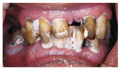
Figure 20-17: Chronic alcoholism is a habit that can produce various intraoral problems. This retired gentleman is a good example of how loss of self-respect can lead to greater oral disease. He quit caring about his appearance and completely gave up oral hygiene as evidenced in the above picture.
Abuse of specific drugs has been shown to have adverse effects on the
dentition. Individuals who regularly use methylenedioxy-methamphetamine
("Ecstasy") may have excessive wear of the teeth.17,66
This occurs through a dual mechanism of decreased salivation and hyperactivity
of the muscles of mastication. In essence, this drug evokes a form of bruxism
that occurs in a dry mouth. Although not reported, other amphetamines may cause
similar conditions. Cocaine has been reported to cause dental erosions because
of its acidic nature.43 Some abusers obtain their high by wetting
the tip of their finger, dipping it into the cocaine, and wiping the drug into
the buccal vestibule or onto the gingival tissues. When the acidic drug comes
in contact with the tooth surface, erosive lesions can develop.
FOREIGN OBJECTS IN THE MOUTH
Another habit that can produce permanent damage to the dentition relates to
placing foreign objects in the mouth. The resultant damage is caused by
abrasion, which is a term used to describe wear or defects in tooth tissues
resulting from contact with a foreign object. The following are some of the
more common types of habits that can cause this damage.
Fingernails
Since the fingernail is an extension of the finger, one may wonder if the common
practice of placing the fingernails between the teeth is a continuation of a
previous thumb- or finger-sucking habit. It may also begin suddenly, well into
adult life, because of a chipped or spaced tooth or some roughness in the mouth
that acts like a magnet for some people, perhaps in an effort to smooth out the
roughness.
The most destructive of the fingernail habits involves the patient's wedging
the fingernail in an interdental area that eventually becomes a space (Figures 20-18A 20-18B 20-18C, and 20-18D). Treatment involves closure of the
space. It is important that the patient be aware that continuation of this
habit can quickly reopen the space. In some cases, it may be necessary to
restrain the individual by constructing a vacuform matrix to cover the entire
arch or an orthodontic retaining appliance (see Figures 20-18C, and 20-18D). The patient should wear this
appliance full time for 6 weeks. Closure of the space during this period,
together with a 3-month retaining period, should be sufficient to break the
habit. You can help ensure that the habit does not recur by cosmetically
contouring the teeth to remove any rough or sharp edges.
Nail biting is also a learned habit that may provide a physical mechanism for
stress relief. Encourage your patients to have short, well-manicured nails.
Rough edges in the nail may cause the patient to smooth the nail unconsciously
by rubbing it in the incisal embrasure. Also, advise the nail biter to carry a
fingernail clipper at all times so that "nervous energy" can be
converted into self-manicuring the nails when a possible urge to bite the nails
exists. Behavioral techniques to reduce stress levels will aid in eliminating
this type of habit.
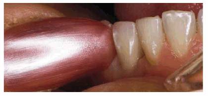
Figure 20-18A: This 30 year old developed a habit of putting her nail between her lower incisors.
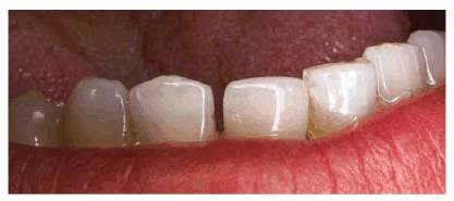
Figure 20-18B: Note the space created between the lateral and central incisors on the lower right side due to fingernail pressure.
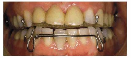
Figure 20-18C: A removable Hawley-type orthodontic appliance was constructed to reposition the lower anteriors. Because of the nature of this appliance, it also helped the patient to break the habit since she could not put the fingernail into the same space.
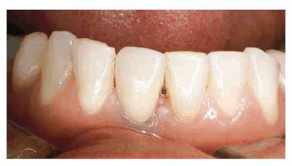
Figure 20-18D: The final result after approximately 6 months of treatment.
Pins Placed between the Teeth
Placing various types of pins, needles, or even bobby pins in one's mouth is
not an uncommon habit, particularly among people who knit and sew. People
suffering tooth deformity from this habit usually hold the pin or needle
between their anterior teeth (Figures 20-19A 20-19B 20-19C and D). Diagnosis can often be made by
checking the patient's protrusive end-to-end relationship to see if a perfect
matching groove is present. It is helpful to ask patients about their work and
hobbies. Taking a thorough habit history, such as the one in Figure 20-20, is helpful. Treatment follows the
pattern of other habits described in this section, that is, appearance is
restored and whatever appliances and means necessary to discourage continuance
of the habit are used. With this problem, it is also useful to tell the patient
to at least vary the location where the pins are held.
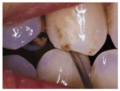
Figure 20-19A: This 39-year-old interior designer had a habit of holding sewing needles and pins between her cuspids.
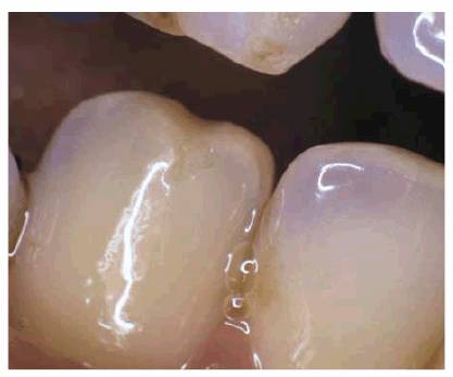
Figure 20-19B: The patient had actually worn a small groove in the biting edge of the teeth that exactly fit the sewing needle she used. In addition to cosmetic contouring or composite resin bonding to add to the worn spot, be sure to have the patient avoid consistently placing any foreign objects between the teeth.
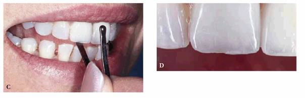
Figure 20-19C and D: This patient wore a small groove in her tooth from constant use of bobby pins.
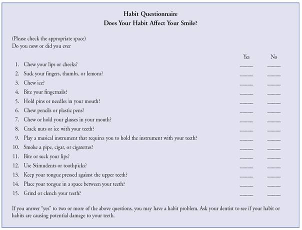
Figure 20-20: The first step in either preventing or stopping a destructive oral habit is to help patients to discover their habits. (Reproduced with permission from Goldstein RE. Change your smile. 3rd edn. Carol Stream, IL: Quintessence, 1997:284.)
Thread Biting
Thread biting may produce notches in the incisal edges of anterior teeth. This
is an occupational habit. Patients who are seamstresses should be warned
against this behavior. Sharp edges of enamel that produce irritation should be
eliminated by careful rounding or restorative treatment (Figures 20-21A, and 20-21B
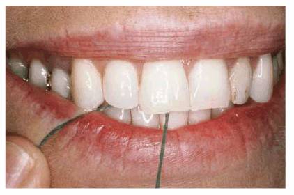
Figure 20-21A: This 55-year-old woman developed a habit of cutting sewing thread with her incisors.
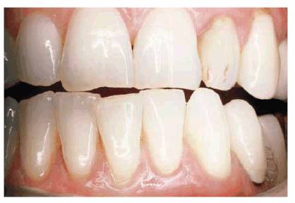
Figure 20-21B: The patient eventually wore a groove in the maxillary right central incisor.
Stimudents or Toothpicks Used as Wedges
Toothpicks or Stimudents can provide an effective means of cleaning tooth
surfaces. If the object is forced between the teeth, however, it can create
unwanted spaces. Patients should be told to use the toothpick or Stimudent like
a soft brush to clean plaque or debris from the smooth surface of the tooth (Figures 20-22A
and B
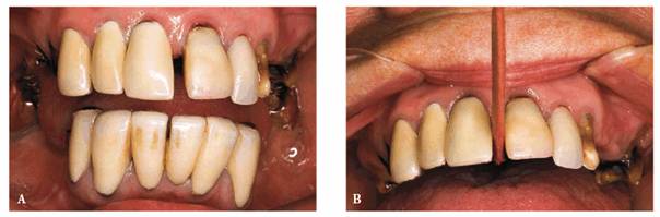
Figure 20-22A and B: These photographs show a patient who constantly placed Stimudents between her front teeth. Although these teeth were originally together with no spaces, the patient quickly separated the teeth to create a space. Her goal was to expand her arch to give more fullness to her face. She had previously been referred for orthodontic treatment but rejected this treatment plan.
Incorrect Use of Dental Floss and
Toothbrush
Abnormal tooth wear may result from improper oral hygiene procedures. Misuse of dental floss may cause abnormal tooth wear. Excessive and strenuous use of dental floss apical to the cementoenamel junction may result in notching of the root surfaces. In addition, tooth abrasion may occur from incorrect use of a toothbrush. Toothbrush abrasion can be extreme, particularly if related to obsessive or compulsive behavior (Figures 20-23A to C
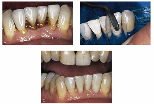
Figure 20-23A to C: This is severe toothbrush abrasion and gingival recession seen in a 37-year-old male with a known obsessive-compulsive disorder. The aggressive brushing of his teeth was one of his extreme habits (see also Chapter 17, Figures 17-9A and B). Composite resin bonding was done to restore the cervical deformities.
Finally, incorrect use of dental floss can lead to abnormal loss of interdental
space. Figures 20-24A 20-24B 20-24C, and 20-24D show a patient who has had bonding
of her anterior teeth and unfortunately developed an incorrect method of
flossing.
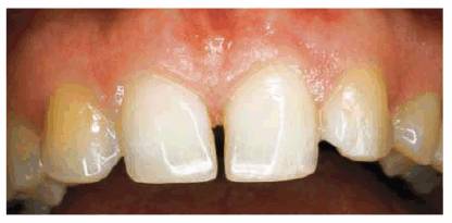
Figure 20-24A: This 25 year old originally presented with a maxillary diastema.
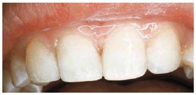
Figure 20-24B: Composite resin bonding was chosen to close the space between the central incisors and convert her canines into laterals. Note that the interdental space looks good between the central incisors.

Figure 20-24C: After questioning the patient about habits, she was requested to demonstrate exactly how she flossed her teeth. What she was doing was pushing the floss into the teeth on one side and then going straight across to the other side without coming back into the contact area; thus, she was "guillotining" her interdental papilla away.
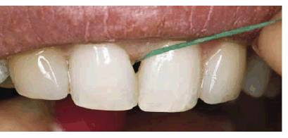
Figure 20-24D: Treatment involved new oral care instructions to prevent further tissue loss and provide an opportunity for the gingiva to regenerate and fill in the interdental space.
Pen/Pencil Chewing
This habit became considerably more destructive when pencils changed from wood
to the newer plastic types (Figures 20-25A 20-25B 20-25C 20-25D, and 20-25E). It is not uncommon to see this
habit in business people who spend a great deal of time at their desks working
figures. Treatment involves wearing an appliance that prevents the patient from
placing the pen or pencil between the teeth.

Figure 20-25A: This 21 year old developed a habit of chewing pencils.
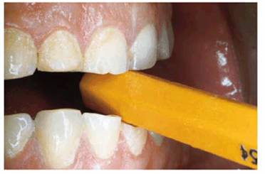
Figure 20-25B: Eventually, wooden pencils were replaced with plastic ones, which he began to turn with his fingers, causing incisal wear.
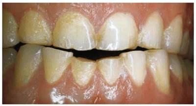
Figure 20-25C: Note the amount of damage caused by the pencil chewing. Composite resin bonding was used as an economic and immediate esthetic replacement for the missing tooth structure. The patient was also given a plastic bite appliance for the maxillary arch that he would wear anytime he felt the need to put a pencil in his mouth.
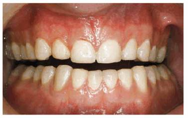
Figure 20-25D: He successfully broke the habit, and the bonding held up for approximately 12 years, when, unfortunately, the patient restarted his previous habit.
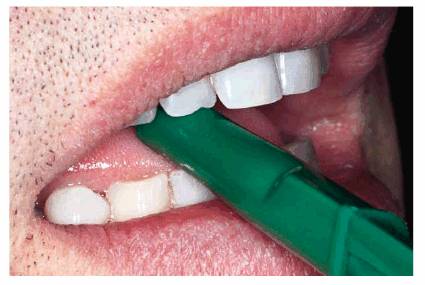
Figure 20-25E: This 46-year-old man had an unconscious habit of not only placing plastic pens in his mouth but also sliding the pen in and out. This wore a groove in the center of the incisal edge.
Pipe Smoking
Any kind of smoking creates its own unesthetic results, staining the teeth and
affecting the health of the oral soft tissues. Pipe smoking has the most
potential for changing tooth relationships. Continually holding a pipe in one
location may cause large notches in several teeth. The problem is that the
patient usually places the pipe stem in the same position, most often the
premolar area. These teeth then become worn or submerged (Figures 20-26A, and 20-26B).
Treatment involves correcting the deformity and helping the patient break the
habit. In this case, it may be easier to teach the patient to change the
pattern of holding the pipe rather than give up the habit altogether. At the
very least, this would make certain that the occlusal forces are distributed
among many teeth. Although the submerged teeth can usually be orthodontically
repositioned, restorative means may provide an easier solution. However, some
tooth structure would probably have to be removed so that the bonded
restoration could be attached using the etched occlusal surface enamel.
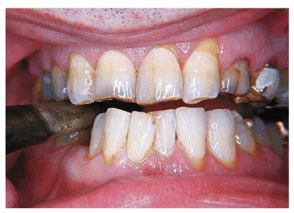
Figure 20-26A: This 56 year old had a long-standing habit of pipe smoking.
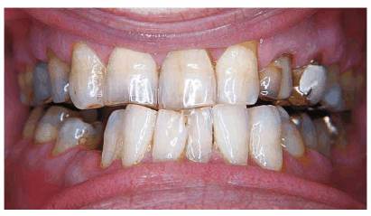
Figure 20-26B: After the pipe was removed, and the patient bit down, the amount of damage caused by the pipe being held in the premolar positions was evident. Particularly note how he favored the right side, which showed a greater amount of abnormal space.
Eyeglasses or Other Objects Placed
between the Teeth
Most persons who consistently place eyeglasses, plastic swizzle sticks, or
other objects in their mouths are not aware of the habit, much less the
resulting functional and esthetic deformity (Figures 20-27A, and 20-27B). Treatment is the same as noted
above.
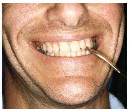
Figure 20-27A: This 31 year old had a habit of holding his eyeglasses with his teeth.
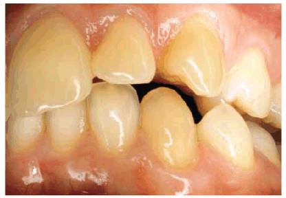
Figure 20-27B: He always held them in the same position, causing the left canine to flare out, thus creating an unattractive and unnecessary space between his front teeth. Patients should always be advised never to hold anything but food between their teeth.
Ice Chewing
Patients seldom realize the damage that chewing ice, a seemingly innocuous
habit, can cause. Chewing ice can fracture teeth and can also produce
microcracks in the enamel. The microcracks themselves are usually not visible
to the eye, but they stain much more readily than normal, especially with
coffee, tea, and soy sauce. If the person chewing ice has defective
restorations, a sliver of ice can also act as a wedge that can split the tooth.
Nut Cracking
Using teeth to open the shell of a nut may be the handiest way to reach the
nutmeat, but it may also be the quickest way to break a tooth. A common
offender is the pistachio nut. In general, the harder the nut is, the more
chance there is of a fractured tooth. The only preventive measure you can take
is to warn patients of the potential damage, particularly if any incisal edge
has been bonded with composite resin.
This chapter could continue for pages with examples of habits of a similar
nature, but the principles of diagnosis and treatment are similar. The first
and most important of these principles is that early diagnosis means
preventive esthetics.
The second principle to remember is that treating the signs of oral habits
is only a temporary measure if the patient continues the habit. Helping
patients break these habits may well be the most difficult part. Important keys
include (1) precise diagnosis of the exact nature of the habit, (2) helping the
patient recognize the habit and the tactful suggestion that the patient learn
through counseling or other methods to better deal with stress and tension, and
(3) correction of damage caused by the habit so that rough edges, open gaps,
tissue changes, or other signs do not contribute to resumption of the habit
that caused them. Appliances to physically prevent the habit may also be useful
in making the patient more aware of the tendency and be a turning point in
breaking the cycle of habit-sign-habit.
The treatment of children's oral habits, particularly digit sucking, is
somewhat easier because there is usually an adoring adult to reinforce changed
behavior. The therapists who participated in a convention of speech
pathologists concerned with such behavior almost uniformly suggested methods
that require commitment on the part of the patient, removal of guilt about the
habit, and a willingness of the parent to give the child some extra attention
as the habit is relinquished. Charts with checkmarks or gold stars are often
used as reinforcement, as are kisses and praises.25
Adults can modify these methods for their own habits:
1. An extremely precise diagnosis of the problem is an essential first step
since many adults may not realize that they have a destructive habit.
2. Once the patient has become aware of the habit, he or she can monitor the
behavior, writing down when, how intensely, and under what circumstances it
occurs. This will reinforce the patient's awareness of the habit, provide some
clues as to what evokes it (such as cheek biting occurring most often in
stressful situations or when tired), and provide many occasions in which not
using the habit can be reinforced. Suggest to your patient that he or she
enlist the assistance of someone else to help identify various aspects of the
habit. A colleague would be a good source if the habit occurs primarily during
working hours. Spouses, relatives, or friends may provide assistance if the
habit takes place during nonworking hours.
3. Some behavioral scientists believe that one habit replaces another.
Certainly, it is easier to create a habit than to break one, so it could be
suggested to patients that they attempt to temporarily replace the destructive
oral habit with a less destructive one such as chewing sugarless gum.
4. Finally, the use of orthodontic devices such as those described in this
chapter will help the patient recognize and break this habit.
An important part of treatment, as well as the record of each new patient,
should be a completed thorough habit questionnaire, such as the one in Figure 20-20. The patient can fill this out
unless he or she is so young that a parent or guardian would be a more
appropriate source of information. This questionnaire should be updated as
regularly as a patient's medical history. Destructive habits can start anytime,
and it is the dentist's responsibility-and opportunity as a professional with
diagnostic ability and an inquisitive nature-to identify these habits and stop
them before more damage is done.
REFERENCES
1. Allen JD, Rivera-Morales WC, Zwemer JD. Occurrence of temporomandibular
disorder symptoms in healthy young adults with and without evidence of bruxism.
Cranio
1990;8:312-8.
2. Andrianopoulos MV, Hanson ML. Tongue-thrust and the stability of overjet
correction. Angle Orthod
1987;57:121-35.
3. Bailey DR. Tension headache and bruxism in the sleep disordered patient. Cranio
1990;8:174-82.
4. Barber TK. Lip habits in preventive orthodontics. J Prev Dent
1978;5:30-6.
5.
6. Bayardo RE, Mejia JJ, Orozco S, Montoya K. Etiology of oral habits. ASDC J Dent
Child 1996;63:350-3.
7. Becker W, Ochsenbein C, Becker BE. Crown lengthening: the
periodontal-restorative connection. Compend Cont
Educ Dent 1998;19:239-40, 242, 244-6.
8. Bishop K, Bell M, Briggs P, Kelleher M. Restoration of a worn dentition
using a double-veneer technique. Br Dent J 1996;180:26-9.
9. Bouquot JE, Seime RJ. Bulimia nervosa: dental perspectives. Pract
Periodont Aesthet Dent 1997;9:655-63.
10. Bowden BD. A longitudinal study of the effects of digit- and dummy-sucking.
Am J Orthod
1966;52: 887-901.
11. Bowles WH, Wilkinson MR, Wagner MJ, Woody RD. Abrasive particles in tobacco
products: a possible factor in dental attrition. J Am Dent
Assoc 1995;126: 327-31.
12. Brown S, Bonifazi DZ. An overview of anorexia and bulimia nervosa, and the
impact of eating disorders on the oral cavity. Compend Cont
Educ Dent 1993;14:1594, 1596-1602, 1604-8.
13.
14. Coccaro PJ, Coccaro PJ Jr. Dental development and the pharyngeal lymphoid
tissue. Otolaryngol
Clin North Am 1987;20:241-57.
15. Cooper BC. Nasorespiratory function and orofacial development. Otolaryngol
Clin North Am 1989;22: 413-41.
16. da Silva AM, Oakley DA, Hemmings KW, et al. Psychosocial factors and tooth
wear with a significant component of attrition. Eur J Prosthodont Restor Dent 1997;5:51-5.
17. Duxbury AJ. Ecstasy-dental implications. Br Dent J
1993;175:38.
18. Ellingsen R, Vandevanter C, Shapiro P, Shapiro G. Temporal variation in
nasal and oral breathing in children. Am J Orthod
Dentofac Orthop 1995;107:411-7.
19. Faulkner KD. Bruxism: a review of the literature.
20. Faulkner KD. Bruxism: a review of the literature. Part II. Aust Dent J 1991;36:355-61.
21. Fombonne E. Increased rates of psychosocial disorders in youth. Eur Arch
Psychiatry Clin Neurosci 1998;248:14-21.
22. Fukuta O, Braham RL, Yokoi K, Kurosu K. Damage to the primary dentition
resulting from thumb and finger (digit) sucking. ASDC J Dent
Child 1996;63:403-7.
23. Gellin ME. Digit sucking and tongue thrusting in children. Dent Clin
North Am 1978;22:603-18.
24. Glass LF, Maize JC. Morsicatio buccarum et labiorum (excessive cheek and
lip biting). Am J
Dermatopathol 1991;13:271-4.
25. Greenly L. Suggestions for combating digit sucking as offered by members at
the 1982 I.A.O.M. Convention. Int J Orofac Myol 1982;8:22-3.
26. Gross AM, Kellum GD, Franz D, et al. A longitudinal evaluation of open mouth posture
and maxillary arch width in children. Angle Orthod
1994;64:419-24.
27. Gross AM, Kellum GD, Michas K, et al. Open-mouth posture and maxillary arch
width in young children: a three-year evaluation. Am J Orthod
Dentofac Orthop 1994;106:635-40.
28. Gross AM, Kellum GD, Morris T, et al. Rhinometry and open-mouth posture in
young children. Am J Orthod Dentofac Orthop 1993;103:526-9.
29. Harris CK, Warnakulasuriya KA, Johnson NW, et al. Oral health in alcohol misusers. Community Dent
Health 1996;13:199-203.
30. Hartgerink DV, Vig PS. Lower anterior face height and lip incompetence do
not predict nasal airway obstruction. Angle Orthod
1989;59:17-23.
31. Haryett RD, Hansen FC, Davidson PO, Sandilands ML. Chronic thumb sucking:
the psychologic effects and the relative effectiveness of various methods of
treatment. Am J Orthod
1967;53:569-85.
32. Hicks RA, Conti PA. Changes in the incidence of nocturnal bruxism in college
students: 1966-1989. Percept Mot
Skills 1989;69:481-2.
33. Hicks RA, Conti PA, Bragg HR. Increases in nocturnal bruxism among college
students implicate stress. Med Hypotheses
1990;33:239-40.
34. Hublin C, Kaprio J, Partinen M, Koskenvuo M. Sleep bruxism based on
self-report in a nationwide twin cohort. J Sleep Res
1998;7:61-7.
35. Infante PF. An epidemiological study of finger habits in preschool children
as related to malocclusion, socioeconomic status, race, sex, and size of
community. J Dent Child
1976;43:33-8.
36. Johnson ED, Larson BE. Thumb-sucking: literature review. ASDC J Dent Child
1993;60:385-91.
37. Kampe T, Edman G, Bader G, et al. Personality traits in a group of subjects with long-standing bruxing
behaviour. J Oral Rehabil
1997;24:588-93.
39. Kellum GD, Gross AM, Walker M, et al. Open mouth posture and cross-sectional nasal
area in young children. Int J Orofac
Myol 1993;19:25-8.
40. Kerr WJ, McWilliam JS, Linder-Aronson S. Mandibular form and position
related to changed mode of breathing-a five-year longitudinal study. Angle Orthod
1989;59:91-6.
41. Kieser JA, Groeneveld HT. Relationship between juvenile bruxing and
craniomandibular dysfunction. J Oral Rehabil
1998;25:662-5.
42. Klein JC. Nasal respiratory function and craniofacial growth. Arch Otolaryngol Head Neck Surg 1986;112:843-9.
43. Krutchkoff DJ, Eisenberg E, O'Brien JE, Ponzillo JJ. Cocaine-induced dental erosions. N Engl J Med
1990;322:408.
44. Langsjoen OM. Dental
effects of diet and coca-leaf chewing on two prehistoric cultures of northern
45. Larsson E. Dummy- and finger-sucking habits with special attention to their
significance for facial growth and occlusion. Swed Dent J
1978;2:23-33.
46. Larsson E. The effect of finger-sucking on the occlusion: a review. Eur J Orthod
1987;9:279-82.
47. Larsson E. Artificial sucking habits: etiology, prevalence and effect on
occlusion. Int J Orofac
Myol 1994;20:10-21.
48. Lavigne GL, Lobbezoo F, Rompe PH, et al. Cigarette smoking as a risk factor
or an exacerbating factor for restless legs syndrome and sleep bruxism. Sleep
1997;20:290-3.
49. Levitas TC. Examine the habit-evaluate the treatment. ASDC J Dent
Child 1970;37:122-3.
50. Linder-Aronson S, Woodside DG, Lundstrom A. Mandibular growth direction
following adenoidectomy. Am J Orthod 1986;89:272-84.
51. Lobbezoo F, Lavigne GJ. Do bruxism and temporomandibular disorders have a
cause-and-effect relationship? J Orofac Pain
1997;11:15-23.
52. Luke LS, Howard L. The effects of thumb sucking on orofacial structures and
speech: a review. Compend Cont Educ Dent 1983;4:575-9.
53. Macaluso GM, Guerra P, Di Giovanni G, et al. Sleep bruxism is a disorder related to periodic
arousals during sleep. J Dent Res
1998;77:565-72.
54.
55. Magnusson T. Is snuff a potential risk factor in occlusal wear? Swed Dent J
1991;15:125-32.
56. Massler M. Oral habits: development and management. J Pedod
1983;7:109-19.
57. McSherry PF. Aetiology and treatment of anterior open bite. J Irish Dent
Assoc 1996;42:20-6.
58. Melsen B, Attina L, Santuari M, Attina A. Relationships between swallowing
pattern, mode of respiration, and development of malocclusion. Angle Orthod
1987;57:113-20.
59. Mikell B. Recognizing tongue related malocclusion. Int J Orthod
1985;23:4-7.
60. Molina OF, dos Santos J Jr, Nelson SJ, Grossman E. Prevalence of modalities
of headaches and bruxism among patients with craniomandibular disorders. Cranio
1997;15:14-25.
61. Ngan P, Fields HW. Open bite: a review of etiology and management. Pediatr Dent
1997;19:91-8.
62. Okeson JP, Phillips BA, Berry DT, et al. Nocturnal bruxing events in subjects
with sleep-disordered breathing and control subjects. J Craniomandib
Disord 1991; 5:258-64.
63. Popovich F, Thompson GW. Thumb and finger-sucking-its relation to
malocclusion. Am J Orthod
1973;63:148-55.
64. Quinn JH. Mandibular exercises to control bruxism and deviation. Cranio
1995;13:30-4.
65. Rayan GM, Turner WT. Hand complications in children from digit sucking. J Hand Surg
1989;14:933-6.
66. Redfearn PJ, Agrawal N, Mair LH. An association between the regular use of
3,4 methylenedioxy-methamphetamine (Ecstasy) and excessive wear of the teeth. Addiction
1998;93:745-8.
67. Reichart PA, Phillipsen HP. Betel chewer's mucosa-a review. J Oral Pathol
Med 1998;27:239-242.
68. Ressler A. "A body to die for": eating disorders and body-image
distortion in women. Int J Fertil
Womens Med 1998;43:133-8.
69. Robb ND, Smith BG. Prevalence of pathological tooth wear in patients with
chronic alcoholism. Br Dent J
1990;169:367-9.
70. Roberts MW, Li SH. Oral findings in anorexia nervosa and bulimia nervosa: a
study of 47 cases. J Am Dent
Assoc 1987;115:407-10.
71. Robertson PB, DeRouen TA, Ernster V, et al. Smokeless tobacco use: how it
affects the performance of major league baseball players. J Am Dent
Assoc 1995;126:1115-21.
72. Ruffs JC,
73. Rugh JD, Harlan J. Nocturnal bruxism and temporomandibular disorders. Adv Neurol 1988;49:329-41.
74. Rytomaa I, Jarvinen V, Kanerva R, Heinonen OP. Bulimia and tooth erosion. Acta Odontol
Scand 1998;56:36-40.
75. Sapiro SM. Tongue indentations as an indicator of clenching. Clin Prev Dent
1992;14:21-4.
76. Schwitzer AM, Bergholtz K, Dore T, Salimi L. Eating disorders among college
women: prevention, education, and treatment responses. J Am Coll
Health 1998;46:199-207.
77. Seligman DA, Pullinger AG. A multiple stepwise logistic regression analysis
of trauma history and 16 other history and dental cofactors in females with
temporomandibular disorders. J Orofac Pain
1996;10:351-61.
78. Shapiro PA. Effects of nasal obstruction on facial development. J Allergy Clin
Immunol 1988;81:967-71.
79. Smith BGN, Robb ND. Dental erosion in patients with chronic alcoholism. J Dent
1989;17:219-21.
80. Stokes N, Della Mattia D. A student research review of the mouthbreathing
habit: discussing measurement methods, manifestations and treatment of the
mouthbreathing habit. Probe
1996;30:212-4.
81. Subtelny JD, Subtelny J. Oral habits-studies in form, function and therapy.
Angle Orthod
1973;43: 349-83.
82.
83. Tsolka P, Walter JD, Wilson RF, Preiskel HW. Occlusal variable, bruxism and
temporomandibular disorders: a clinical and kinesiographic assessment. J Oral Rehabil
1995;22:849-56.
84. Vanderas AP. Relationship between craniomandibular dysfunction and oral
parafunctions in Caucasian children with and without unpleasant life events. J Oral Rehabil
1995;22:289-94.
85. Vanderas AP, Manetas KJ. Relationship between malocclusion and bruxism in
children and adolescents: a review. Pediatr Dent
1995;17:7-12.
86. Van Norman RA. Digit-sucking: a review of the literature, clinical
observations and treatment recommendations. Int J Orofac
Mycol 1997;23:14-34.
87. Venetikidou A. Incidence of malocclusion in asthmatic children. J Clin Pediatr
Dent 1993;17:89-94.
88. Vickers PD. Respiratory obstruction and its role in long face syndrome. Northwest Dent
1998;77:19-22.
89.
90. Weideman CL, Bush DL,
91. Yustin D, Neff P, Rieger MR, Hurst T. Characterization of 86 bruxing
patients with long-term study of their management with occlusal devices and
other forms of therapy. J Orofac Pain 1993;7:54-60.
92. Zain RB, Gupta PC, Warnakulasuriya S, et al. Oral lesions associated with betel quid and
tobacco chewing habits. Oral Dis 1997;3:204-5.
93. Zain RB, Ikeda N, Gupta PC, et al. Oral mucosal lesions associated with
betel quid, areca nut and tobacco chewing habits: consensus from a workshop
held in Kuala Lumpur, Malaysia, November 25-27, 1996. J Oral Pathol
Med 1999;28:1-4.
ADDITIONAL RESOURCES
Aanestad S, Poulsen S. Oral conditions related to use of the lip plug (ndonya)
among the Makonde tribe in
Abreu Tabarini HS. Dental attrition of Mayan Tzutujil children-a study based on
longitudinal materials. Bull Tokyo Med
Dent Univ 1995;42:31-50.
al-Hiyasat AS, Saunders WP, Sharkey SW, Smith GM. The effect of a carbonated
beverage on the wear of human enamel and dental ceramics. J Prosthodont
1998;7:2-12.
al-Hiyasat AS, Saunders WP, Sharkey SW, et al. The abrasive effect of glazed,
unglazed, and polished porcelain on the wear of human enamel, and the influence
of carbonated soft drinks on the rate of wear. Int J
Prosthodont 1997;10:269-82.
al-Hiyasat AS, Saunders WP, Sharkey SW, et al. Investigation of human enamel
wear against four dental ceramics and gold. J Dent
1998;26:487-95.
Altshuler BD. Eating disorder patients. Recognition and intervention. J Dent Hyg
1990;64:119-25.
Attanasio R. Nocturnal bruxism and its clinical management. Dent Clin
North Am 1991;35:245-52.
Attin T, Zirkel C, Hellwig, E. Brushing abrasion of eroded dentin after
application of sodium fluoride solutions. Caries Res 1998;32:344-50.
Bader GG, Kampe T, Tagdae T, et al. Descriptive physiological data on a sleep
bruxism population. Sleep 1997;20:982-90.
Bader JD, McClure F, Scurria MS, et al. Case-control study of non-carious cervical
lesions. Community Dent
Oral Epidemiol 1996;24:286-91.
Bartlett D, Smith B. Clinical investigations of gastro-oesophageal reflux: part
1. Dent Update
1996;23:205-8.
Bauer W, van den Hoven F, Diedrich P. Wear in the upper and lower incisors in
relation to incisal and condylar guidance. J Orofac
Orthop 1997;58:306-19.
Beckett H. Dental abrasion caused by a cobalt-chromium denture base. Eur J
Prosthodont Restor Dent 1995;3: 209-10.
Beckett H, Buxey-Softley G, Gilmour AG, Smith N. Occupational tooth abrasion in
a dental technician: loss of tooth surface resulting from exposure to porcelain
powder-a case report. Quintessence
Int 1995;26:217-20.
Behlfelt K. Enlarged tonsils and the effect of tonsillectomy. Characteristics
of the dentition and facial skeleton. Posture of the head, hyoid bone and
tongue. Mode of breathing. Swed Dent J
Suppl 1990;72:1-35.
Berge M, Johannessen G, Silness J. Relationship between alignment conditions of
teeth in anterior segments and incisal wear. J Oral Rehabil
1996;23:717-21.
Bigenzahn W, Fischman L, Mayrhofer-Krammel U. Myofunctional therapy in patients
with orofacial dysfunctions affecting speech. Folia Phoniatr
1992;44:238-44.
Bishop K, Kelleher M, Briggs P, Joshi R. Wear now? An update on the etiology of
tooth wear. Quintessence
Int 1997;28:305-13.
Blair FM, Thomason JM, Smith DG. The traumatic anterior overbite. Dent Update
1997;24:144-52.
Bohmer CJ, Klinkenberg-Knol EC, Niezen-de Boer MC, et al. Dental erosions and
gastro-oesophageal reflux disease in institutionalized intellectually disabled
individuals. Oral Dis
1997;3:272-5.
Brenchley ML. Is digit sucking of significance? Br Dent J 1992;171:357-62.
Burke FJ, Whitehead SA, McCaughey AD. Contemporary concepts in the pathogenesis
of the Class V non-carious lesion. Dent Update
1995;22:28-32.
Carlson-Mann LD. Recognition and management of occlusal disease from a
hygienist's perspective. Probe
1996;30:196-7.
Cash RC. Bruxism in children: a review of the literature. J Pedodont
1988;12:107-12.
Champagne M. Upper airway compromise (UAC) and the long face syndrome. J Gen Orthod
1991;2:18-25.
Cook DA. Using crayons to educate patients about front-tooth wear patterns. J Am Dent
Assoc 1998;129:1149-50.
de Cuegas JO. Nonsurgical treatment of a skeletal vertical discrepancy with a
significant open bite. Am J Orthod Dentofac Orthop 1997;112:124-31.
Delcanho R. Screening for temporomandibular disorders in dental practice. Aust Dent J
1994;39:222-7.
Djemal S, Darbar
Dodds AP, King D. Gastroesophageal reflux and dental erosion: case report. Pediatr Dent 1997;19:409-12.
Donachie MA, Walls AW. Assessment
of tooth wear in an ageing population. J Dent
1995;23:157-64.
Douglas WH. Form, function and strength in the restored dentition. Ann R Austr
Coll Dent Surg 1996; 13:35-46.
Edwards M, Ashwood RA, Littlewood SJ, et al. A videofluoroscopic comparison of
straw and cup drinking: the potential influence on dental erosion. Br Dent J
1998; 185:244-9.
Ehrlich J, Hochman N, Yaffe A. Contribution of oral habits to dental disorders.
Cranio
1992;10:144-7.
Evans RD. Orthodontics and the creation of localised inter-occlusal space in
cases of anterior tooth wear. Eur J
Prosthodont Restor Dent 1997;5:69-73.
Formicola V. Interproximal grooving: different appearances, different
etiologies. Am J Phys
Anthropol 1991;86:85-7.
Frayer DW. On the etiology of interproximal grooves. Am J Phys
Anthropol 1991;85:299-304.
Friman PC, Schmitt BD. Thumb sucking: pediatrician's guidelines. Clin Pediatr
1989;28:438-40.
Gilmour AG, Beckett HA. The voluntary reflux phenomenon. Br Dent J
1993;175:368-72. Goldstein RE. Esthetics in
dentistry. J Am Dent Assoc
1982;104:301-2.
Goldstein RE. Diagnostic
dilemma: to bond, laminate, or crown? Int J Periodont Restor Dent
1987;87(5):9-30.
Goldstein RE. The difficult patient stress syndrome: Part 1. J Esthet Dent
1993;5:86-7.
Goldstein RE. Change your smile. 3rd edn.
Goldstein RE, Garber DA, Schwartz CG, Goldstein CE. Patient maintenance of
esthetic restorations. J Am Dent
Assoc 1992;123:61-6.
Goldstein RE, Parkins F. Air-abrasive technology: its new role in restorative
dentistry. J Am Dent
Assoc 1994;125:551-7.
Gregory-Head B, Curtis DA. Erosion caused by gastroesophageal reflux:
diagnostic considerations. J Prosthodont
1997;6:278-85.
Grippo JO. Abfractions: a new classification of hard tissue lesions of teeth. J Esthet Dent
1991;3:14-9.
Grippo JO. Noncarious cervical lesions: the decision to ignore or restore. J Esthet Dent
1992;4(Suppl):55-64.
Hacker CH, Wagner WC, Razzoog ME. An in vitro investigation of the wear of
enamel on porcelain and gold in saliva. J Prosthet
Dent 1996;75:14-7.
Harris EF, Butler ML. Patterns of incisor root resorption before and after
orthodontic correction in cases with anterior open bites. Am J Orthod
Dentofac Orthop 1992;101:112-9.
Hazelton LR, Faine MP. Diagnosis and dental management of eating disorder
patients. Int J
Prosthodont 1996;9:65-73.
Hertzberg J, Nakisbendi L, Needleman HL, Pober B. Williams syndrome-oral
presentation of 45 cases. Pediatr Dent
1994;16:262-7.
Heymann HO, Sturdevant JR, Bayne S, et al. Examining tooth flexure effects on
cervical restorations: a two year clinical study. J Am Dent
Assoc 1991;122:41-7.
Hicks RA, Conti P. Nocturnal bruxism and self reports of stress-related
symptoms. Percept Mot Skills
1991; 72:1182.
Hicks RA, Lucero-Gorman K, Bautista J, Hicks GJ. Ethnicity and bruxism. Percept Mot
Skills 1999;88:240-1.
Horsted-Bindslev P, Knudsen J, Baelum V. 3-year clinical evaluation of modified
Gluma adhesive systems in cervical abrasion/erosion lesions. Am J Dent
1996;9:22-6.
Hsu LK. Epidemiology of the eating disorders. Psychiatr Clin
North Am 1996;19:681-700.
Hudson JD, Goldstein GR, Georgescu M. Enamel wear caused by three different
restorative materials. J Prosthet
Dent 1995;74:647-54.
Hugoson A, Ekfeldt A, Koch G, Hallonsten AL. Incisal and occlusal tooth wear in
children and adolescents in a Swedish population. Acta Odontol Scand 1996;54: 263-70.
Ikeda T, Nishigawa K, Kondo K, et al. Criteria for the detection of sleep-associated bruxism in humans. J Orofac Pain
1996;10:270-82.
Imfeld T. Dental erosion. Definition, classification and links. Eur J Oral Sci
1996;104:151-4.
Imfeld T. Prevention of progression of dental erosion by professional and
individual prophylactic measures. Eur J Oral Sci
1996;104:215-20.
Ingleby J, Mackie IC. Case report: an unusual cause of toothwear. Dent Update
1995;22:434-5.
Jagger DC, Harrison A. An in vitro investigation into the wear effects of
selected restorative materials on enamel. J Oral Rehabil
1995;22:275-81.
Jagger DC, Harrison A. An in vitro investigation into the wear effects of
selected restorative materials on dentine. J Oral Rehabil
1995;22:349-54.
Jarvinen VK, Rytomaa II, Heinonen OP. Risk factors in dental erosion. J Dent Res
1991;70:942-7.
Johansson A. A cross-cultural study of occlusal tooth wear. Swed Dent J
Suppl 1992;86:1-59.
Josell SD. Habits affecting dental and maxillofacial growth and development. Dent Clin
North Am 1995;39:851-60.
Josephson CA. Restoration of mandibular incisors with advanced wear. J Dent Assoc S
Afr 1992;47:419-20.
Kaidonis JA, Richards LC, Townsend GC, Tansley GD. Wear of human enamel: a
quantitative in vitro assessment. J Dent Res
1998;77:1983-90.
Kampe T, Hannerz H, Strom P. Ten-year follow-up study of signs and symptoms of
craniomandibular disorders in adults with intact and restored dentitions. J Oral Rehabil
1996;23:416-23.
Kelleher M, Bishop K. The aetiology and clinical appearance of tooth wear. Eur J Prosthodont Restor Dent 1997;5:157-60.
Khan F, Young WG, Daley TJ. Dental erosion and bruxism. A tooth wear analysis from south
east
Kidd EA, Smith BG. Toothwear histories: a sensitive issue. Dent Update
1993;20:174-8.
Kiliaridis S, Johansson A, Haraldson T, et al. Craniofacial morphology,
occlusal traits, and bite force in persons with advanced occlusal tooth wear. Am J Orthodont Dentofac Orthop 1995;107:286-92.
Kleinberg I. Bruxism: aetiology, clinical signs and symptoms. Aust Prosthodont J 1994;8:9-17.
Knight DJ, Leroux BG, Zhu C, et al. A longitudinal study of tooth wear in orthodontically treated patients. Am J Orthod
Dentofacial Orthop 1997;112:194-202.
Kokich VG. Esthetics and vertical tooth position: orthodontic possibilities. Compend Cont Educ Dent 1997; 18:1225-31.
Lambrechts P, van Meerbeek B, Perdigao J, et al. Restorative therapy for
erosive lesions. Eur J Oral Sci 1996;104:229-40.
Lavigne GL, Rompre PH, Montplaisir JY. Sleep bruxism: validity of clinical
research diagnostic criteria in a controlled polysomnographic study. J Dent Res
1996; 75:546-52.
Lee CL, Eakle WS. Possible role of tensile stress in the etiology of cervical
erosive lesions of teeth. J Prosthet
Dent 1984;52:374-80.
Lee CL, Eakle WS. Stress-induced cervical lesions: review of advances in the
past 10 years. J Prosthet Dent 1996;75:487-94.
Leinfelder KF, Yarnell G. Occlusion and restorative materials. Dent Clin North Am 1995;39:355-61.
Leung AK, Robson WL. Thumb sucking. Am Fam
Physician 1991;44:1724-8.
Levine RS. Briefing paper: oral aspects of dummy and digit sucking. Br Dent J
1999;186:108.
Lussi A. Dental erosion clinical diagnosis and case history taking. Eur J Oral Sci
1996;104:191-8.
Lussi A, Portmann P, Burhop B. Erosion on abraded dental hard tissues by acid
lozenges: an in situ study. Clin Oral
Investig 1997;1:191-4.
Mair LH, Stolarski TA, Vowles RW, Lloyd CH. Wear: mechanisms, manifestations
and measurement. Report of a workshop. J Dent
1996;24:141-8.
Marchesan IQ, Krakauer LR. The importance of respiratory activity in
myofunctional therapy. Int J Orofac
Myol 1996;22:23-7.
Maron FS. Enamel erosion resulting from hydrochloride acid tablets. J Am Dent
Assoc 1996;127:781-4.
Matis BA, Cochran M, Carlson T. Longevity of glass-ionomer restorative
materials: results of a 10-year evaluation. Quintessence
Int 1996;27:373-82.
McCoy G. The etiology of gingival erosion. J Oral
Implantol 1982;10:361-2.
McCoy G. On the longevity of teeth. J Oral
Implantol 1983;11:248-67.
McIntyre JM. Erosion. Aust
Prosthodont J 1992; 6:17-25.
Mehler PS, Gray MC, Schulte M. Medical complications of anorexia nervosa. J Womens Health 1997;6:533-41.
Menapace SE, Rinchuse DJ, Zullo T, et al. The dentofacial morphology of bruxers
versus non-bruxers. Angle Orthod 1994;64:43-52.
Mercado MD. The
prevalence and aetiology of craniomandibular disorders among completely
edentulous patients. Aust
Prosthodont J 1993;7:27-9.
Mercado MD, Faulkner KD. The prevalence of craniomandibular disorders in
completely edentulous denture-wearing subjects. J Oral Rehabil
1991;18:231-42.
Metaxas A. Oral habits and malocclusion. A case report. Ont Dent
1996;73:27.
Millward A, Shaw L, Smith AJ. Dental erosion in four-year-old children from
differing socioeconomic backgrounds. ASDC J Dent
Child 1994;61:263-366.
Millward A, Shaw L, Smith AJ, et al. The distribution and severity of tooth
wear and the relationship between erosion and dietary constituents in a group
of children. Int J Paediatr
Dent 1994;4:151-7.
Milosevic A. Tooth wear: an aetiological and diagnostic problem. Eur J Prosthodont Restor Dent 1993;1:173-8.
Milosevic A, Brodie DA, Slade PD. Dental erosion, oral hygiene, and nutrition
in eating disorders. Int J Eat
Disord 1997;21:195-9.
Milosevic A, Dawson LJ. Salivary factors in vomiting bulimics with and without
pathological tooth wear. Caries Res
1996;30:361-6.
Milosevic A, Lennon MA, Fear SC. Risk factors associated with tooth wear in
teenagers: a case control study. Community Dent
Health 1997;14:143-7.
Morley J. The esthetics of anterior tooth aging. Curr Opin
Cosmet Dent 1997;4:35-9.
Moses AJ. Thumb sucking or thumb propping? CDS Rev
1987;80:40-2.
Moss RA, Lombardo TW, Villarosa GA, et al. Oral habits and TMJ dysfunction in facial pain
and non-pain subjects. J Oral Rehabil 1995;22:79-81.
Murray CG, Sanson GD. Thegosis-a critical review. Aust Dent J 1998;43:192-8.
Nel JC, Bester SP, Snyman WD. Bruxism threshold: an explanation for successful treatment of
multifactorial aetiology of bruxism. Aust Prosthodont J 1995;9: 33-7.
Nel JC, Marais JT, van Vuuren PA. Various methods of achieving restoration of tooth structure loss due to
bruxism. J Esthet Dent 1996;8:183-8.
Nemcovsky CE, Artzi Z. Erosion-abrasion lesions revisited. Compend Cont
Educ Dent 1996;17:416-8.
Neo J, Chew CL. Direct tooth-colored materials for noncarious lesions: a 3-year
clinical report. Quintessence Int 1995;27:183-8.
Neo J, Chew CL, Yap A, Sidhu S. Clinical evaluation of tooth-colored materials
in cervical lesions. Am J Dent
1996;9:15-8.
Nunn JH. Prevalence of dental erosion and the implications for oral health. Eur J Oral Sci
1996;104:156-61.
Nunn J, Shaw L, Smith A. Tooth wear-dental erosion. Br Dent J
1996;180:349-52.
Nystrom M, Kononen M, Alaluusua S, et al. Development of horizontal tooth wear in
maxillary anterior teeth from five to 18 years of age. J Dent Res
1990;69:1765-70.
Okeson JP. Occlusion and functional disorders of the masticatory system. Dent Clin
North Am 1995;39:285-300.
Osborne-Smith KL, Burke FJ, Farlane TM, Wilson NH. Effect of restored and
unrestored non-carious cervical lesions on the fracture resistance of
previously restored maxillary premolar teeth. J Dent 1998;26:427-33.
O'Sullivan EA, Curzon ME, Roberts GJ, et al. Gastroesophageal reflux in children and its
relationship to erosion of primary and permanent teeth. Eur J Oral Sci 1998;106:765-9.
Owens BM, Gallien GS. Noncarious dental "abfraction" lesions in an
aging population. Compend Cont Educ Dent 1995;16:552-62.
Paterson AJ, Watson IB.
Case report: prolonged match chewing: an unusual case of tooth wear. Eur J
Prosthodont Restor Dent 1995;3:131-4.
Pavone BW. Bruxism and its effect on the natural teeth. J Prosthet
Dent 1985;53:692-6.
Pierce CJ, Chrisman K, Bennett ME, Close JM. Stress, anticipatory stress, and
psychologic measures related to sleep bruxism. J Orofac Pain
1995;9:51-6.
Pintado MR, Anderson GC, DeLong R, Douglas WH. Variation in tooth wear in young
adults over a two-year period. J Prosthet
Dent 1997;77:313-20.
Powell LV, Johnson GH, Gordon GE. Factors associated with clinical success of
cervical abrasion/erosion restorations. Oper Dent
1995;20:7-13.
Principato JJ. Upper airway obstruction and craniomandibular morphology. Otolaryngol
Head Neck Surg 1991;104:881-90.
Ramp MH, Suzuki S, Cox CF, et al. Evaluation of wear: enamel opposing three
ceramic materials and a gold alloy. J Prosthet
Dent 1997;77:523-30.
Ribeiro RA, Romano AR, Birman EG, Mayer MP. Oral manifestations of Rett
syndrome: a study of 17 cases. Pediatr Dent
1997;19:349-52.
Ritchard A, Welsh AH, Donnelly C. The association between occlusion and
attrition. Aust Orthod J
1992;12:138-42.
Rivera-Morales WC, McCall WD Jr. Reliability of a portable electromyographic unit
to measure bruxism. J Prosthet Dent
1995;73:184-9.
Robb ND, Cruwys E, Smith BG. Regurgitation erosion as a possible cause of tooth
wear in ancient British populations. Arch Oral Biol
1991;36:595-602.
Rogers GM, Poore MH, Ferko BL, et al. In vitro effects of an acidic by-product
feed on bovine teeth. Am J Vet Res
1997;58:498-503.
Schmidt U, Treasure J. Eating disorders and the dental practitioner. Eur J Prosthodont Restor Dent 1997;5: 161-7.
Schneider PE. Oral habits-harmful and helpful. Update Pediatr Dent 1991;4:1-4, 6-8.
Schwartz JH, Brauer J, Gordon-Larsen P. Brief communication: Tigaran (Point
Hope, Alaska) tooth drilling. Am J Phys
Anthropol 1995;97:77-82.
Seligman DA, Pullinger AG. The degree to which dental attrition in modern
society is a function of age and of canine contact. J Orofac Pain
1995;9:266-75.
Seow WK. Clinical diagnosis of enamel defects: pitfalls and practical
guidelines. Int Dent J
1997;47:173-82.
Sherfudin H, Abdullah A, Shaik H, Johansson A. Some aspects of dental health in
young adult Indian vegetarians. A pilot study. Acta Odontol
Scand 1996;54:44-8.
Silness J, Berge M, Johannessen G. A 2-year follow-up study of incisal tooth
wear in dental students. Acta Odontol
Scand 1995;53:331-3.
Silness J, Berge M, Johannessen G. Longitudinal study of incisal tooth wear in
children and adolescents. Eur J Oral Sci
1995;103:90-4.
Silness J, Berge M, Johannessen G. Re-examination of incisal tooth wear in
children and adolescents. J Oral Rehabil
1997;24:405-9.
Smith BG, Bartlett DW, Robb ND. The prevalence, etiology and management of
tooth wear in the United Kingdom. J Prosthet
Dent 1997;78:367-72.
Smith BG, Robb ND. The prevalence of toothwear in 1007 dental patients. J Oral Rehabil
1996;23:232-9.
Sognnaes RF, Wolcott RB, Xhonga FA. Dental erosion I. Erosion-like patterns
occurring in association with other dental conditions. J Am Dent
Assoc 1972;84:571-6.
Speer JA. Bulimia: full stomach, empty lives. Dent Assist
1991;60:28-30.
Spranger H. Investigation into the genesis of angular lesions at the cervical
region of teeth. Quintessence Int 1995;26:183-8.
Steiner H, Lock J. Anorexia nervosa and bulimia nervosa in children and
adolescents: a review of the past 10 years. J Am Acad
Child Adolesc Psychiatry 1998;37:352-9.
Stewart B. Restoration of the severely worn dentition using a systematized
approach for a predictable prognosis. Int J
Periodont Restor Dent 1998;18:46-57.
Suzuki S, Suzuki SH, Cox CF. Evaluating the antagonistic wear of restorative
materials when placed against human enamel. J Am Dent
Assoc 1996;127:74-80.
Taylor G, Taylor S, Abrams R, Mueller W. Dental erosion associated with
asymptomatic gastroesophageal reflux. ASDC J Dent
Child 1992;59:182-5.
Teaford MF, Lytle JD. Brief communication: diet-induced changes in the rates of
human tooth microwear: a case study involving stone-ground maize. Am J Phys
Anthropol 1996;100:143-7.
Teo C, Young WG, Daley TJ, Sauer H. Prior fluoridation in childhood affects
dental caries and tooth wear in a south east Queensland population. Aust Dent J
1997;42:92-102.
Thompson BA, Blount BW, Krumholz TS. Treatment approaches to bruxism. Am Fam
Physician 1994;49: 1617-22.
Timms DJ, Trenouth MJ. A quantified comparison of craniofacial form with nasal
respiratory function. Am J Orthod
Dentofac Orthop 1988;94:216-21.
Touyz LZ. The acidity (pH) and buffering capacity of Canadian fruit juice and
dental implications. J Can Dent Assoc 1994;60:448-54.
Turp JC, Gobetti JP. The cracked tooth syndrome: an elusive diagnosis. J Am Dent
Assoc 1996;127:1502-7.
Tyas MJ. The Class V lesion - aetilogy and restoration. Aust Dent J
1995;40:167-70.
Ung N, Koenig J, Shapiro PA, et al. A quantitative assessment of respiratory patterns and their effects on
dentofacial development. Am J Orthod
Dentofac Orthop 1990;98:523-32.
Villa G, Giacobini G. Subvertical grooves of interproximal facets in Neandertal
posterior teeth. Am J Phys
Anthropol 1995;96:51-62.
Waterman ET, Koltai PJ, Downey JC, Cacace AT. Swallowing disorders in a population
of children with cerebral palsy. Int J Pediatr
Otorhinolaryngol 1992;24:63-71.
West NX, Maxwell A, Hughes JA, et al. A method to measure clinical erosion: the
effect of orange juice consumption on erosion of enamel. J Dent
1998;26:329-35.
Westergaard J, Moe D, Pallesen U, Holmen L. Exaggerated abrasion/erosion of
human dental enamel surfaces: a case report. Scand J Dent
Res 1993;101:265-9.
Woodside DG, Linder-Aronson S, Lundstrom A, McWilliam J. Mandibular and
maxillary growth after changed mode of breathing. Am J Orthod
Dentofac Orthop 1991;100:1-18.
Yaacob HB, Park AW. Dental abrasion pattern in a selected group of Malaysians. J Nihon Univ Sch Dent 1990;32:175-80.
Yamaguchi H, Tanaka Y, Sueishi K, et al. Changes in oral functions and muscular behavior
due to surgical orthodontic treatment. Bull Tokyo Dent Coll 1994;35:41-9.
Young DV, Rinchuse DJ, Pierce CJ, Zullo T. The craniofacial morphology of
bruxers versus nonbruxers. Angle Orthod 1999;69:14-8.
|
