ALTE DOCUMENTE
|
||||||||||
ESTHETIC REMOVABLE PARTIAL DENTURES - Roman M. Cibirka,
INTRODUCTION
The patient who has lost a number of teeth has several treatment alternatives.
The patient may remain partially edentulous until esthetics or function is
compromised, or treatment in the form of a fixed partial denture (FPD),
removable partial denture (RPD), or implant(s) may be pursued. Orthodontics may
be indicated for partially edentulous regions of limited size or to enhance the
prognosis of the rehabilitation through other modalities.
The highly esthetic demands of contemporary dental patients compel dental
practitioners to satisfy their requests. Removable partial dentures designed
without prudence and skillfulness might result in functional or esthetic
insufficiency. Esthetic deficiencies may be shrouded by functional criticisms.
Patients may present with frequent functional complaints of unaccountable pain
or inability to chew when, in fact, they are discontented with the appearance.
Unesthetic RPDs can be avoided with appropriate diagnosis and design using
conventional clasping or attachment-aided prostheses.
CLASSIFICATION OVERVIEW
Universal classification systems for t 15515c216p he partially edentulous arch have been
devised to enhance communication and aid in design. Although numerous
classification systems exist, the most widely accepted is that proposed by
Kennedy21 and further modified by Applegate.1 There are
four classes in the Kennedy classification system ( ). The Kennedy Class I consists of bilateral
edentulous areas located posterior to the remaining natural teeth and is the
most common of the partially edentulous situations.12 The Kennedy
Class II has a unilateral edentulous area located posterior to the remaining
natural teeth. The Kennedy Class III consists of a unilateral edentulous area
with natural teeth remaining both anterior and posterior to it. The rarest
class of the Kennedy classification is the Kennedy Class IV, which is a single,
bilateral (crossing the midline), edentulous area located anterior to the
remaining natural teeth. Edentulous areas other than those determining the
classification are termed modification spaces.
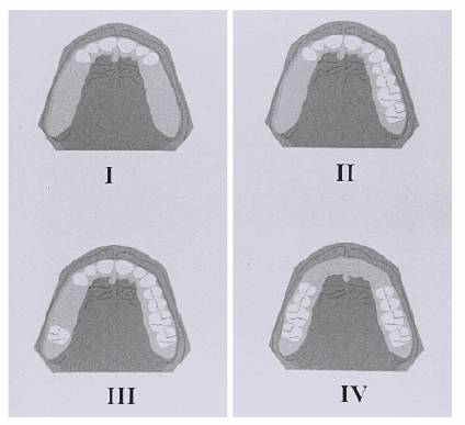
Figure 22-1: Kennedy classification. Kennedy Class I, bilateral distal extension; Kennedy Class II, unilateral distal extension; Kennedy Class III, unilateral edentulous area bounded by natural teeth; and Class IV, single bilateral (crossing the midline) area located anterior to the remaining natural teeth.
PRINCIPLES OF DESIGN
The prudent treatment plan embraces a comprehensive analysis of the patient's
dentition and supportive soft tissues. The health and distribution of the teeth
will influence partial denture component selection and the anticipated
esthetics. Likewise, the quality of the supportive soft tissues dictates the
measure of force transferred to the abutments and guides the component
selection for the tooth-tissue-supported RPD. The greater the tissue support
required, the more likely it is that the forces imparted to the abutment teeth
will increase. The most destructive force is that of torque in the distal
extension design. Minimization of torque should be considered of paramount
importance in the design of the RPD.
Therefore, RPD design should be based on the available support. Kennedy Class I,
II, and large IV RPDs are considered tooth tissue supported. In general,
flexible direct retainer assemblies, mesio-occlusal rests on posterior distal
extension abutments, and indirect retainers to limit rotation are indicated for
tooth-tissue-supported RPDs.4 Kennedy Class III and small IV are
considered tooth-supported RPDs. In these situations, no additional support
from the tissue is generally needed. For these designs, clasp assemblies may be
more rigid, and indirect retainers are usually not indicated.
Examination of the patient requires clinical and radiographic diagnosis of the
teeth and soft tissues for judgment of the support available for the partial
denture. Radiographic interpretation should include (1) periodontal stature,
(2) responses of the teeth to previous stress, (3) vitality of the remaining
teeth, and (4) pathosis. The quantity or height and quality of bone support
often predict the prognosis of an abutment tooth or may influence the design of
a partial denture component. Proper diagnosis necessitates high-quality
radiographs, devoid of angulation errors or processing blemishes. Vertical bone
heights will provide a measure of clinical crown:root ratios. A clinical
crown:root ratio greater than 1:1 should be considered an endangered abutment
with a poor prognosis for RPD support. Stress-breaking direct retainers and
contingency planning should be included in the design of partial dentures to
use an abutment with marginal support.
Bone indices have been described;32 however, they may be difficult
to discern on certain radiographs. A 25% error in actual bone calcification
levels may be found with normal radiographs. Optimum bone qualities are
expressed as normal-sized interdental trabecular spaces that tend to decrease
in size slightly near the coronal portion of the root. Normal bone responds
favorably to stresses within clinical limits. Favorable reaction to stresses
from an existing partial denture may be considered indicative of a future
reaction to stress. Teeth that have experienced previous heavy stress from RPD
support or in conjunction with abnormal occlusal forces and demonstrate normal
to slightly condensed trabeculation, a dense lamina dura, and a heavy cortical
layer are designated as having a positive bone index or factor. Abnormal
stresses will be evidenced as a reduction in the size of the trabeculae being
most pronounced adjacent to the lamina dura. The reduced trabeculae size may be
termed bone condensation and should be indicative of aberrant forces that may
lead to bone loss if the patient becomes less resistant. A compaction of
trabecular spaces and significant alterations to the cortical layer or lamina
dura may be considered a negative bone index or bone factor.
Lamina dura is considered a radiographic measure of abutment tooth health. The
structure is hard cortical bone lining the sockets of the teeth with a primary
function of withstanding mechanical strain. The lamina dura should be intact
and cross interdental spaces to adjacent teeth as a fine, radiopaque white line.
The supportive elements will generally respond to build support where needed
and predict the degree of future response. Mechanical insults from poorly
designed RPDs may overload the remodeling capacity of the body, resulting in
tissue destruction. Bone is approximately 30% organic and stores little
protein; therefore, any alterations in body health will be reflected in the
ability to maintain support. Systemic diseases that alter the reparative
capacity of the body should be strongly considered with partial denture design.
The patient's future health status and manifestations of aging should be
considered in the selection of abutment teeth for loading.
USE OF A SURVEYOR
The dental surveyor is a fundamental instrument for RPD design and treatment
planning. Additionally, the dental surveyor is indispensable for the laboratory
technician to construct a partial denture and fabricate supportive elements
such as surveyed, telescopic, or attachment restorations.
The surveyor may be used for diagnostic cast analysis, contouring abutment
tooth restorations, placement of attachment retainers, milling internal rests,
and reciprocal elements. Survey objectives include (1) determination of an
acceptable path of insertion to eliminate interference with placement or removal,
either hard or soft tissues; (2) identification of proximal tooth surfaces to
be made parallel to act as guiding planes for placement and removal; (3)
location and measurement areas of teeth for undercut and suitable esthetic
clasp placement; (4) delineation of heights of contour; and (5) recording of
cast position, or tripod, for future reference.32,42
An esthetic determinant of the survey is establishing one path of placement to
minimize the retentive element and acrylic resin or denture base display.
Retentive areas may influence the placement of retentive elements, so areas of
retention should be selected to enhance the esthetic value of the RPD. When an
anterior modification space is present, a path of placement should be selected
to minimize excessive modification of adjacent abutment teeth and eliminate
placement interferences. Anterior tissue undercuts may dictate a posteriorly
directed path of placement to avoid excessive need for tissue blockout and
inherent lip fullness from the overcontoured denture base flange. Restoration
of highly esthetic anterior regions should be accomplished through fixed
prosthodontics whenever possible or when the path of placement required for
accomplishment of esthetics might limit the functional efficacy of the partial
denture.
BIOMECHANICS
The design of an RPD must value the mechanics and the biologic considerations.
Maxwell stated that "Common observation clearly indicates that the ability
of things to tolerate force is largely dependent upon the magnitude or
intensity of the force."32 The structures supporting a partial
denture, teeth, and residual ridges are "living things" subjected to
forces. The attributes, frequency, and magnitude of the force will foretell the
success or failure of the RPD and remaining dentition.
Forces applied to an RPD are generally classified into three cranial planes:
vertical, sagittal, and coronal. However, it should be recognized that
functional forces are a summation of individual vector forces in the three
cranial planes. Hence, the actual force encountered by an abutment may be the
result of two differing planar vector forces of varied intensity. Knowledge of
the functional movements patients generate should be considered in the
selection of abutment teeth, retainers, and partial denture design. Widely
distributed abutment teeth with poor periodontal support in a patient with a
parafunctional bruxism habit whose native diet includes nuts will obligate the
dentist to develop a different design than for a patient with sound periodontal
support and few other potentially damaging functional considerations.
A lever is a rigid rod supported somewhere between its two ends at a point,
termed a fulcrum, which allows movement around that point.32 The
lever system allows magnification of force applied at one end of the rod
proportional to the length of the rod from the fulcrum. Consequently, a small
magnitude of force remote to the fulcrum will amplify to potentially
destructive levels, depending on the design of the prosthesis. This is most
apparent in distal extension designs where the length of the lever arm predicts
the degree of force applied to the abutment teeth. Likewise, the dissimilar
characteristics of support from the teeth and soft tissues yield rotation in
three cranial planes.
The tooth:tissue dissimilarity of support is a preeminent concern in distal
extension and Class I, II, or large IV partial denture designs. Class I, II, or
large IV partial dentures derive a great deal of their support from the
residual ridges and a limited amount from the abutment teeth. These types of
RPDs generate the most potentially destructive lever forces. The fulcrum is
generally established through a line connecting the most distal abument teeth
or the rests on those teeth. The Class III or small IV partial denture design
is generally tooth supported with the fulcrum positioned between the abutment
teeth bordering the edentulous space.
The residual ridge has a fibrous connective tissue covering the bone and
underlying the mucosa. The thickness of subepithelial tissue will define the
displaceability of the tissue overlying the residual bone. The displaceability
and the amount of keratinized mucosa overlying the residual ridge will
distinguish the amount of support anticipated from the edentulous regions. The
periodontal ligament is comprised of collagenous fibers, blood vessels, and
interstitial fluid to act as a shock absorber for the dentition. This ligament
or membrane may vary in composition or thickness depending on the amount of
force applied to the tooth. However, the compressibility of the residual ridge
tissues and tooth ligament is not comparable. In fact, a tissue:tooth ratio of
approximately 13:1 exists in healthy tissues.32 This phenomenon
requires careful deliberation when designing and constructing a distal
extension partial denture.
Occlusion is of primary interest in the distal extension prosthesis.
Accentuated occlusal forces or aberrant, parafunctional occlusal forces on the
most remote portion of the distal extension base will impart a greater degree
of leverage force to the supportive elements. Formation of a precise occlusal
scheme will ensure harmonious function and enhance the prognosis of the
abutment teeth.
Tooth morphology should be considered when evaluating potential abutment teeth.
Clinical crown contours and occlusion will often direct retainer, major and
minor connector selection, and rest seat placement.18,35 Root
anatomy is frequently overlooked as a critical component of the supportive
element for a removable prosthesis. In general, single-rooted teeth are less
favorable abutments than multirooted abutments. Divergent roots render more
support than fused roots. Circular roots offer the least resistance to
rotational forces than do oblong root contours. For this reason, premolars,
particularly mandibular premolars, are poor choices to serve as solitary
abutments for distal extension RPDs. Ideally, an FPD should be provided from
the second premolar to the canine to avoid using the second premolar as a
solitary abutment. Periodontally weakened roots provide disproportionately less
surface area for anchorage owing to their conical shape.
PROBLEM SITUATIONS
Perhaps the most difficult situation is the distal extension RPD. This is
complicated when the missing teeth are located unilaterally since functional
requirements make it more difficult to esthetically mask the abutment
attachments. However, if the entire arch is to be restored, then the situation
becomes amenable to either an overdenture or precision attachment. If this is
not the case, then the determination of the lower lip when smiling will help
determine the type of attachment or clasp assembly to use.
SPECIFIC CLASP TYPES AND ESTHETIC CONSIDERATIONS
The use of conventional clasping in esthetic regions of the mouth can present
difficulties with patient acceptance. Proper surveying and mouth preparation
may circumvent complications. Clasps may approach undercuts from a suprabulge
or infrabulge region. Proper abutment tooth selection for clasps and placement
of the clasps far enough into the infrabulge or distal region will maximize the
esthetic benefit. Ideally, suprabulge clasps should be placed in the middle
one-third of the tooth in the region of the proximal plate. The retentive tip
should be located in the gingival one-third but not encroach on the free
gingival margin (Figures 22-2A
and B).
Placing the suprabulge clasp in this manner will improve the esthetic result
and diminish the torquing forces applied to the tooth by the clasp. Infrabulge
clasps will generally provide more enhanced esthetics, although they may have
limitations to their use owing to anatomic considerations. The height of the
vestibule, position of frena and soft tissue, or bony prominences may limit
their application or necessitate preprosthetic surgery.
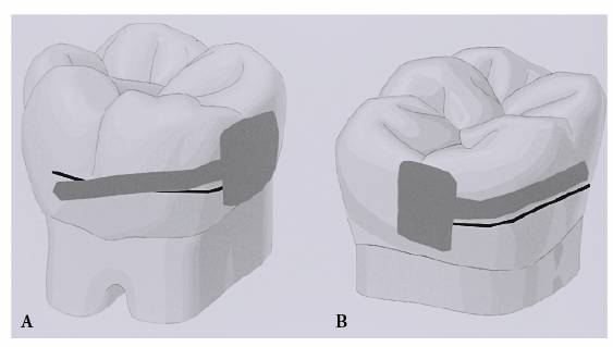
Figure 22-2A and B: Proper placement of the retentive and reciprocal arms. (A) The retentive arm exits the abutment tooth in the middle one-third and terminates in the gingival one-third; only the retentive tip (terminal one-third) is placed below the height of contour. (B) The reciprocal arm exits the abutment tooth in the middle one-third and remains completely above the height of contour.
Circumferential Clasp
Owing to its rigidity, this suprabulge clasp is generally reserved for
tooth-supported abutments in posterior regions of the mouth. It is a cast clasp
of either a round or half-round configuration, both of which provide little
flexibility. When serving as a retentive element, the clasp should only engage
a 0.025-mm undercut to avoid excessive torquing of the tooth. This clasp may
also serve as a bracing or reciprocal element and is positioned above the
height of contour. Due to the relative size (thickness and diameter) of this
clasp, use of the clasp above the height of contour for reciprocation should be
limited in esthetic regions of the mouth. In situations where increased flexibility
is necessary, but there is no place to remote solder a wrought wire clasp, such
as the tooth-supported side of a Kennedy Class II arch, a cast round clasp may
be used. A 20-gauge cast round clasp has been shown to have the same
flexibility as a 19-gauge wrought wire clasp.16
I-, Y-, T-, or Modified T-Bar Clasp
The infrabulge approach of this clasp optimizes esthetics for patients with
reasonably high lip lines or in situations where clasping of maxillary first or
second premolars is indicated (Figure 22-3). It is generally cast as part of
the framework and should exit the meshwork approximately one tooth distal to
the abutment tooth. This allows for optimal tooth positioning without excessive
grinding of the replacement tooth, which would reduce the cosmetic value of the
denture tooth. In Figure 22-4, correct positioning of the
approach arm of the I-bar allows the clasp to traverse from the framework
through the interproximal embrasure region of the first and second replacement
tooth. This will minimize the need to shorten the most anterior denture tooth
to allow for the clasp to traverse from the framework more anteriorly.
Figure 22-3: The use of the infrabulge bar (I-bar) clasp optimizes esthetics, particularly in the maxillary arch.
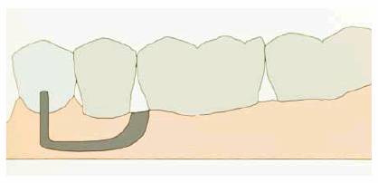
Figure 22-4: The approach arm of the I-bar is placed approximately one tooth distal to the abutment tooth. It exits the meshwork in the interdental area between the replacement teeth to minimize grinding of the replacement teeth.
The T- or Y-bar configuration achieves undercut engagement of 0.25 mm on either
the mesial or distal surfaces of the tooth. A common error is to place both
tips of the T- or Y-bar clasp into an undercut (Figures 22-5A
and B). The
esthetic value may be diminished if the anterior arm of the T- or Y- bar
remains while using a distal undercut. Removal of the anterior arm should be
considered, and a modified T-bar clasp should be selected (Figures 22-6A
and B). A
functional advantage of the modified T-bar is elimination of the mesial arm,
limiting mesial undercut engagement of the clasp during a seating movement of
the denture base toward the residual ridge. This will reduce the torque and
distal tipping of the tooth. As a general rule, clasps should disengage during
denture base movements toward the residual ridge and become active only on
dislodging movements away from the residual ridge. If the height of contour is
located high on the tooth, this clasp design should not be used because of the
space created under the approach arm.
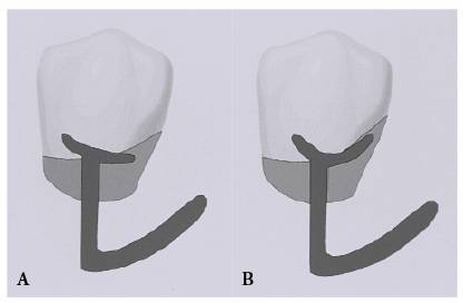
Figure 22-5A and B: Only one tip of the T- or Y-bar clasp should be placed in the retentive undercut. The other tip provides support only.
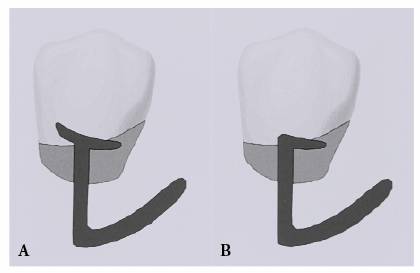
Figure 22-6A and B: The anterior tip of the T-bar clasp may be eliminated, producing the modified T-bar clasp.
Rest-Proximal Plate-I-Bar Clasp
The rest-proximal plate-I-bar (RPI) clasp, described by Kratochvil24,25
and later modified by Krol,26,29 consists of the following components:
(1) mesio-occlusal rest, (2) proximal plate, and (3) I-bar clasp. The retentive
tip of the I-bar should engage a 0.25-mm midfacial undercut (Figure 22-7). As for the T- or Y-bar clasps,
the approach arm should traverse from the meshwork approximately one tooth
distal from the abutment tooth. Esthetically, the RPI clasp fulfills all
requirements of a conventional clasp yet demonstrates minimal tooth coverage,
relatively limited metal display, and an infrabulge approach. The
mesio-occlusal rest stabilizes the tooth and resists distal tipping. The design
is indicated for distal extension situations and allows for disengagement of
the clasp under occlusal force to the denture base. As with the T- or Y-bar,
this infrabulge approach may not be desirable if adequate vestibular height is
not present or anatomic structures, such as frena, are present. Infrabulge clasps
may be more esthetically pleasing for patients with a low lip line.
Figure 22-7: The RPI clasp design consists of a mesio-occlusal rest, proximal plate, and midfacial I-bar clasp (courtesy of Dr. John R. Ivanhoe).
Mesial Groove Reciprocation Clasp
The mesial groove reciprocation (MGR) clasp, described by McCartney,31
is indicated for maxillary distal extension RPDs when canines serve as the
abutment teeth (see Figure 22-3). Facial bracing is important
because, unlike premolars, the mesiolingual contour of the canine does not
usually present enough surface to resist distal movement. Adequate bracing is
necessary to resist distal movement that would disengage the retentive portion
of a distally placed clasp from the surface of the canine and result in a loss
of retention.20
When necessary, the labial surface should be prepared so that its height of
contour is at the same occlusogingival level as that of the lingual surface. A
distal guide plane is not prepared. A 1-mm depression is prepared in the center
of the distal half of the labial surface, gingival to its height of contour (Figures 22-8 , and ). Retention is attained with a
19-gauge cast or wrought wire I-bar engaging a 0.25-mm undercut on this
surface. The MGR clasp incorporates a prepared mesial groove to provide
reciprocation. A vertical mesial groove guiding plane 1 to 2 mm in length is
prepared in the mesiolingual surface within the mesial marginal ridge enamel.
To complete the abutment modification, the mesial reciprocation groove is
extended over the mesial marginal ridge to terminate in a spoon-shaped mesial
rest seat. Occasionally, a small amalgam restoration may be required when
dentin is exposed while preparing sufficient depth for lateral force
resistance.
Figure 22-8: Mesial groove reciprocation clasp natural tooth preparation. A distal guide plane is not prepared. A 1-mm depression is prepared in the center of the distal half of the labial surface, gingival to the height of contour. A mesial groove that provides reciprocation extends over the mesial marginal ridge to a mesial rest seat.
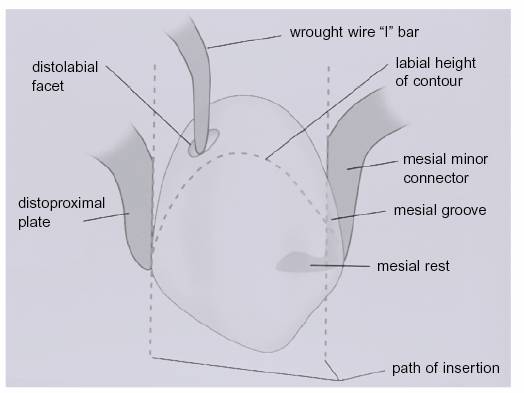
Figure 22-9: Mesial groove reciprocation clasp framework design. An I-bar engages a 0.25-mm undercut in the prepared depression on the distal surface. The mesial minor connector contacts the mesial groove and terminates in the mesial rest seat.
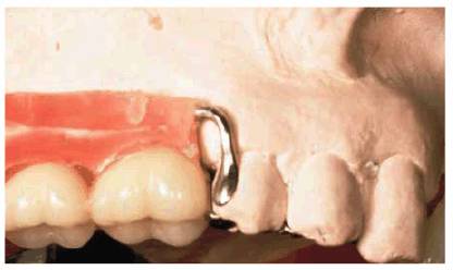
Figure 22-10: The mesial groove reciprocation clasp is indicated for maxillary teeth where esthetics is a concern.
Ring Clasp
This clasp is used for inclined maxillary or mandibular molars with natural
undercuts on the mesiobuccal or mesiolingual surface, respectively. The ring
clasp should never be used as an unsupported ring, known as a back-action clasp,
as it cannot provide both reciprocation and stabilization.32 It is
usually designed with an additional bracing arm to prevent excessive flexing.
An additional rest seat placed on the opposite side of the tooth enhances the
rigidity of the clasp assembly and may aid in resisting further mesial
migration of the tooth. All of the clasp assembly, except for the retentive
tip, must lie above the height of contour. Consequently, it is not an esthetic
clasp assembly and is reserved for molar abutments.
Embrasure Clasp
This clasp will be used in posterior regions of the mouth in the quadrant
without an edentulous space, as in Class II situations. This clasp avoids
excessive distal extension of the major connector. The embrasure clasp is a
suprabulge clasp that should have an adequate sluiceway prepared through the
embrasure of the abutment teeth to allow for proximal rests and emergence of
the suprabulge clasp arm elements near the height of contour (Figures 22-11A
and B).
Adequate sluiceway depth will also provide for proper metal thickness to ensure
rigidity and avoid occlusal interference from the opposing dentition.
Figure 22-11A and B: The embrasure clasp is used on posterior teeth where no modification space is present.
Combination Clasp
The combination clasp consists of a wrought wire clasp arm and cast reciprocal
arm (Figure 22-12).23 It is most
frequently used adjacent to a distal extension base to promote stress-breaking
characteristics to the abutment tooth. The wrought wire, being more flexible
(less brittle), may be used in smaller diameter with less danger of fracture.
Nineteen-gauge wrought wire in a 0.5-mm mesial undercut is generally indicated
for canine and premolar distal extension abutments. Remote soldering of the
clasp to the framework provides increased flexibility.8 Due to its
round form, light refraction is decreased, making the metal display less
noticeable than with the broader surface of a cast clasp.
Figure 22-12: The combination clasp consists of a wrought wire retentive arm with a cast reciprocating arm or plated surface (courtesy of Dr. John R. Ivanhoe).
Retention Enhancement
Traditionally, enamelplasty or a cast restoration has been indicated for an
abutment tooth with an inadequate undercut. The improvements in resin
composites have made them a conservative, cost-effective, and minimally
invasive method for enhancing retention. However, variable results have been
reported from the studies using resin composite to enhance retention. In vitro
studies have shown that cast I-bars produced wear of the resin composite,43
whereas stainless steel round clasps did not cause a noticeable loss of
retention.13 The use of a partial-coverage porcelain laminate bonded
to a tooth to enhance retention is a viable alternative.14
Rest Seats
In general, mesio-occlusal rest seats are indicated for posterior distal
extension abutments when the occlusion permits.24,49 For
tooth-supported RPDs, rest seats are placed on either side of the modification
space to prevent tissueward movement of the RPD and for ease of fabrication.
Cingulum rest seats are indicated for anterior teeth. However, the lack of
adequate enamel often precludes placement of a positive cingulum rest seat on
the mandibular anterior teeth. Traditionally, incisal rests have been advocated
for mandibular anterior teeth. Unfortunately, they are unesthetic, may
interfere with the occlusion, and may increase torquing forces on the teeth.
Bonded resin composite or metal rest seats have been shown to provide a
satisfactory and esthetic alternative to the incisal rest (Figure 22-13
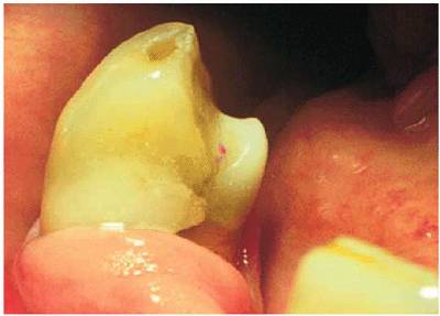
Figure 22-13: Bonded resin composite rest seat.
Flange Design
A labial flange in the anterior region is indicated when residual ridge
resorption has occurred and additional lip support is needed. The flange should
extend to the junction of the attached and unattached mucosa and should be
contoured to blend in with the adjacent teeth. Also, the flange should not
extend into an undercut apical to the adjacent teeth.38
Occasionally, tinting of the denture base to match the pigmentation of the
patient may be indicated.7,17,19
Replacement Teeth
Teeth should be selected to match the size, shape, shade, and contour of the
adjacent teeth. In some instances, it will be necessary to contour the tooth,
and, occasionally, it may be necessary to stain the artificial tooth or place a
restoration in the tooth to match adjacent teeth. A technique to modify the
shade, contour, and occlusal contacting surfaces of denture teeth with
light-polymerized resin composite has been described.46 Microfilled
resins for veneering facial surfaces are advocated because these are more
easily polished and provide an improved esthetic appearance. These changes are
most easily accomplished when the artificial tooth is fabricated from acrylic
resin. The acrylic denture base resin should be contoured to match the size and
contour of those of the adjacent teeth. The artificial teeth should be
positioned to simulate the position of the natural teeth. If natural teeth
remain, they may be used as a guide for placing the artificial teeth in a
harmonious arrangement.
Other Esthetic Considerations
The patient should be assessed in totality rather than as an aggregate of
singular entities. The potential consequence that one treatment has on another
region of the mouth and the overall result requires careful appraisal. Although
it is the intent of most practitioners to maximize the esthetic value of
treatment for the patient, the esthetic awareness and desire of the patient
merit consideration. The implementation of complex components, potentially
increasing cost, maintenance, or difficulties with hygiene for a patient
unconcerned with esthetics, is not prudent. However, the assessment of patient
awareness needs to be bona fide. The apathetic patient can create postinsertion
obstacles if a genuine esthetic concern is not detected. This type of patient
will frequently respond to queries of esthetics with "Do whatever you
think would look good, Doctor," or "I don't care about the
appearance, as long as I can chew." Great caution should be exercised when
managing the prosthetic care of these patients.
Skeletal anomalies that may effect esthetics should be brought to the patient's
attention prior to treatment. Any discussion following the completion of care
may often be interpreted as an excuse. Particular examples would include
patients who believe that the RPD will correct skeletal discrepancies, overt
facial wrinkling, or other cosmetic concerns normally requiring surgical
intervention. A skeletal Class II patient or a patient with vertical maxillary
excess will be particularly aware of a maxillary anterior modification space
for the RPD. The excessive resin display or lip displacement justifies
consultation prior to RPD construction, allowing the patient the opportunity to
consider alternative treatment options to meet his or her esthetic needs.
Tooth morphology and anticipated placement require evaluation of presurgical
diagnostic casts. Most patients will request replacement of the missing
dentition to maintain their previous esthetic situation. This should be readily
accomplished, although if a suitable replacement is not feasible, the
limitations should be discussed with the patient prior to commencing treatment.
Encumbrances may be owing to tooth size or shape limitations or positioning
difficulties, which may detract from the function of the partial denture.
Examples may include the patient with natural anterior teeth that were much
larger than the commercially available artificial dentition or the request to
maintain the anterior tooth display in a patient demonstrating an excessive
vertical overlap of the maxillary incisors. Clearly, esthetic and functional
concerns may create the need for investigation of alternative treatment options
or acceptance of the limitations by compromising either the esthetics or functional
design. Any of these situations should remain well documented and explained to
the patient completely.
ALTERNATIVE TREATMENT MODALITIES
In situations demanding maximal esthetics, alternatives to conventional RPD
design must be in the practitioner's armamentarium. Alternative treatment
modalities will often produce a result in prudent design with function and
esthetics. The use of dental attachments is discussed in this chapter; however,
finances, as well as dexterity or the ability to complete or maintain complex
care, often dictate the need for conventional alternatives.
Adjunctive Mechanisms for Minimizing
Metal Display
Camouflaging of RPD clasps, including the addition of acrylic resin and resin
composite, has been reported in the literature.33,39 The difficulty
with the use of acrylic resins or resin composite to veneer to RPD metals lies
in the differences between their abilities to flex and their coefficients of
thermal expansion. Non-noble metals possess strength and resist significant flexure.
However, resins are subjected to greater deformation from physical and thermal
conditions. The resin composite matrix also tends to be brittle beyond its
elastic limit. As a result, the abilities of the metals and resins to deform
plastically are incompatible. Other concerns include the effect of the
intraoral forces of mastication, the adjustability of veneered clasps, and the
additional bulk of the clasp created by the addition of the veneering material.
Excessive shortening and thinning of the clasp should be avoided to ensure
rigidity and minimize the breakage potential of the clasp.34
Rotational Path Removable Partial
Dentures
The rotational path RPD is a relatively uncomplicated method that eliminates
the use of esthetically objectionable clasping in the anterior region of the
mouth (Figures 22-14A
and B).22,27,28,50
It uses an anterior rigid portion of the framework and a conventional flexible
posterior retentive clasp as the retentive components. The main advantage of
this design is the minimal use of clasps. The esthetic result is enhanced, and
the tendency toward plaque accumulation is reduced. However, both the clinical
and laboratory procedures required for the rotational path RPD are technique
sensitive.
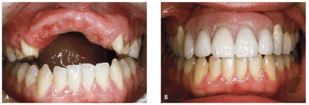
Figure 22-14A and B: (A) The maxillary anterior teeth were lost as a result of a traumatic injury. The bone loss in the anterior maxilla is significant. (B) The rotational path removable partial denture allows the elimination of anterior clasp arms to improve esthetics.
The rotational path RPD should be limited to tooth-supported situations to
prevent torquing of abutment teeth. This design also requires that positive
rest seats be used. Cingulum and extended occlusal rest seats are indicated for
canine and premolar abutments, respectively (Figures 22-15A
and B, and ). For premolars, the rest seats
should be extended to 1.5 to 2.0 mm deep occlusogingivally with nearly parallel
facial and lingual walls. A restoration may be indicated to adequately contour
the rest seat.
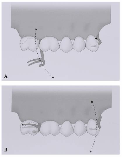
Figure 22-15A and B: (A) The rotational path removable partial denture uses an anterior rigid portion of the framework that engages an undercut and a conventional flexible posterior retentive clasp. After engaging the anterior undercut, the prosthesis is rotated into the fully seated position along an arc. (B) This arc demonstrates the arc along which the anterior rigid retainer would have to move for the prosthesis to be dislodged.
Figure 22-16: The rotational path design uses extended rests on the anterior abutments.
The cast is first surveyed at a 0-degree tilt to determine the adequacy of
undercuts on the mesial surfaces of the anterior abutments and the distofacial
surfaces of the posterior abutments (Figure 22-17). The amount of undercut needed for
the anterior teeth is 0.25 to 0.5 mm. This position is registered using tripod
marks. The cast is then tilted until the undercuts of the anterior abutments
are eliminated. The analyzing rod is then used to determine whether access
exists for the rests to be seated. There must be no interferences for the
anterior segment to go to place (Figure 22-18). If it is satisfactory, the second
cast tilt should be registered on the cast with a second set of tripod marks (Figures 22-19A
and B).
Major connectors with minimal palatal or lingual tooth contact are indicated to
avoid interferences to seating of the framework. It is important that during the
framework try-in appointment, there is minimal adjustment of the anterior
proximal plate; otherwise, the anterior retentive component may be lost. The
rotational path RPD is not indicated for distal extension RPDs, arches with
lingually inclined teeth, severely tapered arches, and arches with multiple
edentulous areas.
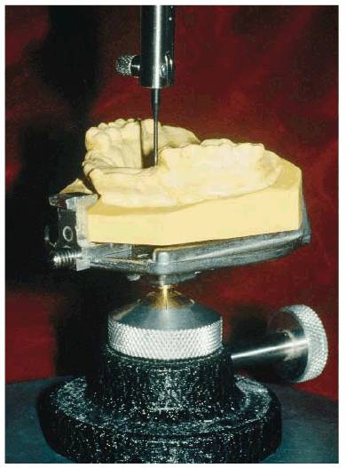
Figure 22-17: The cast is first surveyed with a 0-degree tilt to determine the adequacy of undercuts on the mesial surfaces of the anterior abutments and on the distal facial surfaces of the posterior abutments. This position is registered using tripod marks.
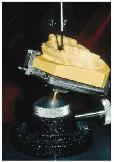
Figure 22-18: The cast is then tilted to eliminate the undercuts of the anterior abutments. This tilt is registered with a second set of tripod marks.
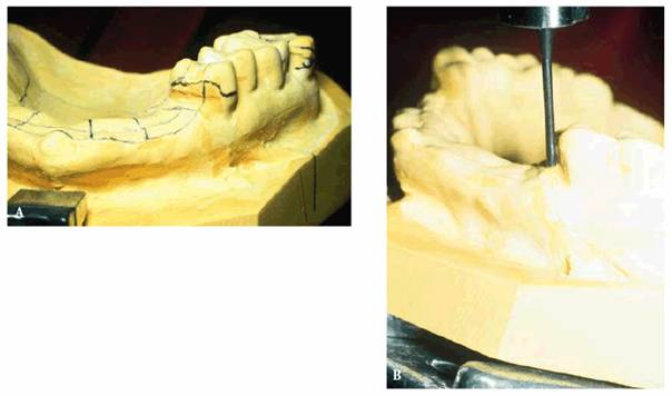
Figure 22-19A and B: The heights of contour made at the two paths of insertion. The superior height of contour is made at the 0-degree tilt. The inferior height of contour represents the path of insertion whereby the undercuts of the anterior abutments are eliminated. The area between the two lines represents the undercut into which the anterior rigid section of the framework is seated. Care must be taken during finishing and fitting of the framework in this area; otherwise, retention may be lost.
ATTACHMENTS FOR REMOVABLE PARTIAL
DENTURES
Diagnosis and Treatment Planning
The demands for highly esthetic dental restorations provide the catalyst for
the attachment RPD. The esthetic expectations of a patient should be the
primary directive for attachment use. The psychological component of treatment
planning of the RPD remains crucial to the success or failure of the
rehabilitation. Meeting the patient's esthetic and functional expectations
while not exceeding the biomechanical attributes of the supportive structures
will result in successful therapy. The anticipated function of the prostheses
by the patient must not exceed the physiologic capacity of the teeth and
tissues.
Proper treatment planning of the attachment RPD encompasses similar concepts to
the conventional RPD. Fundamental biologic tenets must be adhered to for
successful treatment. The components of guiding planes, rigid major and minor
connectors, and indirect retention remain important in the philosophy of
design. Suitable tissue preparation, accurate border extension, and tissue
coverage without impingement are important adjuncts. Correct prosthetic
planning will reduce the possibility of tissue abuse and enhance the prognosis
for success.
Definition. An attachment is a connector consisting of two or more
parts.40 One part is connected to a root, tooth, or implant and the
other part to a prosthesis. Attachment RPDs have been empirically termed
"precision attachments" for years. The terminology of precision
attachment partial dentures is frequently misused. Attachment partial dentures
should be classified by the nature of the attachment fabrication, location, and
biomechanical properties. Attachments used in RPDs are most commonly classified
in the following manner: (1) precision, (2) semiprecision, (3) intracoronal:
nonresilient and resilient, and (4) extracoronal: nonresilient and resilient.3,36,40
Attachments are subdivided into two general categories: precision and
nonprecision.9,40 Precision attachments consist of machined
components of special alloys under precise tolerances within 0.01 mm. The
metallurgic properties of the alloys are controlled to minimize the
intra-attachment wear and are designed in a manner that affords most wear to
occur on interchangeable elements. The intra- abutment portion of the attachment
will generally evidence little to no wear, allowing accurate replacement while
maintaining the specific tolerances designed. These systems allow ease of
replacement interchangeability of the standard components.
Semiprecision attachments require the direct casting of plastic, wax, or
refractory patterns. They are considered semiprecision because they are subject
to inconsistent water:powder ratios, burnout temperatures, and other variables.
The resulting components may dimensionally change and reduce the preciseness of
their accuracy of fit. The primary advantages of the semiprecision attachments
are economy, ease of fabrication, and ability to be cast in a wide variety of
alloys without the problem of coefficiency differences between the casting alloy
and the attachment alloy.9,40
General Considerations for Attachments
The variability in the circumstances for use of attachments and the variety of
attachments available preclude the establishment of a standard model. Selection
should be based on the functional and physiologic requirements of the
restoration. Consideration of the laboratory expertise in using particular
attachments must be contemplated. Selection of an attachment with specific
biomechanical and functional attributes may be finalized by the technician's
ability to use the attachment and fabricate the prosthesis.
Attachment Use. A significant consideration in the selection of an
attachment should be the long-term maintenance. Retrievability should be
regarded with equality to function in the design and selection of an attachment
for the esthetic RPD. Repeated use of similar attachments increases the
knowledge of the practitioner and technician alike. This repetition will prove
beneficial for efficacious delivery of care, management of difficult
situations, and postoperative maintenance. The dental team should limit the
application of dental attachments to a selection that meets the functional and
esthetic requirements of the majority of patients and the level of expertise of
the team. Other attachments may be considered periodically; however, use of
other attachments may prove to be the rarity rather than the norm. This
self-imposed limitation will ensure correct fabrication of the partial denture,
untroubled delivery of care, and unrestricted maintenance of the prosthesis.
Periodic planned or unplanned maintenance of the attachment prosthesis will be
required. Consistent use of an attachment selection may safeguard adequate
supply of replacement parts in the event of accidental breakage.37
Indications and Contraindications. The overwhelming indication for the
attachment RPD is esthetics. Numerous skillfully designed conventional RPDs are
not worn simply because the patient does not like the appearance. Elimination
of the buccal or labial direct retainer or clasp arm is a key factor in
establishing an esthetically acceptable design. Once the need for an
attachment-assisted RPD is established, the selection of the attachment type
should be based on the biomechanical, physiologic, and functional attributes of
the patient or technical expertise of the dental team.9,10
The contraindications to the use of attachments in RPDs are numerous. One must
consider anatomic, biomechanical, personal, and physiologic factors in
determining the selection of attachments. The health and morphology of the
abutment teeth remain a preeminent factor in the selection of an attachment.
Short clinical crowns prove to be the foremost contraindication to the use of
attachments in the construction of RPDs. The tooth must have adequate crown
height to house the attachment components and effectively offset the leverage
forces exerted on the crown. The leverage forces are most often observed in
distal extension RPDs. In addition, adequate height must be present for the
corresponding attachment components to be housed within the RPD framework or
supportive acrylic resin while allowing proper artificial tooth placement.3,9,10,36,37,40
Adjunctive Procedures. Gingivectomy, or crown-lengthening procedures,
may overcome the clinical disadvantage of short clinical crown height. This
preprosthetic procedure will generally improve fixed prosthesis retention and
resistance form and may increase the effective undercut, thereby enhancing the
retention for a conventional clasping mechanism. This may avoid the need for
placement of a surveyed crown when attachments are not a feasible treatment
modality. Gingival crown-lengthening procedures may be required to provide
adequate occlusal cervical space for attachment positioning while maintaining
the functional attributes of the selected attachment to be used.
Orthodontic therapy should be considered with the presence of tipped or
malpositioned teeth. The orthodontic correction of malpositioned teeth will
avoid excessive tooth preparation, enhance vertical loading, avert the need for
endodontics, and provide easier development of a common path of placement for
the attachment partial denture. A particular degree of parallelism is required
of all attachments. Orthodontic correction of malpositioning will allow proper
attachment orientation. A nonresilient precision attachment requires the higher
degree of parallelism.
Teeth with large pulps will not allow for incorporation of an internal box
within the crown preparation to accommodate certain attachments.3,9,10
The result of improper preparation would be an excessively overcontoured tooth
leading to a periodontal liability. Endodontic therapy may be required in
certain instances for the use of attachments. Endodontics
should also be considered when preparation of a tooth with a large core
restoration might provide little resistance to fracture. The placement of an
intraradicular core might offer enhanced resistance to fracture under the
functional loading of an attachment RPD.
The placement of attachments in pontics is an option that can avoid possible
violations of biologic principles during tooth preparation or the need for
adjunctive procedures (Figures 22-20A and B). The use of attachments lingually
positioned in a traditional pontic or distally located in a cantilevered pontic
has been described.30
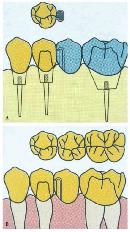
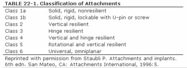
Figure 22-20A and B: Intracoronal attachment types
such as the Score-BR, PDC, Omega-M, Beyeler, etc. (Attachments International,
Dexterity. Poor patient dexterity remains a strong contraindication for
the placement of an attachment RPD. Patients lacking adequate hand coordination
may encounter significant difficulty manipulating the prosthesis in the mouth.
For some, it may be a virtual impossibility. While the average life expectancy
of the population increases, more patients become potential candidates for RPD
treatment. Debilitating diseases affecting neuromuscular control and joint
mobility are likely to correspondingly increase. Arthritis, Parkinson's
disease, cerebrovascular accidents, and other situations that influence fine
motor skills might preclude efficacious attachment partial denture use or, at
least, direct the attachment selection. Consequently, dexterity should remain a
strong diagnostic consideration with all potential attachment RPD patients.
Patients demonstrating average dexterity will generally be able to manipulate
placement and removal with relative ease over time. A resilient attachment will
generally be more easily accommodated rather than a rigid intracoronal attachment
with a precise path of placement.
Cost. The design and construction of the complex attachment RPD
treatment are costly. Cost in terms of time, effort, and resource commitment
can be anticipated. The economic factors may predict the feasibility of using attachments.
The prudent practitioner should anticipate an increased amount of diagnostic
effort, laboratory expense, chair time, and maintenance in this form of
therapy. These factors should be explained to the patient. The patient should
anticipate charges for periodic attachment maintenance or replacement.
Subsequently, these considerations support the use of a limited number of
different attachments for efficacious delivery of care and reduced chair time.37
Oral Hygiene Maintenance. A final factor to be considered in the
possible exclusion of attachment use for patients is the long-term maintenance
of the prosthesis. It must be anticipated that periodic evaluation, adjustment,
or replacement of attachment components will be required. The inability of patients
to travel or return on a regular or periodic basis should be considered
contraindications to the use of attachments. Oral hygiene may also be
considered a parameter of attachment selection. Attachments will accumulate
plaque and calculus, limiting the effectiveness or intended function of the
attachment. Additionally, attachment use implicates the fabrication of full- or
partial-coverage castings. Patients with high caries rates may experience a
diminished prognosis with rehabilitations consisting of multiple fixed
restorations.
Biomechanics and Support
Once a decision has been made to restore a region with an attachment
prosthesis, the manner in which the vertical and horizontal forces are to be
supported requires consideration. A partial prosthesis may be toothborne or
tooth-tissueborne. The forces imparted to the prosthesis and its supportive
elements should be as widely distributed as possible.
The periodontal health and support of the natural teeth should be considered in
the selection of an attachment design. The forces should be equitably
distributed over as many teeth as possible within the biologic and physiologic
capacity of the supportive dentition. The denture bases should offer the
broadest support possible for mucosal coverage.
Distal extension situations raise the dilemma of load distribution between the
teeth and mucosa. The amount of soft-tissue compressibility over the distal
extension residual ridge remains disproportionate to the abutment teeth. This
phenomenon will create unharmonious movement of the partial denture, imparting
leverage forces to the abutment teeth, possibly resulting in harm to the
abutment teeth, mucosa, and residual ridge, if not considered in the selection
of an attachment. Only teeth with suitable clinical crown height and
periodontal stature should be considered for attachment use. The presence of
excessive tissue compressibility or unsupported tissue might prescribe the need
for preprosthetic surgical intervention.3,9,10,36
Path of Insertion
With the aid of a surveyor, the anticipated path of insertion must be
considered to develop appropriate guiding planes and attachment placement
within the confines of the natural dentition. A less resilient attachment will
generally dictate a smaller degree of tolerance or more parallelism relative to
the path of insertion. Rigid and intracoronal attachments must closely
accommodate nonsurgically correctable tissue/anatomic limitations or undercuts.
For example, distal extension situations may require a distally inclined path
of placement to accommodate extension into the retromylohyoid fossa, whereas an
anterior modification space may require a labially inclined path of insertion
and attachment orientation.3,36
Knowledge of the anticipated path of insertion may guide the attachment
selection to a more resilient, universal design that can offer a greater
tolerance to the path of placement. The path of insertion of the abutment
crowns may be determined at this time and may indicate the need for
preprosthetic endodontics or surgery.
Once a prosthesis has been placed along its path of insertion, anterior,
posterior, and lateral forces alone or in combination influence the stability
of the prosthesis. The tendency of the forces to dislodge the prosthesis must
be counteracted through direct and indirect retainers. Direct retention may
occur through friction of the attachment components, framework components with
the teeth, or mucosal coverage of the denture bases. The forces of adhesion,
cohesion, and surface tension between the base, saliva, and mucosa cause a
pressure reduction on compression and further inhibit denture base movement.
Indirect Retention
Resistance to lateral displacing forces must be provided through rigid bracing
components and the vertical height of the residual ridges. Bilateral distal
extension bases use the mucosa and teeth of both sides of the dental arches for
resistance to lateral forces. A force on one side of the arch is resisted by
the components or tissue/base integrity of the contralateral side. This
supports the increased stability usually found in bilateral distal extension
bases as compared with unilateral designs. The design of certain attachments
will provide indirect retention; however, the effectiveness of the indirect
retention will vary. In attachment systems that offer little or no indirect
retention, it must be incorporated in the framework design. In general, the
more precise or rigid the attachment design is, the greater is the degree of
indirect retention inherent in the design. Additionally, the more widely spaced
the retainers are, the greater the support and stability are when compared with
a design with retainers placed closely together.3,9,10,36,37,40,41
As attachment designs increase in the degree of indirect retention, generally,
a greater amount of force to the supportive elements will be generated. Because
of this increase in leverage forces transferred to the abutment teeth by the
prosthesis, many teeth treated with castings incorporating attachments must be
splinted to adjacent teeth. This concept safeguards the functional and
biomechanical overloading of the supportive elements.5,6,11,15
Tooth Preparation
Preparation design should anticipate an increased degree of the forces to be
applied to the teeth by the attachment mechanism. Avoidance of excessive taper,
replacement of suspicious or weakened core restorations, and adequate axial
wall height will reduce the risk of tooth fracture or decementation of the
restoration. Therefore, most teeth will require full crown coverage for
adequate retention and resistance form.
The preparations should consider the morphology of the tooth as related to the
attachment selection. Adequate tooth structure must be present in all
dimensions to allow incorporation of the attachment pattern yet retain the
emergence profile and clinical crown contours of the tooth. Buccolingual,
incisocervical, and mesiodistal space must be considered before a bur is placed
to the tooth tissue. Alternative attachment selection or adjunctive procedures
should be planned prior to preparation to allow for completion of the intended
restoration and to enhance the functional and periodontal success of the
restoration.
Attachment Selection Considerations
Proper attachment selection requires evaluation of five factors: location,
function, retention, available space, and cost. Location can be subdivided into
intracoronal, extracoronal, radicular, and bar type of attachments.40
Location. Intracoronal attachments are incorporated entirely within the
contours of the cast crown for the tooth. It is imperative that adequate space
exists in all three dimensions for both incorporation of the attachment and the
maintenance of natural tooth contours to ensure proper use of the attachment
and a positive prognosis of the restoration and the tooth. If it is not
possible to place a box in the preparation to accommodate the matrix component
of the attachment, an alternative attachment selection should be made. The
advantage of the intracoronal attachment is that the forces exerted by the
prosthesis are applied more closely to the long axis of the tooth. All
intracoronal attachments are nonresilient and may require double abutting or
splinting of the adjacent teeth. This form of attachment offers indirect
retention and a more precise path of placement. Most wear will occur on
placement and removal. In situations with diminished attachment length as a
result of reduced interocclusal height, milled lingual bracing arms should be
considered (Figures 22-21A to C). Careful consideration should be given
to the amount of reduction in attachment length that will also allow for
maintenance of the functional aspects of the attachment. Most manufacturers
will state the optimal and minimal lengths of the attachment.
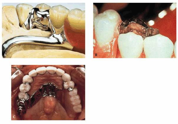
Figure 22-21A to C: (A) Milled lingual bracing arm on an RPD framework. The design allows development of normal crown contours with placement of the RPD. (B) The Biloc and Plasta attachment (Attachments International) allows the bracing arm to be incorporated into the crown contours. (C) A traditional lingual bracing or reciprocal arm may create bulk or result in tongue irritation.
Extracoronal attachments are situated external to the developed contours of the
crown. Normal emergence profile and tooth contours may be maintained while
minimizing the amount of tooth structure preparation. The more conservative
preparation reduces the risk of or need for devitalization.
The majority of extracoronal attachments have resilient attributes. This will
improve the ability of patients demonstrating dexterity problems when inserting
the prosthesis. However, the extracoronal positioning will increase the
likelihood of hygiene difficulties. Patients will require fastidious hygiene
instruction using floss and adjunctive periodontal aids to prevent food
entrapment and calculus accumulation. Inadequate hygiene will generally result
in hyperplastic tissue inflammation subjacent to the attachment apparatus.
Function. The functional attributes of an attachment require
differentiation between the intention of the prosthesis as being solid or
resilient. Kennedy Class III and small to moderate-size (replacement of less
than seven teeth) Class IV tooth-supported prostheses should be considered
solid, whereas large Class IV and distal extension I or II prostheses are
increasingly tissue supported and should be considered resilient.
Rigid attachment mechanisms may include locking pins. Locking and nonlocking
attachments allow for virtually no movement between the prosthesis and the
abutment tooth. Resilient attachments allow for a spectrum of movement ranging
from limited uniplanar to universal. Staubli has categorized rigid and
resilient attachments into six classifications, from rigid to universal
resiliency.40 The higher classification number correlates with a
greater degree of resiliency and suggests less torque transfer to the root or
implant abutment. The classifications are shown in Table 22-1.
Retention. Retention of the attachment components may be based on frictional,
mechanical, frictional-mechanical, magnetic, and suction characteristics.
Frictional retention is developed by the resistance to the relative motion of
two or more surfaces in contact. Greater intimate surface contact will usually
correlate with an increase in the amount of retention. Mechanical retention
implies the resistance to relative motion by means of a physical undercut. The
degree of undercut and the ability to adjust the physical component will
predict retention. Frictional and mechanical retention combines parameters
previously discussed and should be considered in situations necessitating
increased retention with appropriate abutment support. Magnetic retention is
created by attraction of certain materials to a surrounding field of force
produced by the motion of electrons and atomic alignment. This type of
retention is not largely used and may be diminished by corrosion of the
elements. Suction is created by a negative pressure similar to the intaglio
surface of a denture to the supportive residual ridge.
Space. Space is a principal consideration for the selection of an
attachment. Vertical space is measured from free gingival margin to the
marginal ridge of the abutment. Avoidance of tissue impingement and maintenance
of a proper emergence profile is paramount at the cervical region. Cautious
placement of the superior aspect of the attachment will circumvent occlusal
interferences. The length of attachments that rely on frictional retention
should be maximized to maintain resistance to dislodgment. Placement of the
attachment should be as low on the tooth as possible to reduce the tipping or
leverage forces applied. Buccolingual space is equally important to avoid
overcontouring the crown. Additional bulk will be required buccal and lingual
to the attachment for the casting alloy. Proper analysis of mesiodistal
measurement ensures proper proximal contour and will provide an indication of a
need for boxes in the development of the preparation. The largest attachment
possible should be selected. This requires careful prepreparation analysis that
includes the arrangement of denture teeth in a diagnostic wax-up. This will
help ensure the highest functional and esthetic value to the reconstruction.
Cost. Cost is related to the complexity of the attachment and the
material components. In general, precision attachments are precision machined
from known, possibly noble alloys. The accuracy, manufacturing, and precious
nature of the composition will demand a higher cost. Semiprecision attachments are
made of plastic or other refractory materials subject to variables in the
casting procedure, possibly leading to inaccuracies in the preciseness of fit.
The greater simplicity in the manufacturing techniques significantly reduces
the cost of using these attachments.
Intracoronal Attachments
Advantages. Intracoronal attachments, if used correctly, are
incorporated entirely within the contours of the crown. This is advantageous
for maintenance of tooth dimension and morphology. The positioning of the attachment
near the long axis of the tooth allows force direction to be located along the
long axis of the tooth. This creates a more advantageous biomechanical loading
and force transfer to the tooth with a reduction in adverse leverage forces.
Maintenance of natural tooth contours and the ability to properly place an
adjacent replacement tooth without excessive recontouring or alteration for
adaptation around an external attachment generally make intracoronal
attachments more esthetic. Less possibility of food entrapment near the
gingival tissues will enhance long-term prognosis and comfort.3,9,10,36,40,41
Disadvantages. A disadvantage of intracoronal attachments is the more
excessive tooth reduction required for proper positioning of the attachment.
Teeth with large pulps or young patients often contraindicate the use of
intracoronal attachments or necessitate endodontic therapy for attachment use.
The three-dimensional size of the tooth will predict the functional or
biomechanical success with this attachment. Large clinical crowns (at least 4
mm) are usually required for intracoronal attachments. Decreasing the length by
one half reduces the retention by a factor of eight. This may be overcome by
using a mechanical type of retentive element. The cost and precision of
intracoronal attachments may be a limiting factor. Patient dexterity,
maintenance, and repair are disadvantages or possible contraindications to the
use of this type of attachment. Attachment alignment is critical due to the
limited resilience and finite path of placement possible. This creates a
limited path of placement for the prosthesis.3,9,10,36,40,41
It is our intention to present commonly used attachments that meet the
considerations previously described. However, we recognize that other
attachments similar in design and meeting the functional and biomechanical
criteria for use may be prescribed. The intracoronal attachment obligates a
sound abutment tooth and demand for high esthetic value. A clinical crown of
greater than 4 mm is generally required with a similar faciolingual width. A
preparation depth of the internal box will be approximately 2 mm. The
frictional retention attachments must maximize clinical length to offer the
greatest degree of retention. Generally, in situations where the clinical crown
will be 3.5 mm or less, a mechanical retention attachment type should be
considered.
Types of Intracoronal Attachments
Stern G/A, Stern G/L, and Stern Type 7 (Sterngold, Attleboro, MA). The
Stern G/A, Stern G/L, and Stern Type 7 are intracoronal precision attachments
providing frictional retention and allowing for some degree of adjunctive
mechanical retention.41 The Stern G/A attachment may be considered
for segmenting an FPD, which may require modification to an RPD in the future.
The gold alloy patrix offers an expansion slot on the gingival edge for
enhancement of frictional retention. The Stern Type 7 does not offer conversion
from an FPD to an RPD, although it has similar adjustment of the frictional
retention through the use of expansion slots. The Stern G/A expansion slot
design (Figure 22-22) allows for the patrix faceplate to remain flat
against the matrix wall, thereby reducing wear. The Stern G/L employs a
gingival latch mechanism to provide mechanical retention in addition to the
frictional retention of the similar Stern G/A and Type 7 (Figure 22-23). The Stern G/L patrix is produced in two
designs, the flat-back and ESI, and in two faciolingual widths, 0.70 and 0.96
inches. The width characteristics are axiomatic, although the shape
characteristics predict the method of attachment to the RPD framework. The
flat-back design requires soldering to the framework or casting a retentive arm
to the attachment for resin retention within the denture base. The ESI offers
greater versatility, allowing soldering, electrosoldering, and acrylic resin
attachment to the RPD framework (Figure 22-24). Due to the presence of the mechanical gingival
lock, this type of attachment allows one of the shortest clinical crown height
requirements of 2.7 mm.
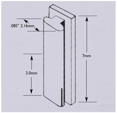
Figure 22-22: Stern G/A dimensions and illustration of expansion slot to allow for frictional retention adjustment. (Reproduced with permission from Sterngold, International. Advanced restorative products catalog. Attleboro, MA: Sterngold, 1998:14.)
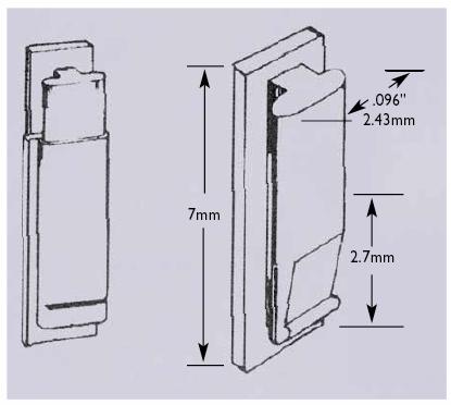
Figure 22-23: Stern G/L dimensions and illustration of an expansion slot to allow for frictional retention adjustment and gingival latch component. (Reproduced with permission from Sterngold, International. Advanced restorative products catalog. Attleboro, MA: Sterngold, 1998:15.)
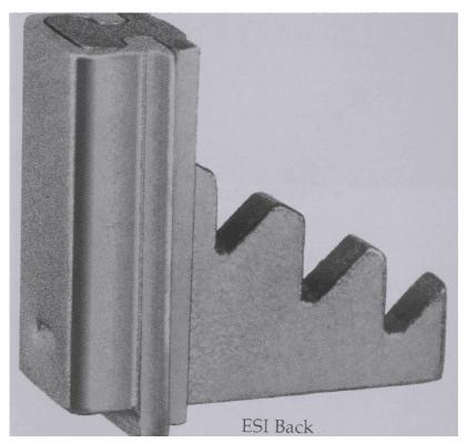
Figure 22-24: Stern G/L ESI back allows for resin retention to the RPD framework during attachment relation. Resin retention allows for retrievability of the attachment for ease of maintenance. (Reproduced with permission from Sterngold, International. Advanced restorative products catalog. Attleboro, MA: Sterngold, 1998:15.)
Swiss McCollum and Crismani. The Swiss McCollum attachment (Sterngold) (Figure 22-25) offers conversion characteristics similar to
the Stern G/A and an adjustable retention flange.40 This retention
flange must be oriented to face buccally and ordered from the manufacturer
appropriately. The attachment may be soldered to the framework, or retention
elements may be cast to the patrix portion of the attachment for luting with
acrylic resin to the denture base. A milled lingual ledge should be developed
in the abutment crown for bracing arm construction. The Stern McCollum
(Sterngold) attachment (Figure 22-26) offers an adjustment slot on the face of the
patrix that allows access when the slot is situated lingually for cross-arch
stabilization.41 The Crismani attachment (Sterngold) has a similar
adjustment slot design, although in cross-section resembles an inverted
triangular shape rather than the T-shape of the Stern McCollum attachment.40
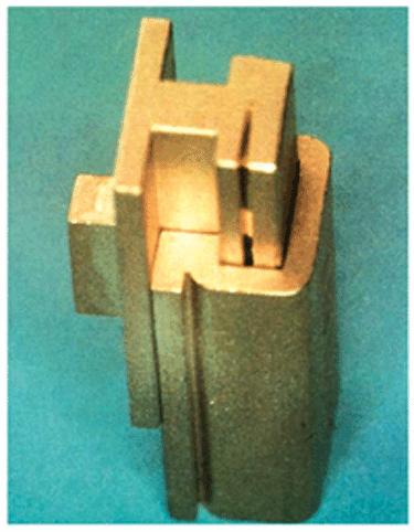
Figure 22-25: Swiss McCollum attachment (Sterngold). Note that the expansion slot must be positioned to face buccally.
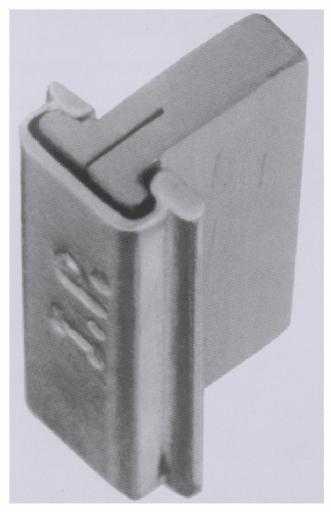
Figure 22-26: Stern McCollum (Sterngold) attachment. Note that the expansion slot is positioned on the face of the attachment oriented along the ridge crest. (Reproduced with permission from Sterngold, International. Advanced restorative products catalog. Attleboro, MA: Sterngold, 1998:16.)
Schatzmann, Biloc and Plasta, and Score. The Schatzmann, Biloc and Plasta,
and Score attachments (Attachments International, San Mateo, CA) offer
conversion possibilities from FPDs to RPDs.40 The Schatzmann
attachment is an adjustable slide attachment providing frictional and
mechanical retention. The mechanical plunger element is easily replaced
chairside at minimal cost in time or expense. The Biloc and Plasta attachment,
an intracoronal semiprecision attachment, offers a machined patrix in two alloy
possibilities and a castable plastic matrix. A lingual bracing arm is highly
recommended and is indicated in fixed, Kennedy Class I or II situations (Figure 22-27). The Score system offers multiple application
options interchanging three semiprecision castable plastic patrix
configurations with one castable matrix: the Score-PD, Score-BR, and Score-UP.
The PD version combines frictional and mechanical retention, whereas the UP
version incorporates a U-pin to lock the patrix and matrix segments together.
This version (UP) allows for interchangeability to extracoronal resilient
restorations using Dalbo (Cendres & Metaux SA, Biel, Switzerland), ASC 52
(Attachments), or Ceka-type attachments.
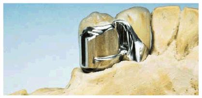
Figure 22-27: Biloc and Plasta attachment (Attachments International) allows for construction of an intracoronal attachment with a milled bracing arm. This design offers incorporation of the RPD bracing arm into the proper clinical crown contours. The mesial portion of the bracing arm is similar in orientation and function to the intracoronal portion of the attachment on the distal of the crown.
The patrix portion of the attachment types
described are either similar in metallurgic properties or possess
characteristics allowing a greater degree of wear when compared with the
matrix. Consequently, the frictional wear of the patrix reduces retention and
supports the adjustment capacity of the components. When the amount of wear or
loss of retention exceeds the adjustment capacity, replacement of the patrix
component is necessary. This clarifies the advantage of a precision-milled
component. For replacement, a new patrix is purchased and replaced into the RPD
without concern for casting inaccuracies or difficulties retrofitting the
patrix portion to the abutment matrix, as might be experienced with
semiprecision attachments.
Attachment connection to the RPD may be accomplished in a variety of ways, as
previously described. Soldering to the framework remains the most permanent and
possibly the most common method. However, acrylic attachment of the patrix or
patrix portion of the attachment to the RPD provides the highest degree of
retrievability. In acrylic attachment patrices, the worn patrix component is
retrieved from the RPD, and the new patrix is luted into place with
autopolymerizing acrylic resin, often without disturbing the artificial teeth (Figure 22-28). The disadvantages of this technique are the
discoloration and potential weakness of the resin. However, this technique
remains more time and resource efficient than rebasing the RPD to retrieve a soldered-to
attachment. A soldered technique requires artificial tooth removal and
replacement owing to the excessive heat generated from the retrieval and
resoldering of the patrix to the framework.
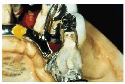
Figure 22-28: Patrix attachment with autopolymerizing resin to the RPD framework allows for easy retrievability and attachment replacement. This type of patrix placement increases the accuracy of the framework relation to the tissues and the abutment teeth.
Extracoronal Attachments
Advantages. The advantages of extracoronal attachments include
resiliency in certain designs and less abutment tooth preparation. The
conservative nature of the preparation required would suggest less harm to the
pulp and reduced risk of potential endodontic intervention. The resiliency in
design provides advantageous stress-breaking characteristics in distal
extension situations (ie, Class I or II arches). Attachment alignment is not as
critical in highly resilient extracoronal attachments due to the omniplanar
motion possible. This creates the advantage of multiple paths of placement for
the prosthesis. Patients with biomechanical limitations not withstanding a
rigid attachment apparatus or anatomic limitations precluding a finite path of
placement are strong candidates for resilient attachments.3,9,10,36,40,41
Disadvantages. The adverse aspects of extracoronal classification
include the potential for torque imparted by the attachment to the tooth and
hygiene maintenance. Careful recall evaluation is necessary to ensure proper base-tissue
relationships and fastidious oral hygiene. Tooth positioning around the
attachment apparatus is often difficult and diminishes functional or esthetic
value if adequate space is not available. Some resilient extracoronal
attachments do not allow for "locking" to a rigid state. This may
create difficulties with relining and rebasing procedures. Indirect retention
and bracing are not incorporated into most extracoronal attachment designs and
will necessitate the addition of components to provide these functions.3,9,10,36,40,41
As with intracoronal attachments, it was our intention to present commonly used
attachments while understanding that other attachments similar in design and
meeting the functional and biomechanical criteria for use may be prescribed.
Types of Extracoronal
Attachments
Dalbo Attachment System (Cendres & Metaux SA). This attachment is
one of the oldest and most successful extracoronal attachments and is
classified as an adjustable, directed-hinge distal extension attachment.40,41
This system features lateral stability, vertical resiliency, and hinge movement
(Figures 22-29A to D). The advantages of the Dalbo system are
the intrinsic direct retainer and excellent stability owing to the vertical
beam. The attachment may be used in unilateral or bilateral applications (Figures 22-29E and F, 22-29G, 22-29H, 22-29I and J). The unilateral configuration provides a larger
vertical bar for enhanced lateral stability. The attachment is offered in two
sizes, although the mini version lacks vertical resiliency (see Figure 22-29D). The vertical resiliency is rendered through
the presence of a spring and found only in the standard unilateral and
bilateral designs. The difference between the standard and the mini is
approximately 2 mm in clinical crown height requirement, 1.7 to 2.0 mm in
preparation depth, and 1 mm in faciolingual width requirement. As in all
extracoronal attachments, the amount of space required in the denture base is
approximately 5.5 to 6.0 mm. This often creates difficulty with tooth placement
and inadequate strength for the resin. The minimum amount of resin recommended
should be strictly adhered to so as not to compromise the strength of the
denture base in the region of the attachment. This extracoronal retainer offers
a mechanism to "lock" the attachment for reline procedures.
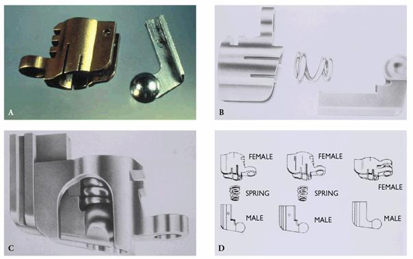
Figure 22-29A to D: (A) Dalbo attachment (Cendres & Metaux). (B) A spring allows for vertical resiliency, and a ball allows for horizontal rotation. (C) A compressed spring allowing for vertical resiliency. (D) Bilateral application, unilateral application, and mini (left to right).
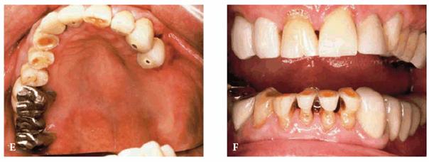
Figure 22-29E and F: This 75-year-old man wanted to improve both function and esthetics. Note the considerable wear in both the maxillary and mandibular dentitions.
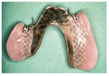
Figure 22-29G: A removable prosthesis with Dalbo attachments in place.
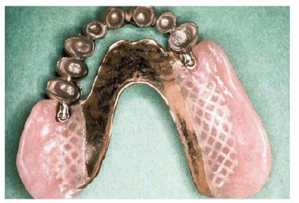
Figure 22-29H: The metal-ceramic framework is easily fixed to the Dalbo attachments.
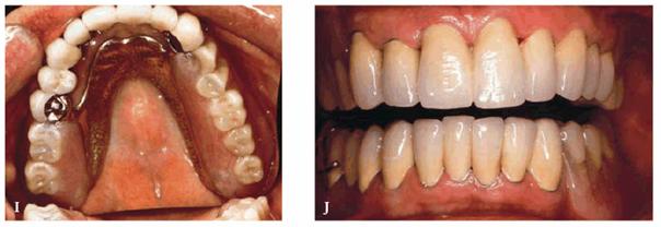
Figure 22-29I and J: Fixed metal-ceramic prostheses combined with Dalbo attachments provided maximum function and esthetics and were easy for this patient to insert and remove.
Octolink. The Octolink System (Attachments International), an
extracoronal precision/semiprecision attachment, furnishes a large degree of
movement and is classified as a universal hinge with vertical resiliency (Figures 22-30A to D).41 The patrix button is
adjustable and is screwed into a metal keeper, or retention nut, which is
retained in the acrylic denture resin or may be spot-welded to the RPD
framework. The matrix becomes incorporated into the crown through either a
cast-to technique or a castable plastic technique (Figures 22-30E, 22-30F, 22-30G, 22-30H, and 22-30I). A minimum of 4.0 mm of vertical abutment tooth
height is necessary, although a minimum of 6.0 mm of space is mandatory for the
retentive keeper and patrix component in the denture base.
Figure 22-30A to D: (A) Octolink attachment (Attachments International). (B) Spacer used to allow for vertical resiliency. (C) and (D) Note the vertical and omniplanar resiliency of the attachment.
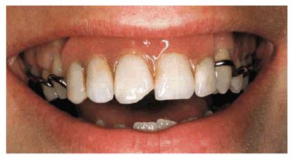
Figure 22-30E: This 30-year-old man was embarrassed because the clasps of his removable prosthesis showed when he smiled.
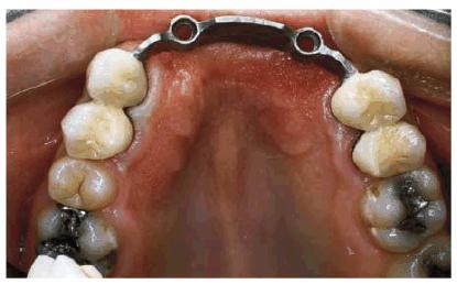
Figure 22-30F: A four-unit metal-ceramic splint combined with an Octolink (Attachments International) framework would better support the removable prosthesis.
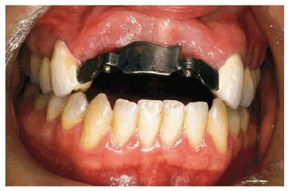
Figure 22-30G: This frontal view shows the adaptation of the metal framework to the alveolar ridge.
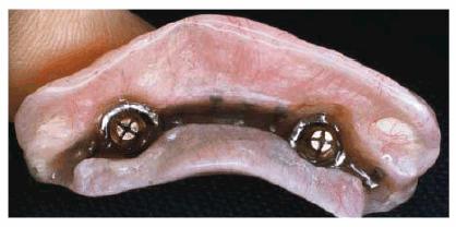
Figure 22-30H: This view shows how the Octolink (Attachments International) attachments will fit into the removable prosthesis.
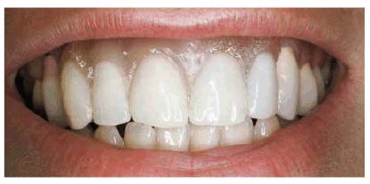
Figure 22-30I: The final smile shows the esthetic improvement offered by the combination of a secure attachment and a natural-looking acrylic flange.
SA SwissAnchor. The SA SwissAnchor (Attachments International) is a
semiprecision castable pattern providing either rigid or simultaneous vertical
and hinge resiliency, depending on the design selected. A spacer will provide
vertical resiliency. This system requires a vertical abutment tooth height of 3.5
mm. The patrix is screw retained in a keeper that is retained in the denture
acrylic resin. This system is interchangeable with the regular Ceka.40,41
Stern ERA and Stern-RV. The Stern ERA and Stern-RV (Sterngold) are
commonly used semiprecision attachment providing universal hinge and vertical
resiliency (Figures 22-31A to E).41 Retention may be varied
through use of four color-coded nylon patrices indicating four levels of
retention (Figures 22-31F and G). An optional metal jacket serves as a
keeper for the patrix retentive element, which may be alternatively retained
within the acrylic resin (Figure 22-31H). However, this is more difficult to change
once retention is diminished. Patrices may be easily changed without the use of
acrylic resin. The Stern ERA requires a minimum of 4 mm of vertical height,
whereas the Stern-RV requires 3.5 mm of vertical space (Figure 22-32). No additional preparation depth is required
for matrix incorporation to the crown restoration. A large space of 6.5 mm is
required within the denture base for the patrix component and an additional 0.3
to 0.5 mm for the optional ERA Metal Jacket. The manufacturer recommends an
additional 1.0 mm of denture base resin for patients demonstrating parafunction
or "habitually strong occlusions." The patrix component is the
variable between the Stern ERA and the Stern-RV. Both patrices fit with the
selected matrix.41,47
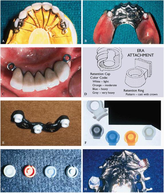
Figure 22-31A to H: (A) Maxillary bilateral
distal extension application of Stern ERA attachments. (B) Male
placement within an RPD framework (courtesy of Dr. Steven K. Nelson). (C)
The need for parallelism with each other in the sagittal plane is not required.
Note the splinting of fewer than six remaining anterior teeth. (D)
Diagram of matrix and patrix components. The color-coded retentive cap has four
different levels of retention. (Reproduced with permission from Sterngold,
International. Advanced restorative products catalog.
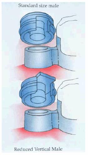
Figure 22-32: Comparison of the Stern ERA and the ERA-RV. The retentive cap varies in vertical height. (Reproduced with permission from Sterngold, International. Advanced restorative products catalog. Attleboro, MA: Sterngold, 1998:26.)
Hader Vertical. This extracoronal semiprecision attachment (Attachments
International) is compatible with conventional clasping on the contralateral
side.41 The resilience of the attachment allows slight hinging
movement, although it will load abutment teeth more strongly than other
resilient attachments. This attachment requires a 4.5-mm vertical tooth height
without internal preparation limitations.
ASC 52 (Attachments International). The ASC 52 ball attachment functions
well in limited space.40 It is a universal attachment offering
rotational and vertical movement. A spring-loaded connector allows for
adjustable retention. The matrix is offered as a platinum cast-to alloy or a
semiprecision plastic pattern. The precision patrix is constructed of stainless
steel.
ORS-DE (Attachments). Superior stress-breaking characterizes this attachment.40
This semiprecision castable plastic patrix pattern requires a minimum of 4.0-mm
vertical height or nearly 6.0 mm of tooth height. The matrix will require a
nearly identical amount of space within the denture base. A matrix O-ring may
be retained in a metal retainer ring and provides easy retrieval and
maintenance (Figures 22-33A and B).
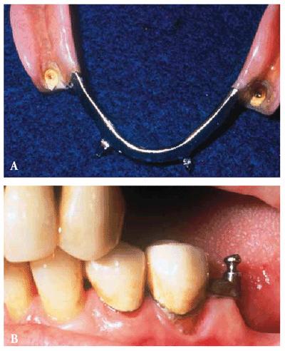
Figure 22-33A and B: (A) An ORS-DE matrix O-ring in an RPD framework. (B) An ORS-DE attachment in a distal extension application. Note that adequate vertical space is necessary for interocclusal clearance (courtesy of Dr. Steven K. Nelson).
Special Use Attachments
Special use attachments should be considered for limited use based on the
esthetic, functional, or anatomic needs of the patient. These types of
attachments augment the armamentarium of the practitioner, although they may
often increase the complexity and expense. Plunger-type or pawl attachments are
an excellent adjunct for esthetic anterior teeth with required function as
retentive abutments.2 Classified as an intracoronal attachment, the
spring-loaded plunger allows for a full range of motion, mimicking a
universal-type extracoronal design. This attachment may be used with
conventional or attachment partial dentures. A reciprocal lingual arm should
encompass 180 degrees and terminate in a rest seat. Matrix components or
concavities are incorporated into the natural tooth or crown dependent on the
situation or attachment, whereas patrix components are luted with acrylic to
the framework and not soldered. Examples of plunger attachments are the Hannes
anchor, the IC attachment, and the SwissTac/Tach E-Z (Attachments
International) (Figures 22-34A to C).40,41
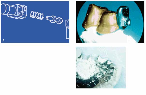
Figure 22-34A to C: (A) Diagram of an IC plunger attachment. (B) Female cast into abutment restoration. (C) Plunger and spring apparatus incorporated into resin or RPD.
Splint bar designs incorporate Hader/EDS, Dolder, CM Bar & Rider or Ackermann clips, ABS, CBS, or PPM Bar Systems.40,41 Selection is based on the degree of resilience, anatomic limitations, or convenience. The primary indication would be the splinting of abutment teeth while providing retention for the RPD. Careful consideration for the degree of resilience and interocclusal space for tooth arrangement must be provided (Figures 22-35A, 22-35B and C, 22-35D, 22-35E, and 22-35F).
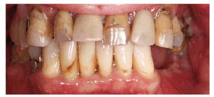
Figure 22-35A: This well-known 60-year-old attorney complained about his appearance. Examination revealed posterior bite collapse, extreme breakdown of his remaining teeth, and periodontal disease.
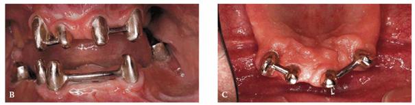
Figure 22-35B and C: Following periodontal surgery, maxillary and mandibular overdentures were constructed using Dolder bar attachments. Note the access that provides the patient with the ability for proper hygiene.
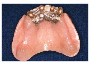
Figure 22-35D: This removable bridge provides excellent ridge adaptation plus Dolder bar secure retention for an ideal overdenture.
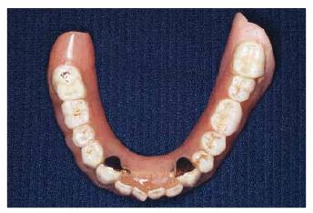
Figure 22-35E: This man wanted some exposed gold and a somewhat crowded anterior tooth arrangement for what he considered a "natural look."
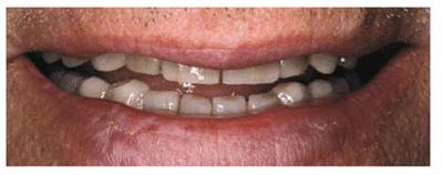
Figure 22-35F: A custom tooth staining and natural tooth arrangement gave him the appearance he thought appropriate.
Dental implant placement offers a highly predictable treatment approach for
overpartial dentures. Long tooth-bounded modification spaces requiring a
significant amount of tissue support for the partial denture bases can benefit
from application of overdenture abutment attachments on endosseous dental
implants. Class I and II RPD classifications may have significant improvement in
retention and stability by dental implant placement in the distal region of the
edentulous residual ridge. The support offered by the overdenture concept using
a dental implant would dramatically reduce the tipping and leverage forces
imparted to the distal abutment teeth. This treatment modality may offer
support comparable to the Class III RPD.
Milled lingual ledges have been described as an adjunctive component that will
accommodate placement of lingual bracing arms, generally providing compensation
for short attachment length.40,41 A technique of providing
frictional retention of components through precision milling has been
described. Spark erosion technology to create precision milled fit has proved
to be successful.45 Adjunctive elastoclips may offer additional
retention. The intimacy of fit developed with spark erosion or precision
milling provides frictional retention, nearly eliminating any resilience. This
type of attachment technology must be considered nonresilient and should be
applied to the appropriate supportive elements. Additionally, the cost of this
treatment approach may be prohibitive. A preprosthetic laboratory analysis of
the anticipated cost may be useful in establishing the degree of remuneration
required for the success of the rehabilitation.
REFERENCES
1. Applegate OC. The rationale of partial denture choice. J Prosthet Dent
1960;10:891-907.
2. Bagley D. Versatile uses for plunger attachments. Trends Tech Contemp Dent Lab 1993;10:33-5.
3. Baker JL, Goodkind RJ. Precision attachment removable partial dentures. San
Mateo, CA: Mosby, 1981.
4. Becker CM, Kaiser DA, Goldfogel MH. Evolution of removable partial denture
design. J Prosthodont 1994;3:158-66.
5. Berg T, Caputo A. Load transfer by a maxillary distal-extension removable
partial denture with cap and ring extracoronal attachments. J Prosthet Dent 1992; 68:784-9.
6. Berg T, Caputo A. Maxillary distal-extension removable partial denture
abutments with reduced periodontal support. J Prosthet Dent 1993;70:245-50.
7. Berte JJ, Hansen CA. Custom tinting denture bases by visible light cure
lamination. J Prosthodont 1995; 4:129-32.
8. Brudvik JS, Wormley JH. Construction techniques for wrought wire retention
arms as related to clasp flexibility. J Prosthet Dent 1973;30:769-74.
9. Burns DR, Ward JE. A review of attachments for removable partial denture
design: part 1. Classification and selection. Int J Prosthodont 1990;3:98-102.
10. Burns DR, Ward JE. A review of attachments for removable partial denture
design: part 2. Treatment planning and attachment selection. Int J Prosthodont 1990;3:169-74.
11. Chou TM, Eick JD, Moore DJ, Tira DE. Stereophotogrammetric analysis of
abutment tooth movement in distal-extension removable partial dentures with
intracoronal attachments and clasps. J Prosthet Dent 1991;66:343-9.
12. Curtis DA, Curtis TA, Wagnild GW, Finzen FC. Incidence of various classes
of removable partial dentures. J Prosthet Dent 1992;67:664-7.
13. Davenport JC, Hawamdeh K, Harrington E, Wilson HJ. Clasp retention and
composites: an abrasion study. J Dent 1990;18:198-202.
14. Dixon DL, Breeding LC, Swift EJ Jr. Use of a partial-coverage porcelain
laminate to enhance clasp retention. J Prosthet Dent 1990;63:55-8.
15. El Charkawi HG, El Wakad MT. Effect of splinting on load distribution of
extracoronal attachment with distal extension prosthesis in vitro. J Prosthet Dent 1996;76:315-20.
16. Frank RP, Brudvik JS, Nicholls JI. A comparison of the flexibility of
wrought wire and cast circumferential clasps. J Prosthet Dent 1983;49:471-6.
17. Gerhard R, Sawyer N. Dentures to harmonize with heavily pigmented tissues. J Am Dent Assoc 1966;73: 94-5.
18. Goodkind RJ. The effects of removable partial denture design on abutment
tooth mobility: a clinical study. J Prosthet Dent 1973;30:139-46.
19. Haeberle CB, Khan Z. Construction of a custom-shaded interim denture using
visible-light-cured resin. J Prosthodont 1997;6:153-6.
20. Hansen CA, Iverson GW. An esthetic removable partial denture retainer for
the maxillary canine. J Prosthet Dent 1986;56:199-203.
21. Kennedy E. Partial denture construction. Dental Items of Interest.
1928:3-8.
22. King GE, Barco MT, Olson RJ. Inconspicuous retention for removable partial
dentures. J Prosthet Dent 1978;39:505-7.
23. Kotowicz WE, Fisher RL, Reed RA, Jaslow C. The combination clasp and the
distal extension removable partial denture. Dent Clin North Am 1973;17:651-60.
24. Kratochvil FJ. Influence of occlusal rest position and clasp design on
movement of abutment teeth. J Prosthet Dent 1963;13:114-24.
25. Kratochvil FJ. Partial removable prosthodontics. Philadelphia: WB Saunders,
1988.
26. Krol AJ. Clasp design for extension base removable partial dentures. J Prosthet Dent 1973;29:408-15.
27. Krol AJ, Finzen FC. Rotational path removable partial dentures: part 1.
Replacement of posterior teeth. Int J Prosthodont 1988;1:17-27.
28. Krol AJ, Finzen FC. Rotational path removable partial dentures: part 2.
Replacement of anterior teeth. Int J Prosthodont 1988;1:135-42.
29. Krol AJ, Jacobson TE, Finzen FC. Removable partial denture design outline
syllabus. 4th edn. San Rafael, CA: Indent, 1990.
30. Lepe X, Land MF. Use of an intra-pontic attachment as cosmetic support for
a removable appliance. Ill Dent J 1990;59;280-3.
31. McCartney JW. The MGR clasp: an esthetic extracoronal retainer for
maxillary canines. J Prosthet Dent 1982;46:490-3.
32. McGivney GP, Castleberry DJ. McCracken's removable partial prosthodontics.
9th edn. St Louis, MO: Mosby, 1995.
33. Moreno de Delgado M, Garcia LT, Rudd KD. Camouflaging partial denture
clasps. J Prosthet Dent 1986;55:656-60.
34. Morris HF, Brudvik JS. Influence on polishing of cast clasp properties. J Prosthet Dent 1986;55:75-7.
35. Myers RE, Pfeifer DL, Mitchell DL, Pelleu GB. A photoelastic study of rests
on solitary abutments for distal-extension removable partial dentures. J Prosthet Dent 1986;56:702-7.
36. Preiskel HW. Precision attachments in dentistry. St. Louis: Mosby, 1968.
37. Schuyler CH. An analysis of the use and relative value of the precision
attachment and the clasp in partial denture planning. J Prosthet Dent 1953;
3:711-4.
38. Smith BJ. Esthetic factors in removable partial prosthodontics. Dent Clin North Am 1979;23:53-63.
39. Snyder HA, Duncanson MG Jr, Johnson DL, Bloom J. Effects of clasp flexure
on a 4-META adhered light polymerized composite resin. Int J Prosthodont 1991;4:364-70.
40. Staubli PE. Attachments & implants: reference manual. 6th edn. San
Mateo, CA: Attachments International, 1996.
41. Sterngold-ImplaMed, Int. Advanced restorative products catalog. Attleboro,
MA: Sterngold-ImplaMed, 1998.
42. Stewart KL, Rudd KD, Kuebker WA. Clinical removable partial prosthodontics.
2nd edn. St. Louis: Ishiyaku EuroAmerica, 1992.
43. Tietge JD, Dixon DL, Breeding LC, et al. In vitro investigation of the wear
of resin composite materials and cast direct retainers during removable partial
denture placement and removal. Int J Prosthodont 1992; 5:145-53.
44. Toth RW, Fiebiger GE, Mackert JR, Goldman BM. Shear strength of lingual
rests prepared in bonded composite. J Prosthet Dent 1986;56:99-104.
45. Weber H, Frank G. Spark erosion procedure: a method for extensive combined
fixed and removable prosthodontic care. J Prosthet Dent 1993;69:222-7.
46. Weiner S, Krause AS, Nicholas W. Esthetic modification of removable partial
denture teeth with light-cured composites. J Prosthet Dent 1987;57:381-4.
47. Williamson RT. Removable partial denture fabrication using extracoronal
attachments: a clinical report. J Prosthet Dent 1993;70:285-7.
48. Wong R, Nicholls JI, Smith DE. Evaluation of prefabricated linqual rest
seats for removable partial dentures. J Prosthet Dent 1982;48:521-6.
49. Zach GA. Advantages of mesial rests for removable partial dentures. J
Prosthet Dent 1975;33:32-5.
50. Zarb GA, MacKay HF. Cosmetics and removable partial dentures-the Class IV
partially edentulous patient. J Prosthet Dent 1981;46:360-8.
ADDITIONAL RESOURCES
Goldstein RE. Esthetics in dentistry. 1st edn. Philadelphia: JB Lippincott,
1976:110-35.
|
