Insulinoma
- (Tumor
of pancreatic islet beta cell origin; most often benign solitary tumor but
~5% are malignant; 5-10% of patients may have MEN type I)
- See Fig.
13-10 and Tables 13-13 and .
- In
patients with fasting hypoglycemia, insulinoma should be considered the
cause until another diagnosis can be proved. No single test is
certain to be diagnostic; multiple tests may be required.
- Fasting 24-36 hrs provokes hypoglycemia in 80-90% of these patients; 72 hrs of
fasting provokes hypoglycemia in >95% of these patients, especially if
punctuated with exercise. Absence of ketonuria implies surreptitious food
intake or excess insulin effect (differentiate by blood glucose level).
Low serum glucose and high serum insulin establishes the diagnosis, i.e.,
insulin level is inappropriately elevated for the degree of hypoglycemia
(in normal persons, insulin level becomes <5 µU/mL or undetectable).
P.627
Serum insulin rarely reaches these high levels in patients
with reactive hypoglycemia. Serum C-peptide is similarly inappropriately
elevated, in contrast to factitious hypoglycemia. In women, serum glucose can
fall to 20-30 mg/dL during fasting and return to normal without treatment; in
men, a fall in serum glucose to <50 mg/dL is considered abnormal.
- Serum insulin/C-peptide ratio is <1.0 in molarity units.
- Proinsulin level is normally ≤ 20% of total insulin; increased in
insulinoma.
- Proinsulin >30% of serum insulin after overnight fast suggests insulinoma.
(May also be increased in renal disease.) (Proinsulin
is included in the immunoassay of total insulin and separation requires
special technique.)
- Serum insulin values are
not useful in reactive hypoglycemia but should always be measured in cases
of fasting hypoglycemia.
- Occasional patients with
insulinoma have very low serum insulin levels; their serum shows very high
proinsulin level that interferes with the insulin immunoassay, giving
falsely low values.
- Serum insulin/glucose ratio >0.3 when serum glucose >50 mg/dL indicates
inappropriate hyperinsulinism, and this usually indicates insulinoma if
factitious hypoglycemia is ruled out. Ratio may be slightly higher (e.g.,
≤ 0.35 in obese persons). Has no diagnostic value in insulinoma.
- Stimulation tests are not
usually necessary and may be dangerous if serum glucose <50 mg/dL. Too
many false-positive and false-negative results make these tests
unreliable.
- Tolbutamide tolerance
test:.
- Glucagon stimulation
test: Administer 1 mg of glucagon IV during 1-2 mins; measure serum
insulin three times at 5-min intervals and then twice at 15-min
intervals. Patients with insulinoma show an exaggerated response of serum
immunoreactive insulin. Serum insulin >100 µU/mL after glucagon
stimulation in a patient with fasting hypoglycemia and inappropriate
insulin secretion strongly suggests insulinoma.
- Infusion of exogenous
insulin to reduce serum glucose level also suppresses the secretion of
insulin and of C-peptide in normal persons but not in patients with
insulinoma. C-peptide level usually remains elevated if insulinoma is
present (C-peptide is also not suppressed in islet cell hyperplasia and
nesidioblastosis) but falls to very low level if beta cell function is
normal.
- OGTT is useless for
diagnosis; results may be normal, flat (in ~20% of healthy persons), or
show impaired tolerance.
- After overnight fast,
reference ranges are
- Serum insulin: 1-25
µU/mL.
- Serum proinsulin:
<20% of total measurable insulin.
- Serum C-peptide: 1-2
ng/mL.
- Ratio of insulin to
glucose: <0.3 (up to 0.35 in obese persons). During fasting, ratio
decreases in healthy persons and increases in insulinoma patients.
Ketoacidosis,
Diabetic
- See Tables
13-11, .
- Blood glucose is increased (usually >300 mg/dL); range from slightly
increased to very high. Very increased glucose (>500-800 mg/dL)
suggests nonketotic hyperosmolar hyperglycemia (because glucose levels
become very high only when extracellular fluid volume is markedly
decreased). Glucose level of <200 mg/dL may occur, especially in
alcoholics or pregnant women with insulin-dependent diabetes. Glucose
concentration is not related to severity of diabetic ketoacidosis.
- Plasma acetone is increased (4+ reaction when plasma is diluted 1:1 with water).
(Acetone is usually 3-4× the concentration of acetoacetate but does not
contribute to acidosis.) Nitroprusside reagent tests (e.g., Acetest,
Ketostix, Chemstrip) react with acetoacetate, not with beta-hydroxybutyrate,
weakly with acetone; therefore weak positive reaction with ketone does not
rule out ketoacidosis. Beta-hydroxybutyrate/acetoacetate ratio varies from
3:1 in mild cases to 15:1 in severe diabetic ketoacidosis. With correction
of diabetic ketoacidosis, conversion of beta-hydroxybutyrate to
acetoacetate gives a stronger nitroprusside test reaction; do not mistake
this for worsening of diabetic ketoacidosis.
- Urine ketone tests are
not reliable for diagnosing or monitoring diabetic ketoacidosis.
- May be positive in
≤ 30% of first morning specimens in pregnancy.
P.628
- False-positive results
reported in presence of some sulfhydryl drugs (e.g., captopril).
- False-negative results
may occur with highly acidic urine, after large doses of ascorbic acid,
or when test strips are exposed in air for extended time.
- Metabolic acidosis (pH <7.3 and/or bicarbonate <15 mEq/L) is mainly
due to beta-hydroxybutyrate and acetoacetate. Some lactic acidosis may
exist, especially if shock, sepsis, or tissue necrosis is present; suspect
this if pH and AG do not respond to insulin therapy. Whole spectrum of
patterns from pure hyperchloremic acidosis to wide-AG acidosis. May be
obscured by complicating metabolic alkalosis.
- Volume and electrolyte depletion (due to glucose-induced osmotic diuresis)
- Absence of volume
depletion should arouse suspicion of other possibilities (e.g.,
hypoglycemic coma, other causes of coma).
- Very low sodium (120
mEq/L) is usually due to hypertriglyceridemia and hyperosmolality
although occasionally may be dilutional due to vomiting and water intake.
Low in 67%, normal in 26%, increased in 7% of cases. Depleted body stores
are not reflected in these initial values, which reflect relative water
loss and blood glucose level.
- Serum potassium is
normal in 43%, increased in 39% due to potassium exit from cells
secondary to acidosis; initial low potassium in 18% of cases indicates
severe depletion.
- Serum phosphate
decreased in 10% of cases, normal in 18%, and increased in 71%; falls
with onset of therapy due to loss by osmotic diuresis and cellular
uptake. Severe depletion (<0.5 mg/dL) may cause muscle weakness,
rhabdomyolysis, impaired cardiac function, etc. Excessive replacement may
cause hypocalcemia and hypomagnesemia.
- Serum magnesium may be
decreased in 7% (in prolonged ketoacidosis), normal in 25%, increased in
68% of cases.
- Azotemia is present (BUN
is usually 25-30 mg/dL); creatinine may be proportionally increased more
than BUN due to methodologic interference by acetoacetate.
- Serum osmolality is
slightly increased (up to 340 mOsm/L).
- WBC is increased (often
>20,000/cu mm) even without infection; associated with decreased
lymphocytes and eosinophils.
- Hb, Hct, total protein
may be increased due to intravascular volume depletion.
- Serum amylase may be
increased (may originate from salivary glands rather than pancreas) in
≤ 36% of patients; increase from both sources in 16% of cases.
- Serum AST, ALT, LD, and
CK are increased in 20-65% of cases, partly due to methodologic
interference of acetoacetate in colorimetric methods. CK may be increased
due to phosphate depletion and rhabdomyolysis.
- Thyroid function tests
are not reliable (due to sick thyroid syndrome).
- Laboratory findings due
to complications in treatment
- Hypoglycemia
- Hypokalemia
- Alkalosis
- Arterial thrombosis
(e.g., organ infarction, limb ischemia)
- Cerebral edema
- Laboratory findings due
to precipitating medical problem (e.g., infection, myocardial infarction,
vascular disorder, trauma, pregnancy, emotional problem, endocrine
disorder; not found in 25% of cases); these should always be sought.
- See Acidosis,
Metabolic, Acidosis, Lactic, Nonketotic hyperglycemic coma
- Follow-up laboratory
tests every 2-4 hrs initially and less often with clinical improvement.
Bedside fingerstick glucose test can be performed initially every 30-60
mins to determine rate of fall of glucose and when to add glucose to IV
fluids.
- ESR may be increased in
diabetic patients even in absence of infection and when serum protein is
normal, particularly when glycemic control is poor; does not necessarily
indicate underlying infection.
Prader-Willi
Syndrome
- (Mental
retardation, muscular hypotonia, obesity, short stature, and hypogonadism
associated with diabetes mellitus)
- Diabetes mellitus
frequently develops in childhood and adolescence but is insulin resistant,
responds to oral hypoglycemic drugs, and is not accompanied by acidosis.
P.629
Somatostatinoma
- (Rare
condition)
- Diabetes mellitus that
improves after resection of the tumor
- Hypochlorhydria
- Steatorrhea
- Occasionally anemia is
present.
Tumors
of Pancreas (Hormone-Secreting), Primary
|
Cell Type
|
Hormone Secreted
|
Tumor
|
|
B cell
|
Insulin
|
Insulinoma
|
|
D cell
|
Gastrin
|
Gastrinoma
|
|
A cell
|
Glucagon
|
Glucagonoma
|
|
H cell
|
VIP
|
Vipoma
|
|
D cell
|
Somatostatin
|
Somatostatinoma
|
|
HPP cell
|
Human pancreatic polypeptide (HPP)
|
HPP-secreting tumor (very rare tumor)
|
|
- Other rare
hormone-secreting tumors have been identified as causing ectopic ACTH
syndrome, atypical carcinoid syndrome, SIADH, ectopic hypercalcemia
syndrome.
Tumors
of Pancreas (Islet Cell), Classification
- Insulin-secreting beta
cell tumor (may be benign or malignant, primary or metastatic) produces
hyperinsulinism with hypoglycemia.
- Non-insulin-secreting
non-beta cell tumor (benign or malignant, primary or metastatic) may
produce several types of syndromes.
- Z-E syndrome.
- Profuse diarrhea with
hypokalemia and dehydration.
- Profuse diarrhea with
hypokalemia (and sometimes periodic paralysis) may occur as a separate
syndrome without peptic ulceration. (Some patients
have histamine-fast achlorhydria.) Diabetic-type glucose tolerance
curves may occur in some patients because of chronic potassium depletion.
May be associated with MEN.
- Nonspecific diarrhea.
- Steatorrhea (due to
inactivation of pancreatic enzymes by acid pH).
Vipoma
- (Secreted
by specialized endocrine cells of the amine precursor uptake and
decarboxylation system that inhibit gastric acid production and stimulate
gastrointestinal secretion of water and electrolytes; most tumors are
found in pancreas but ~30% are extrapancreatic, e.g., bronchogenic carcinoma,
pheochromocytoma, ganglioneuroblastoma. 60% are malignant.)
- Voluminous watery diarrhea (6-10 L/day) with dehydration
- Hypokalemia that may be
associated with hypokalemic nephropathy
- Metabolic acidosis
- Hypercalcemia in 50% of
cases
- Abnormal OGTT
- Achlorhydria or
hypochlorhydria
- Increased plasma VIP >75 pg/mL. RIA may show cross reactivity with
other gastrointestinal hormones. (Should be collected in special chilled
syringe containing EDTA and a plasma protease inhibitor and frozen
immediately after centrifugation.) Specificity is >88% and positive
predictive value is 86% (varies among laboratories). Increased values may
also occur in patients with cutaneous mastocytoma, severe hepatic failure,
or portocaval shunts.
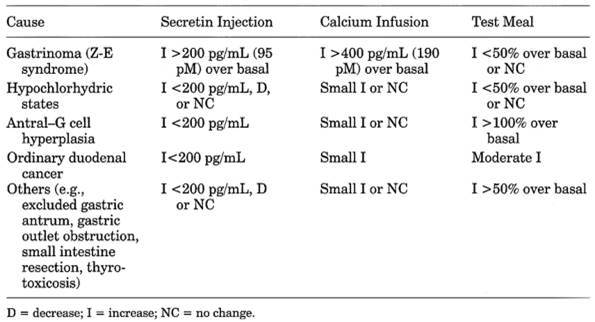
Zollinger-Ellison
(Z-E) Syndrome (Gastrinoma)
- See Table
13-15.
- Due to gastrinomas
(non-beta cell tumors often arising in pancreas)
P.630
|
|
|
Table 13-15. Serum Gastrin Response to
Provocative Tests in Hypergastrinemia
|
- Tumors
are multiple in 28% of patients and may be ectopic (e.g., >50% are in
duodenal wall; 9% are extrapancreatic and extraintestinal; selective
venous sampling for gastrin may be helpful for localizing tumor).
- Tumors
are malignant in 62% of patients; 34% of patients have metastases.
- Diffuse
hyperplasia occurs in 10% of patients.
- Increased basal serum gastrin. Fasting serum gastrin >1000 pg/mL and basal
acid output >15 mEq/hr with recurrent peptic ulcer is virtually
diagnostic. Level of >500 pg/mL (normal <100 pg/mL) is highly
suggestive of gastrinoma in absence of achlorhydria or renal failure.
Level of <100 pg/mL is unlikely to be gastrinoma. 100-500 pg/mL occurs
in ~40% of gastrinoma patients and ~10% of ulcer patients without
gastrinoma. If fasting serum gastrin is increased but is <1000 pg/mL,
secretin-provocative test and acid secretory rate test should be
performed.
- IV injection of secretin (1-2 U/kg body weight) is the most sensitive
and accurate provocative test. This provokes an increase of serum gastrin
≥ 110 pg/mL within 10 mins. Some ulcer patients may have serum
gastrin increase of ≤ 200 pg/mL. Serum gastrin decreases in most
nongastrinoma patients. Negative response occurs in ~5% of gastrinoma
patients. Selective injection of secretin into gastroduodenal artery
causes serum gastrin to increase >50% in 30 secs in hepatic or portal
vein blood (both should be sampled). Postoperative fasting serum gastrin
and secretin levels are both necessary to determine cure.
- Other provocative tests
such as IV injection of calcium gluconate (4 mg of calcium/kg) or
ingestion of a standard test meal are not as sensitive or specific as
secretin test. Response after calcium infusion is positive if serum
gastrin is ≥ 395 pg/mL.
- There is a large volume of highly acidic gastric juice in the absence of pyloric
obstruction. (12-hr nocturnal secretion shows acid of >100 mEq/L and
volume of >1500 mL; baseline secretion is >60% of the secretion
caused by histamine or betazole stimulation.) It is refractory to vagotomy
and subtotal gastrectomy. Hypochlorhydria (basal pH >3) or achlorhydria
excludes diagnosis of Z-E syndrome (see Gastric Analysis).
- Basal acid output is > 15 mEq/hr (normal is <10 mEq/hr) in 90% of
cases if no previous gastric surgery was done or >5 mEq/hr if previous
vagotomy or gastric resection was performed. Ratio of basal acid output to 15515b120p
maximal output >0.6 strongly favors gastrinoma, but false-positive and
false-negative results are common. If basal acid output determination is
not possible, pH >3 excludes Z-E syndrome if patient is not on
antisecretory drugs. Fasting serum gastrin >1000 pg/mL and gastric pH
P.631
<2.5 almost certainly indicates Z-E syndrome; both should be measured
because they may be a poorly correlated in individual patients.
- Hypokalemia is frequently
associated with chronic severe diarrhea, which may be a clue to this
diagnosis.
- Serum albumin may be
decreased.
- Steatorrhea occurs rarely
due to low pH produced in intestine.
- Laboratory findings due
to peptic ulcer (present in 70% of patients) of stomach, duodenum, or
proximal jejunum (e.g., perforation, fluid loss, hemorrhage)
- ○ Clues to Z-E syndrome are ulcers in unusual locations or
giant or multiple ulcerations (25% of these patients), rapid or severe
recurrence of ulcer after adequate therapy, recurrent ulcer after surgery,
prominent gastric folds, gastric acid hypersecretion with
hypergastrinemia, family history of peptic ulcer or ulcers with other
endocrine disorders, duodenal ulcers without Helicobacter pylori.
- ○ MEN type I should be ruled out in all patients with Z-E
syndrome, which may be the initial manifestation of MEN type I. 25% of
cases of this syndrome are associated with MEN type I. 40-60% of cases of
MEN type I have Z-E syndrome.
- Ultrasonography, angiography,
CT scan, and MRI are normal in 50% of patients.
Laboratory
Tests for Evaluation of Adrenal-Pituitary Function
- Complete 24-hr urine
collections may be difficult to obtain in some patients.
- Plasma samples are
simple to obtain but are altered by diurnal variation, episodic pulsatile
secretion, renal and metabolic clearance, stress, protein binding, and
effect of drugs. Therefore abnormal screening tests must be confirmed by
tests that stimulate or suppress the pituitary-adrenal axis.
- Increased function is
tested by suppression tests and decreased function is tested by
stimulatory tests.
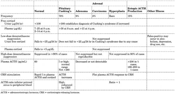
- Cortisol measurements
have largely replaced other steroid determinations in diagnosis of
Cushing's syndrome.
Adrenocorticotropic
Hormone (Acth) Stimulation (Cosyntropin) Test
Use
- Differential diagnosis of
adrenal insufficiency
- Not helpful in diagnosis
of Cushing's syndrome.
Rapid
Screening Test
- Administer 0.25 mg of
synthetic ACTH (cosyntropin) IM or IV and measure baseline and 30-, 60-,
and 90-min plasma cortisol levels. If response is not normal, perform long
test (see below).
- Interpretation
- Normal: baseline plasma
cortisol of >5.0 µg/dL with increase to 2× baseline level ≥ 20
µg/dL is sufficient single criterion of normal adrenal function to
preclude need for further workup; 60- and 90-min levels can be omitted.
Increase in urine 17-OHKS has also been used.
- Addison's disease: ruled
out by a positive response.
- Hypopituitarism: a slight
increase is shown the first day and a greater increase the next day.
- Adrenal carcinoma: little
or no response; marked increase in urine 17-KS.
- Adrenal hyperplasia:
shows increase of 3-5× baseline level.
Long
Test
- Daily infusion of ACTH
for 5 days, with before and after measurement of serum cortisol, 24-hr
urine measurements for cortisol, 17-OHKS. (Protect possible Addison's
disease patient against adrenal crisis with 1 mg of dexamethasone.)
P.632
- Interpretation
- Normal: at least 3× increase
with maximum above upper reference value
- Complete primary adrenal
insufficiency (Addison's disease): no increase in urine steroids or
increase of <2 mg/day
- Incomplete primary
adrenal insufficiency: less than normal increases on all 5 days or slight
increase on first 3 days, which may be followed by decrease on days 4 and
5
- Secondary adrenal
insufficiency (due to pituitary hypofunction): "staircase" response of
progressively higher values each day (delayed but normal response)
- Adrenal insufficiency due
to chronic steroid therapy: may require prolonged ACTH testing to elicit
the "staircase" response; may produce increments only in 17-OHKS but not
in 17-KS
- CAH (21-hydroxylase and
17-hydroxylase deficiency): increase in 17-KGS and 17-KS, little or no
change in 17-OHKS
Aldosterone,
Plasma
Use
- Diagnosis of primary
hyperaldosteronism
- Differential diagnosis of
fluid and electrolyte disorders
- Assessment of adrenal
aldosterone production
Increased
In
- Primary aldosteronism
- Secondary aldosteronism
- Bartter's syndrome
- Pregnancy
- Very low sodium diet
- Urine aldosterone is also
increased in nephrosis.
Decreased
In
- Hyporeninemic
hypoaldosteronism
- CAH
- Congenital deficiency of
aldosterone synthetase
- Addison's disease
- Very high sodium diet
Androstenedione,
Serum
(A major adrenal
androgen in serum; also produced by testes and ovaries)
Use
Diagnosis of virilism and hirsutism
Increased
In
- CAH due to 21-hydroxylase
deficiency; marked increase is suppressed to normal levels by adequate
glucocorticoid therapy. Suppressed level reflects adequacy of therapeutic
control. May be better than 17-hydroxyprogesterone for monitoring therapy
because it shows minimal diurnal variation, better correlation with
urinary 17-KS excretion, and plasma levels are not immediately affected by
a dose of glucocorticoid.
- Adrenal tumors
- Cushing's disease
- Polycystic ovarian
disease
Decreased
In
Addison's disease
P.633
Corticotropin-Releasing
Hormone (CRH) Stimulation Test
1 µg/kg of body weight or 100 µg of CRH is
given IV; blood is then drawn at 15-min intervals for 2-3 hrs to measure ACTH
and cortisol. ACTH concentration from both inferior petrosal sinuses and
peripheral vein is compared after CRH stimulation. For initial test, blood
sampling from jugular veins is simpler and less invasive; negative results can
be confirmed by petrosal sinus sampling. Use of only peripheral plasma ACTH and
cortisol has little value.
Use
- Confirm diagnosis of
Cushing's disease when patient has positive response to dexamethasone
suppression, CRH administration, or metyrapone stimulation.
- Especially useful when
high-dose DST is equivocal or when biochemical data indicate a pituitary
source but radiographic examination is normal.
Interpretation
- Differentiate pituitary
and nonpituitary causes of Cushing's syndrome, especially ectopic ACTH
production. Ratio of ACTH in jugular and peripheral veins of >2 before
and >3 after administration of CRH is diagnostic for Cushing's disease.
Petrosal sampling sensitivity, specificity, and accuracy approach 100%.
Different values obtained from right and left petrosal sinuses suggests
side on which tumor is located.
- Cushing's syndrome due to
pituitary adenoma: positive response is exaggerated increase above
baseline of >50% in plasma ACTH and >20% in cortisol concentrations.
After surgical removal of adenoma, basal concentrations of ACTH and
cortisol are undetectable but response to CRH is normal.
- Hypercorticalism of
adrenal origin: plasma ACTH is low or undetectable before and after CRH
without any cortisol response.
- Ectopic ACTH syndrome: no
ACTH or cortisol response in ~92% of patients; positive response in ~8% of
patients.
- Psychiatric states associated
with hypercorticalism (e.g., depression, anorexia nervosa, bulimia): in
uni- or bipolar depression, both peak and total ACTH response is
decreased; only a normal small decrease in cortisol occurs; after recovery
response is not distinguishable from that of normal persons. Similar
findings may occur in obsessive-compulsive disorders and alcoholism. Manic
patients have response similar to that of controls.
Dehydroepiandrosterone
Sulfate (DHEA-S), Serum
(Produced by
androgenic zone of adrenal cortex)
Use
- Indicator of adrenal
cortical function, especially for differential diagnosis of virilization.
- Replaces 17-KS urine
excretion with which it correlates; shows no significant diurnal
variation, thereby providing rapid test for abnormal androgen secretion.
Increased
In
- CAH: markedly increased
values can be suppressed by dexamethasone. Highest values occur in CAH due
to deficiency of 3-beta-hydroxysteroid dehydrogenase.
- Adrenal carcinoma:
markedly increased levels cannot be suppressed by dexamethasone.
- Cushing's syndrome due to
bilateral adrenal hyperplasia shows higher values than Cushing's syndrome
due to benign cortical adenoma, in which values may be normal or low.
P.634
- Cushing's disease
(pituitary etiology): moderate increase
- In hypogonadotropic hypogonadism,
DHEA-S is usually normal for chronologic age and high for bone age, in
contrast to idiopathic delayed puberty in which DHEA-S is low relative to
chronologic age and normal relative to bone age.
- First few days of life,
especially in sick or premature infants.
Decreased
In
- Addison's disease
- Adrenal hypoplasia
Dexamethasone
Suppression of Pituitary Acth Secretion
Low-Dose
Dexamethasone Suppression Test (DST)
- 0.5 mg of dexamethasone
(synthetic glucocorticoid) is given orally every 6 hrs for eight doses;
specimen collection as for high-dose test below. A rapid overnight
variation for screening uses a single 1-mg dose at 11 p.m. with plasma
cortisol collection the next day at 8 a.m. Following in 2 hrs by CRH
stimulation improves diagnostic accuracy, sensitivity, and specificity to
~100% in diagnosis of Cushing's syndrome.
- Use
- Good screening test to
rule out Cushing's syndrome and to identify cases for further testing
because few (1-2%) false-negative results are seen. Should be reserved
primarily for cases with mildly increased urine cortisol or
pseudo-Cushing's syndrome.
- Interference
- False-positive results
may occur in acute and chronic illness, alcoholism, depression, and use of
certain drugs (e.g., phenytoin, phenobarbital, primidone); estrogens may
cause a false-positive overnight DST.
- Noncompliance (check by
measuring plasma dexamethasone)
- Interpretation
- Normal response is a fall
in urine free cortisol to <25 µg/24 hrs, in plasma cortisol to <5
µg/dL, or in urine 17-OHKS to <4 mg/24 hrs. Fall in urine free cortisol
>90% or in 17-OHKS >64% has 100% reported specificity, i.e., normal
result excludes hypercorticalism.
- Patients with Cushing's
syndrome of any cause almost always have abnormal lack of suppressibility.
Repeat testing is sometimes needed for accurate diagnosis.
High-Dose
DST
- 2 mg of dexamethasone is
given orally every 6 hrs for eight doses; plasma cortisol is measured 6
hrs after last dose and urine free cortisol and 17-OHKS are measured on
the second day; baseline specimens are taken for 2 days before test.
- Use
- The high-dose test is the
basic test to differentiate Cushing's disease (in which only relative
resistance to glucocorticoid negative feedback is seen) from adrenal
tumors or ectopic ACTH production (usually complete resistance).
- Interpretation
- Cushing's disease
(pituitary tumor)
- Suppression of urine
free cortisol to <90% of baseline (59% sensitivity, 100% specificity)
and urine 17-OHKS to <65% of baseline (72% sensitivity, 94%
specificity) strongly differentiates Cushing's disease from ectopic ACTH
production, but not all pituitary tumor patients show such marked
suppression. Some patients with large ACTH-producing pituitary adenomas
have marked resistance to high-dose dexamethasone suppression. In
long-standing cases, nodular hyperplasia of adrenal may develop, causing
autonomous cortisol production and resistance to DST.
- Ectopic ACTH syndrome or
nodular adrenal hyperplasia
- No suppression in 80% of
cases.
- Adrenal adenoma or
carcinoma or ectopic ACTH syndrome
- Urinary 17-OHKS and
urine and plasma cortisol are not decreased after high or low doses of
dexamethasone. Adrenal tumors do not reproducibly suppress.
- Patients with psychiatric
illness may be resistant.
- Interferences
- Atypical or
false-positive responses may occur due to drugs (e.g., alcohol, estrogens
and birth control pills, phenytoin, barbiturates, spironolactone),
pregnancy, obesity, acute illness and stress, severe depression.
P.635
11-Deoxycortisol
(Compound S), Serum
(Present in
blood as an intermediate in synthesis of cortisol from 17-hydroxyprogesterone;
excretion in urine is included in 17-KGS and Porter-Silber 17-OHKS
measurements.)
Use
- In metyrapone test (see
next section), in which blood level parallels changes in urine 17-OHKS
- In functioning
pituitary-adrenal system an increase from <200 ng/dL baseline to
>7000 ng/dL is seen 8 hrs after large dose of metyrapone, whereas in
nonfunctioning system very little increase in blood level is seen.
Increased
In
- CAH (11-Beta-Hydroxylase
deficiency)
- After metyrapone
administration in normal persons
Decreased
In
Adrenal insufficiency
Metyrapone
Test
- Adrenal suppression of
pituitary secretion of ACTH is inhibited by administration of 750 mg of
metyrapone (which blocks cortisol production, leading to increased ACTH
secretion and therefore of 11-deoxycortisol) every 4-6 hrs beginning at
midnight; draw baseline plasma levels at 8 a.m. and following 8 a.m.
- Do not perform metyrapone
test until ACTH test proves that adrenals are sensitive to ACTH.
Use
- To distinguish Cushing's
disease from ectopic ACTH production
- To assess if adrenal
insufficiency is secondary to pituitary disease. Some increase in
11-deoxycortisol indicates that some pituitary reserve exists; in primary
adrenal insufficiency, no rise occurs.
Interpretation
- ACTH deficiency (secondary
Addison's disease):
- Healthy persons and those
with pituitary Cushing's disease: basal plasma 11-deoxycortisol increases
≥ 400× or >10 µg/dL if cortisol falls to <7 µg/dL and plasma
ACTH rises to >100 ng/L or urine 17-OHKS increases by 70% (71%
sensitivity) or to 2.5× the previous baseline concentration or to >10
mg/24 hrs.
- Adrenal tumor with excess
cortisol production: no increase or fall in urinary 17-OHKS and 17-KS.
Test is positive in 100% of cases of adrenal hyperplasia without tumor,
50% of cases of adrenal adenoma, and 25% of cases of adrenal carcinoma.
- Ectopic ACTH syndrome:
may not be accurate in this condition.
Renin
Activity, Plasma (Pra)
See Aldosteronism.
Diseases
of Adrenal Gland
Adrenal
Hyperplasia, Congenital
- (Errors
of metabolism due to specific deficiencies of enzymes needed for normal
steroid synthesis of three main hormone classes [specific abnormalities on
short arm of chromosome 6]: mineralocorticoids [17-deoxy pathway],
glucocorticoids [17-hydroxy pathway], and sex steroids. All forms have
decreased cortisol production; this stimulates compensatory secretion of
pituitary ACTH, which causes the adrenal hyperplasia and hypersecretion of
other pathways.)
P.636
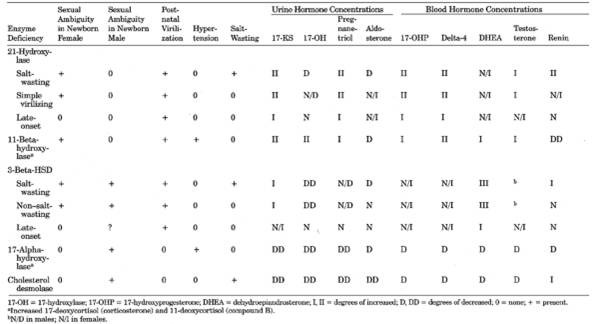
- The most common forms are
summarized in Table 13-16. The synthetic pathways
are shown schematically in Fig. 13-11 to illustrate
the altered hormonal levels.
- Establish diagnoses by increase in specific precursor steroids in blood or urine, which
can be suppressed by administration of glucocorticoids.
- Finding
of increased 17-hydroxyprogesterone or androstenedione in amniotic fluid
permits prenatal diagnosis.
- In the United States,
occurs in 1 in 80,000-100,000 live births.
- CAH should always be
ruled out in infants with
- Ambiguous genitalia and
presence of nuclear sex chromatin
- Continued vomiting after
pyloroplasty
- Siblings affected with
CAH
- Salt loss
21-Hydroxylase
Deficiency
- (>90%
of cases of CAH are of this form; three types are recognized)
- Severe Deficiency
(Salt-Losing) Form
- Severe enzyme deficiency not compensated by increased ACTH secretion; cortisol
levels are decreased.
- Excess production of salt-losing steroids plus inability to secrete aldosterone
causes characteristic acute adrenal crisis.
- Salt-wasting crisis usually occurs 1-2 wks after birth with hyponatremia,
hyperkalemia, acidosis, severe dehydration, and shock.
- Increased ACTH causes hypersecretion of androgens and virilization of female
external (ambiguous) genitalia but internal genitalia are usually normal.
Males do not show abnormal genitals at birth but may show precocious
puberty. Both sexes show rapid early growth, but premature closure of
epiphyses causes shorter stature.
- Moderate Deficiency
(Simple Virilizing) Form
- Salt-wasting is mild or
absent.
- Moderate enzyme deficiency compensated by increased ACTH secretion causing
cortisol secretion close to normal and marked increase in androgens
(characteristic increase in androstenedione and,
to a lesser extent, testosterone) and cortisol precursors, some of which
(progesterone, 17-hydroxyprogesterone) cause
some salt wasting; the latter causes compensatory increase in PRA and
increased aldosterone secretion.
- Androgen ratio in urine of 11-desoxy-17-KS to 11-oxy-17-KS is ~1:1 (normal adult ratio
= 1:4).
- Urinary
excretion of 17-KS and blood steroid secretion can be suppressed by
dexamethasone (1.25 mg/m2/day for 7 days), which
differentiates CAH from virilizing adrenal tumors. Urine 17-OHKS
levels are normal.
- Normal or low cortisol
levels show little or no response to ACTH administration.
- Karyotyping should be
done to establish genetic sex whenever external genitalia are ambiguous.
- Mild Deficiency
(Attenuated; Late-Onset) Form
- At puberty, females show
hirsutism and oligomenorrhea; must be differentiated from polycystic ovary
syndrome.
- Increased
17-hydroxyprogesterone: Extreme increase in salt-wasting form of
21-hydroxylase deficiency (20-500× normal; often >25,000 ng/dL) is
usually diagnostic; 24-hr urinary metabolite (pregnanetriol) is also
increased. Not as increased in simple virilizing form. In mild deficiency
enzyme block is evident only after ACTH stimulation: excessive rise
(5-10×) of 17-hydroxyprogesterone 30 or 60 mins after administration of
ACTH (cosyntropin) 0.25-1.0 mg IV or 6 hrs after 0.40 mg IV (normal
<900 ng/dL; late-onset form >2000 ng/dL; severe form >16,000
ng/dL). Response of exaggerated increase of 17-hydroxyprogesterone is used
to identify carriers.
- Aldosterone deficiency is
also present in salt-wasting form but not in simple virilizing form;
increased in both forms.
- In nonclassical forms,
same biochemical pattern occurs (with lower levels) but symptoms of
virilization, abnormal growth and puberty, infertility, etc., may be
slight or absent.
- 17-hydroxyprogesterone
and delta-4 levels are used to monitor glucocorticoid therapy by reduction
to normal.
- Cortisol levels are usually fixed and unresponsive
- Excess adrenal androgen production (e.g., androstenedione, dehydroepiandrosterone
[DHEA], testosterone, urinary 17-KS), which is suppressed by
glucocorticoids.
P.637
|
|
|
Table 13-16. Comparison of Different
Forms of Congenital Adrenal Hyperplasia
|
P.638
|
|
|
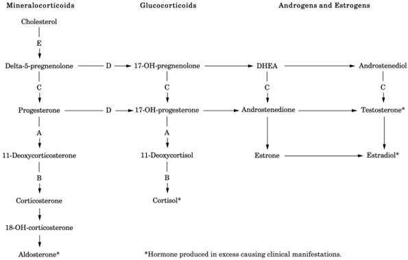
Fig. 13-11. Pathway of adrenal hormone
synthesis. Hormones above the level of the deficient enzyme are present in
increased amount; those below this level are decreased in amount (see Table 13-16). Shunting to other pathways may occur. Findings
depend on completeness of enzyme deficiency, degree of hormone deficiency, or
excessive accumulation. (A = 21-hydroxylase; B = 11-hydroxylase; C =
3-beta-hydroxysteroid dehydrogenase; D = 17-hydroxylase; E = 20,22-desmolase;
DHEA = dehydroepiandrosterone.)
|
P.639
- Genetic testing and prenatal diagnosis are available
- Neonatal screening shows increased 17-hydroxyprogesterone in dried filter paper blood
spot.
- False-positives may
occur due to prematurity and low birth weight, illness, and in infants
<24 hrs old.
11-Beta-Hydroxylase
Deficiency
- (Causes
<3% of cases of CAH; excess mineralocorticoids cause hypertension,
which may not appear until adulthood; excess androgen causes female
pseudohermaphroditism at birth, postnatal virilism in males and females;
males with mild form may have only hypertension or gynecomastia.)
- Increased serum deoxycorticosterone causing hypokalemia and suppression of renin and
aldosterone.
- Increased 11-deoxycortisol and 17-hydroxyprogesterone and increase of their
metabolites in urine: tetrahydro-deoxycorticosterone and
tetrahydro-11-deoxycortisol.
- PRA levels can be used to
monitor therapy.
- Glucocorticoid therapy
returns deoxycorticosterone to normal.
3-Beta-Hydroxysteroid
Dehydrogenase Deficiency
- (Rare
autosomal recessive disorder; complete deficiency causes death)
- Impaired secretion with decrease of cortisol, aldosterone, androstenedione, and sex
steroids.
- Increased plasma 17-hydroxypregnenolone, pregnenolone, DHEA; increased ratio of
delta-5 (pregnenolone, 17-hydroxypregnenolone, DHEA) to delta-4
(progesterone, 17-hydroxyprogesterone, delta-4-androstenedione) causing
mild virilization.
17-Alpha-Hydroxylase
Deficiency
- Decreased serum 17-hydroxylated steroids and androgens
- Decreased urine 17-KS and 17-OHKS
- Increase in serum corticosterone and deoxycorticosterone and their
urinary metabolites in urine, causing hypertension, hypokalemia
- Decreased aldosterone and PRA
- PRA levels can be used
to monitor therapy.
Cholesterol
Desmolase Deficiency
- (Complete
deficiency incompatible with life. Mild condition in females may cause
short stature, virilization, irregular menses, infertility. Affected males
may show short stature, infertility. Early diagnosis and therapy may
prevent this. Ambiguous or female genitalia in male children.)
- Virtually no steroids (cortisol, aldosterone, androgens) are produced.
- Very low urine 17-OHKS levels are not increased by ACTH stimulation.
- Aldosterone is very low in plasma and urine
- Hyponatremia, hyperkalemia, rapid dehydration, shock, and early death if
not recognized at birth.
Corticosterone
Methyloxidase Deficiency
- Type I
- . Hyponatremia and
hyperkalemia
- . Decreased aldosterone and 18-hydroxycorticosterone
- . Increased corticosterone
- Type II
- . Ratio of urinary metabolites of 18-hydroxycorticosterone to
aldosterone is markedly increased (normal is <3.0); is a better marker
than 18-hydroxycorticosterone levels.
17-Beta-Hydroxysteroid
Dehydrogenase Deficiency
- Increased delta-4-testosterone ratios in peripheral and spermatic blood is
diagnostic.
- During first week of
life:
- Urine total 17-KS may be
as high in normal infants as in those with CAH due to maternal steroids.
Normally falls to <1 mg/24 hrs during second week of life;
P.640
therefore serial determinations should be performed in suspected cases. Level
of <1 mg/24 hrs rules out CAH; an increasing level suggests CAH but a
decreasing level does not rule out CAH. Is increased in all virilizing forms
except lipoid type.
|
|
|
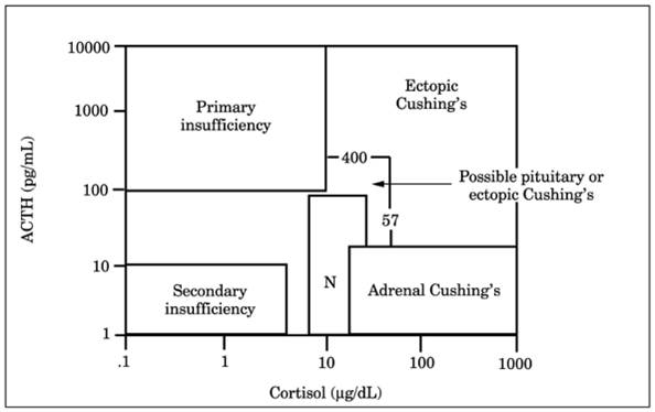
Fig. 13-12. Adrenocorticotropic hormone
(ACTH or corticotropin) and cortisol limits that are useful in diagnosis of
Cushing's syndrome. This figure shows the corticotropin versus cortisol area
ambiguous for the differential diagnosis of pituitary-dependent and ectopic
Cushing's syndrome and the area diagnostic for ectopic Cushing's syndrome. (N
= normal.) (From
Snow K, et al.
Biochemical evaluation of adrenal dysfunction. Mayo Clin Proc
, with permission.)
|
- 17-hydroxyprogesterone
is the most valuable test in 21-hydroxylase or 11-hydroxylase deficiency.
- Detectable amounts of
pregnanetriol in urine or plasma after first week of life is usually
diagnostic of CAH, but in some patients this may not appear until age
>1 mo.
- 17-OHKS in urine is not
particularly useful in diagnosis of CAH.
Adrenocortical
Insufficiency
- See Table
13-17 and Figs. 13-12 and .
Acute
- Primary (e.g.,
Waterhouse-Friderichsen syndrome [hemorrhagic necrosis due to
anticoagulant therapy, coagulopathy], antiphospholipid syndrome, sepsis,
postoperative state)
- Secondary to pituitary or
hypothalamic disorders
- After cessation of
prolonged steroid therapy
- Dehydration
- Azotemia is due to effect
of dehydration and shock on renal function.
- Serum sodium and chloride
are decreased and potassium is increased in some patients.
- Hypoglycemia occurs
regularly.
- Direct eosinophil count
is >50/cu mm (<50/cu mm in other kinds of shock).
- Blood cortisol is markedly decreased (<5 µg/dL).
Chronic
(Addison's Disease)
- Due To
- Chronic
- Primary
- Granulomas (e.g., TB,
sarcoidosis)
P.641
|
|
|
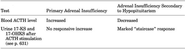
Table 13-17. Laboratory Differentiation
of Primary and Secondary Adrenal Insufficiency
|
|
|
|
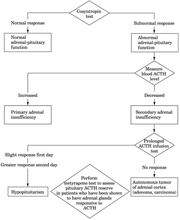
Fig. 13-13. Algorithm for diagnosis of
adrenal insufficiency.
|
P.642
- Metastatic carcinoma
(lung, breast, kidney), lymphoma
- Amyloid
- Autoimmune adrenalitis:
diagnosed by circulating adrenal antibodies; may be associated with
other autoimmune conditions (e.g., Hashimoto's thyroiditis, PA)
- Systemic fungal
infections (e.g., histoplasmosis, cryptococcosis, blastomycosis)
- AIDS (e.g.,
opportunistic infections with CMV, bacteria, protozoa)
- Adrenal hypoplasia
(neonates)
- Adrenoleukodystrophy
- Secondary
- Simmonds' disease
(idiopathic atrophy of pituitary)
- Destruction of
pituitary or hypothalamus by granulomas, tumor, etc.
- Low serum cortisol and increased ACTH are diagnostic of primary adrenal failure. In
primary deficiency both cortisol and aldosterone are deficient, with salt
loss causing increased PRA; in secondary deficiency, aldosterone
production is maintained but other secondary endocrine deficiencies may
appear, e.g., hypothyroidism, hypogonadism.
- Increased blood ACTH (200-1600 pg/mL) with wide variation between morning and evening
levels in primary adrenal hypofunction but decreased or absent ACTH in
pituitary (secondary) hypoadrenalism. Normal value rules out primary but
not mild secondary insufficiency. Increased ACTH level is quickly
suppressed by replacement therapy.
- Decreased ACTH with low cortisol indicates ACTH deficiency
- Decreased blood cortisol (<5 µg/dL in 8-10 a.m. specimen) is useful
screening test. High or high-normal result excludes both primary and
secondary adrenocortical insufficiency. ≤3 µg/dL is said to indicate
adrenal insufficiency and obviate need for further testing. Borderline
result is indication for ACTH stimulation test.
- Long ACTH stimulation test is necessary for diagnosis of secondary adrenal
insufficiency.
- Metyrapone inhibition
test is performed if ACTH test causes some increase in blood cortisol.
- Cortisol treatment
interferes with all of the above tests and must be discontinued for 24-48
hrs before testing. Dexamethasone interferes with metyrapone test and
plasma ACTH levels.
- Urine 17-OHKS is absent
or markedly decreased.
- Urine 17-KS and 17-KGS
are markedly decreased.
- Antiadrenal antibodies are found in most cases of idiopathic Addison's disease and
are said to rule out adrenoleukodystrophy and secondary adrenal
insufficiency. Said to have very high sensitivity and specificity, and are
predictive of impending or compensated adrenocortical failure. Idiopathic
Addison's disease requires ruling out tumor, TB, and other granulomatous
diseases of adrenals.
- Serum potassium is
increased; may be low in secondary adrenal insufficiency.
- Serum sodium and chloride
are decreased. Sodium-potassium ratio is <30:1.
- The Robinson-Power-Kepler
water tolerance test and the Cutler-Power-Wilder sodium chloride
deprivation test have been replaced by the ACTH stimulation tests, which
are more direct and avoid the risk of crisis.
- BUN and creatinine may be
moderately increased; may be decreased in secondary adrenal insufficiency.
Fasting hypoglycemia is present, with a flat oral glucose tolerance curve
and insulin hypersensitivity. IV GTT results show a normal peak followed
by severe prolonged hypoglycemia.
- Neutropenia and relative
lymphocytosis are common.
- Eosinophilia is present
(300/cu mm). (A total eosinophil count of <50 is
evidence against severe adrenocortical hypofunction.)
- Normocytic anemia is
slight or moderate but difficult to estimate because of decreased blood
volume.
- Blood volume is
decreased; Hct level is increased (because of water loss).
- Laboratory tests for
associated conditions
- Primary adrenocortical
insufficiency may be caused by CAH or associated with hypoaldosteronism.
- Secondary (pituitary)
insufficiency may be associated with laboratory findings of
hypothyroidism, hypogonadism, diabetes insipidus.
P.643
Aldosteronism,
Primary
- (Excessive
mineralocorticoid hormone secretion causes renal tubules to retain sodium
and excrete potassium.)
- See Fig.
13-14, and
and Tables 13-18 and .
Due To
- Solitary adrenocortical
adenoma (64% of patients)
- Idiopathic bilateral
adrenal hyperplasia (32% of patients)
- Adrenal carcinoma (<5%
of patients)
- Ectopic production of
aldosterone by adrenal embryologic rest within kidney or ovary (rare)
- Ectopic production of
ACTH or aldosterone by nonadrenal neoplasm (rare)
- Glucocorticoid-suppressible
hyperaldosteronism (<1% of patients)
- Classic
biochemical abnormalities are
- Decreased serum
potassium (see Table 13-18).
- Increased aldosterone
production that cannot be suppressed by volume expansion or increased
sodium intake (sodium loading).
- Suppressed PRA.
- Hypokalemia (usually <3.0 mEq/L) not related to use of diuretics or
laxatives in a hypertensive patient is a strong indicator.
- Present in 80-90% of
cases; is often mild (3.0-3.5 mEq/L). Aldosteronism should be suspected in any hypertensive patient with
spontaneous or easily provoked hypokalemia. May be normal in cases of
shorter duration before classic clinical picture develops (~20% of cases
initially).
- Hypokalemia is usually
less in hyperplasia than in adenoma, but considerable overlap occurs.
- Hypokalemia ≤2.7
mEq/L in a hypertensive patient is usually due to primary aldosteronism,
especially adenoma.
- Intermittent hypokalemia
or normokalemia may occur, especially in adrenal hyperplasia etiologies.
- In patients with
essential hypertension on diuretic therapy, urine potassium decreases to
<30 mEq/L in 2-3 days after cessation of diuretics but continues in
primary aldosteronism patients. (This should be checked several times
after cessation of diuretic use.)
- Hypokalemia is
alleviated by administration of spironolactone and by sodium restriction
but not by potassium replacement therapy. Administration of
spironolactone for 3 days increases serum potassium >1.2 mEq/L. It
also increases urine sodium and decreases urine potassium. Negative
potassium balance reoccurs in 5 days. It increases urinary aldosterone
(this is variable in hypertensive and healthy people).
- Saline infusion causes
significant fall in serum potassium. This hypokalemia is a reliable
screening test.
- Hypokalemia is more
severe with adenoma than with hyperplasia (normal in ~20% of latter).
- Hyperkaluria is present
even with low potassium intake; values <30 mEq/24 hrs essentially
rules out primary aldosteronism. Sodium output is reduced.
- ○ High normal or
increased serum sodium, hypochloremia, and metabolic alkalosis (CO2
content >25 mEq/L; blood pH tends to increase to >7.42); correlates
with severity of potassium depletion. Are clues in all types of primary
aldosteronism.
- Suggestive
screening test results are inappropriate kaliuresis, low PRA
(<3.0 ng/mL/ hr), high plasma aldosterone and aldosterone/PRA ratio
(>30) (morning sample, taken with upright posture).
- Confirm
diagnosis by measuring response of aldosterone and PRA excretion to
sodium loading and depletion. Discontinue interfering drugs (for ≥2
wks).
P.644
|
|
|
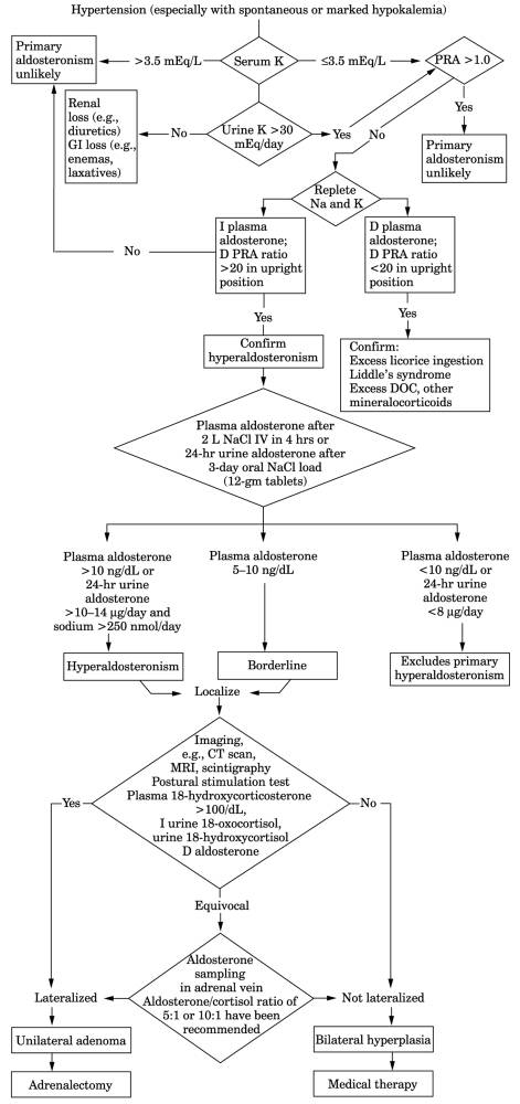
Fig. 13-14. Algorithm for diagnosis of
aldosteronism. (CT = computed tomography; D = decreased; DOC =
deoxycorticosterone; I = increased; MRI = magnetic resonance imaging.)
|
P.645
|
|
|
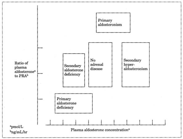
Fig. 13-15. Relationship between plasma
aldosterone concentration and ratio of plasma aldosterone to plasma renin
activity (PRA) in disorders of mineralocorticoid deficiency or excess.
|
- Increased plasma
(reference range = 30-110 ng/L) and/or urinary aldosterone that is
relatively nonsuppressible by salt loading or volume expansion. May be
normal in 30% of cases (due to episodic secretion or chronic potassium
deficiency, which can suppress aldosterone secretion; therefore must
replete potassium before measurement if serum level is <3.0 mEq/L).
Plasma aldosterone level of <8.5 ng/dL after morning saline infusion
rules out primary aldosteronism. Reference values decline by 30-50% with
increasing age. Plasma aldosterone is normal in recumbent hypertensive
and nonhypertensive persons without aldosteronism and increases 2-4×
after 4 hrs of upright posture; increases ≥ 33% in aldosteronism
due to adrenal hyperplasia, but no increase occurs if due to adrenal
adenoma.
- Test for increased
urinary aldosterone (reference range 2-16 µg/24 hrs) is best initial
screening procedure (normal salt intake, no drugs; not detectable on all
days). Cannot be reduced by high sodium intake or deoxycorticosterone
administration. Therefore high sodium chloride intake (10-12 gm/day)
causes 24-hr urine aldosterone level > 14 µg/24 hrs and sodium level
>250 mEq/24 hrs; urine aldosterone level <14 µg/24 hrs rules out
primary aldosteronism except for glucocorticoidremedial type; 96%
sensitivity and 93% specificity.
- Volume expansion (by
high salt intake, infusion of 2 L of sodium chloride in 4 hrs, or
deoxycorticosterone) suppresses aldosterone level by >50-80% of
baseline level in hypertensive patients without primary aldosteronism but
not in patients with primary aldosteronism. (Plasma aldosterone level is
first increased by having patient in upright position for 2 hrs.) Because
plasma aldosterone levels vary from moment to moment, a single specimen
may not properly reflect adrenal secretion.
- PRA fails to rise to
≥ 4 ng/mL 90 mins after stimulus of low-sodium diet,
furosemide-induced volume contraction, and upright posture.
P.646
|
|
|
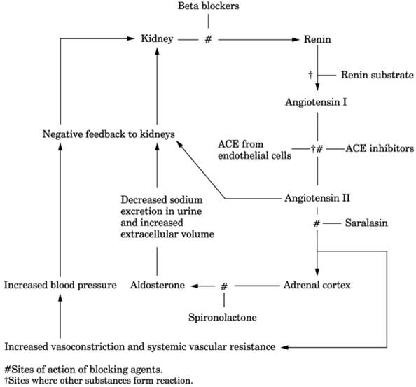
Fig. 13-16. Renin-angiotensin system and
blocking sites. (ACE = angiotensin-converting enzyme.)
|
- Plasma aldosterone/PRA
ratio of ≥ 50 at 8 a.m. or in random blood sample after ambulating
2 hrs in patient not on medication is said to indicate primary
aldosteronism except in cases of chronic renal insufficiency. Does not
distinguish adenoma from hyperplasia.
- Captopril (ACE inhibitor that blocks angiotensin II production) administered as 25 mg IV
at 8 a.m. decreases aldosterone in plasma 2 hrs later in normal persons
and those with essential hypertension but remains elevated in patients
with primary aldosteronism (Fig. 13-17).
|
|
|
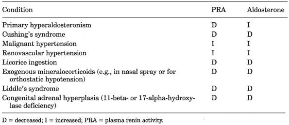
Table 13-18. Differential Diagnosis of
Causes of Hypertension and Hypokalemia
|
|
|
|
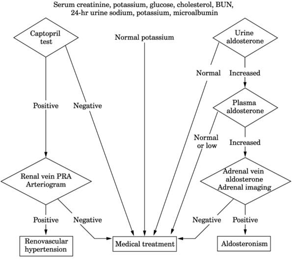
Fig. 13-17. Flow chart for diagnosis of
suspected renovascular hypertension.
|
|
|
|
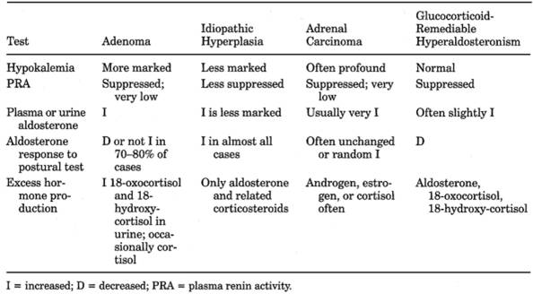
Table 13-19. Differential Diagnosis of
Aldosteronism
|
|
|
Sensitivity (%)
|
Specificity (%)
|
|
Potassium <4.0 mEq/L
|
|
|
|
Stimulated renin <2.5 ng/mL/3 hrs
|
|
|
|
Suppressed aldosterone >10 ng/dL
|
|
|
|
Sequential 1,2 and 3
|
|
|
|
- ○ Basal plasma
18-hydroxycorticosterone level of >100 ng/dL at 8 a.m. supports
diagnosis of aldosteronoma.
- Urine is neutral or
alkaline (pH >7.0) and not normally responsive to ammonium chloride
load.
- Its large volume and low
specific gravity are not responsive to vasopressin or water restriction
(decreased tubular function, especially reabsorption of water).
- Plasma cortisol and ACTH
are normal.
- Urine 17-KS and 17-OHKS
are normal.
- Serum magnesium falls.
- Glucose tolerance is
decreased in ≤ 50% of patients.
- ○ After the
diagnosis of aldosteronism is established, cases due to
tumor (treated surgically) should be distinguished from those due to
idiopathic hyperplasia (treated medically) (see Table
13-19 and Fig. 13-14). Aldosterone
concentration in adrenal vein plasma is higher on side of adenoma,
preferably measured by corticotropin stimulation (90-95% diagnostic
accuracy). Cortisol should also be measured to evaluate accuracy of
adrenal vein sampling. Adenomas can also be localized by CT, MRI, or
scintigraphy with 131I-labeled iodocholesterol after dexamethasone
suppression (uptake increased in adenoma and absent in idiopathic cases
and usually also in carcinoma). Rarely there is unilateral nodular adrenal
hyperplasia similar in function to an adenoma. Patients with adenomas have
higher plasma 18-oxocortisol (>15 µg/d) and 18-hydroxycorticosterone
(>60 µg/d) concentrations, which decrease on standing; plasma
aldosterone also decreases or fails to increase >30% on standing. In
patients with bilateral adrenal hyperplasia and normal persons, plasma
aldosterone increases with upright position. A small subset of hyperplasia
cases mimic adenoma because they are associated with
angiotensin-independent aldosterone overproduction and are cured by
unilateral adrenalectomy.
PRA
- Use
- Particularly useful to
diagnose curable hypertension (e.g., primary aldosteronism, unilateral
renal artery stenosis).
- May help to differentiate
patients with volume excess (e.g., primary aldosteronism) with low PRA
from those with medium to high PRA; if patients in latter group show
marked rise in PRA during captopril test, they should be worked up for
renovascular hypertension, but patients with little or no rise are not
likely to have curable renovascular hypertension.
- Captopril test criteria
for renovascular hypertension: stimulated PRA of ≥12 µg/L/hr,
absolute increase in PRA of ≥ 10 µg/L/hr, increase in PRA of
≥ 150% (or ≥ 400% if baseline PRA is <3 µg/L/hr)
- In
children with salt-losing form of CAH due to 21-hydroxylase deficiency,
severity of disease is related to degree of increase. PRA level may serve
as guide to adequate mineralocorticoid replacement therapy
PRA Is Decreased
(<1.5 ng/ml/3 hrs) in
- 98% of cases of primary
aldosteronism. Usually absent or low and can be increased less or not at all
by sodium depletion and ambulation in contrast to secondary aldosteronism.
PRA may not always be suppressed in primary aldosteronism; repeated
testing may be necessary to establish the diagnosis. Normal PRA does not
preclude this diagnosis; not a reliable screening test.
- Hypertension due to
unilateral renal artery stenosis or unilateral renal parenchymal disease
- Increased plasma volume
due to high-sodium diet, administration of salt-retaining steroids
- 18-25% of essential
hypertensives (low-renin essential hypertension) and 6% of normal controls
- Advancing age in both
normal and hypertensive patients (decrease of 35% from the third to the
eighth decade)
- May also be decreased in
patients with CAH secondary to 11-hydroxylase or 17-hydroxylase deficiency
with oversecretion of other mineralocorticoids
P.649
- Rarely in Liddle's
syndrome and excess licorice ingestion
- Use of various drugs
(propranolol, clonidine, reserpine; slightly with methyldopa)
- Usually cannot be
stimulated by salt restriction, diuretics, and upright posture, which
deplete plasma volume; therefore measure before and after furosemide
administration and 3-4 hrs of ambulation.
- Antihypertensive
and hypotensive drugs should be discontinued for at least 2 wks before
measurement of PRA; spironolactone may cause an increase for up to 6 wks;
estrogens may cause an increase for up to 6 mos
- Blood
should be drawn in an ice-cold tube and the plasma immediately separated
in a refrigerated centrifuge. Renin level should be indexed against 24-hr
level of sodium in urine
PRA May Be
Increased In
- Secondary aldosteronism
(usually very high levels), especially malignant or severe hypertension
(see next section)
- 50-80% of patients with
renovascular hypertension. Normal or high PRA is of limited value to
diagnose or rule out renal vascular hypertension. Very high PRA is highly
predictive but has poor sensitivity. Low PRA using renin-sodium nomogram
in untreated patient with normal serum creatinine argues strongly against
this diagnosis.12
- 15% of patients with
essential hypertension (high-renin hypertension)
- Renin-producing tumors of
the kidney (see Chapter 14)
- Reduced plasma volume due
to low-sodium diet, diuretics, hemorrhage, Addison's disease
- Some edematous
normotensive states (e.g., cirrhosis, nephrosis, congestive heart failure)
- Sodium or potassium loss
due to GI disease, 10% of patients with chronic renal failure.
- Normal pregnancy
- Pheochromocytoma
- Last half of menstrual
cycle (twofold increase)
- Erect posture for 4 hrs
(twofold increase)
- Ambulatory patients
compared to bedridden patients
- Bartter's syndrome
- Use of various drugs
(diuretics, ACE inhibitors, vasodilators; sometimes calcium antagonists
and alpha-blockers, e.g., diazoxide, estrogens, furosemide, guanethidine,
hydralazine, minoxidil, nitroprusside, saralasin, spironolactone,
thiazides)
Aldosteronism,
Secondary
Due To
- Decreased effective blood
volume
- Congestive heart failure
- Cirrhosis with ascites
(aldosteronism 2000-3000 mg/day)
- Nephrosis
- Sodium depletion
- Hyperactivity of
renin-angiotensin system
- Renin-producing renal
tumor (see Chapter 14)
- Bartter's syndrome
- Toxemia of pregnancy
- Malignant hypertension
- Renovascular
hypertension
- Oral contraceptive drug
use
Cushing's
Syndrome
See Table 13-20 and Figs. 13-12 and .
P.650
|
|
|
Table 13-20. Comparison of Different
Causes of Cushing's Syndrome
|
P.651
Due To
- ACTH-dependent
(plasma ACTH is increased): 80%
- Pituitary (Cushing's
disease): 85%
- Pituitary tumor: 70-90%
(may be part of MEN type I;)
- Hyperplasia of pituitary
adrenocorticotropic cells (rare)
- Ectopic CRH syndrome:
<1%
- Ectopic ACTH production:
15%
- Neoplasms (e.g.,
small-cell carcinoma of lung; carcinoids)
- Oat cell carcinoma: 50%
- Tumors of foregut
origin: 35% (e.g., bronchial or thymic carcinoid, medullary thyroid
carcinoma, islet cell tumors)
- Pheochromocytoma: 5%
- Others: 10%
- ACTH-independent
(plasma ACTH is suppressed): 20%
- Adrenal (adrenal cause is
predominant in children)
- Adenoma: >50%
- Carcinoma: <50%; 65%
of patients aged <15 yrs
- Micronodular
hyperplasia: ~1%
- Macronodular
hyperplasia: <1%
- Iatrogenic
- Therapeutic (glucocorticoids
or ACTH)
- Illicit use by athletes
- Factitious
- Pseudo-Cushing's syndrome
- Major depressive
disorder: 1%
- Chronic alcoholism:
<1%
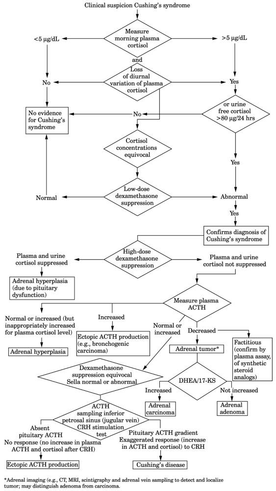
- Definitive diagnosis or exclusion is made only by laboratory tests, which consist of
two parts:
- Establish autonomous
hypercortisolism and loss of diurnal rhythm.
- Determine cause (see Fig. 13-18).
- Diagnosis of excessive cortisol production may include measurement of increased
plasma cortisol (>30 µg/dL at 8 a.m. and >15 µg/dL at 4 p.m.),
measurement of 24-hr urine free cortisol, 17-OHKS, and 17-KS, DST.
Interferences
- More than one test may be
needed because these are misleading in up to one-third of patients for
various reasons:
- Baseline measurements
are increased by stress.
- Baseline measurements
may vary daily, which makes DST difficult to interpret.
- Some drugs alter ACTH
production or interfere with assays.
- Impaired renal function
affects measurements.
- Cortisol production is
somewhat proportional to obesity or large muscle mass.
- Cortisol production is
pulsatile rather than uniform, even in cases of ectopic ACTH production
or Cushing's disease.
- Cortisol secretion may
not be very increased on every determination.
24-Hr Urinary Free Cortisol
Use
- Screening for
- Cushing's syndrome
(increased)
- Adrenal insufficiency
(decreased)
Interpretation
- Increase is most useful
screening test (best expressed as per gram of creatinine, which should
vary by <10% daily; if >10% variation, two more 24-hr specimens
should be collected). Should be measured in three consecutive 24-hr
specimens to ensure proper collection and account for daily variability,
even in Cushing's syndrome. Found in 95% of Cushing's syndrome. <100
µg/24 hrs excludes, and >300 µg/24 hrs establishes, the diagnosis of
Cushing's syndrome. If values are intermediate, low-dose DST is indicated.
P.652
Interferences
- False-positives or
false-negatives are very rare; is more reliable than blood levels, which
vary with time of day, require standardized collection, and are secreted
in pulsatile fashion, making 24-hr urine cortisol preferred test.
- Increased values may
occur in depression or alcoholism but do not exceed 300 µg/24 hrs.
- Alcoholism
- Various drugs (e.g.,
phenytoin, phenobarbital, primidone)
- Acute and chronic
illnesses
- Depression
- Not affected by body
weight.
Plasma Free Cortisol
Use
- Loss of normal diurnal
variation for screening for Cushing's syndrome (normal persons have
highest concentration at 8 a.m. and lowest between 8 p.m. and midnight);
this diurnal variation disappears early and may be absent or reversed in
70% of Cushing's syndrome and 18% of patients without Cushing's syndrome
(due to depression, alcoholism, stress, etc.). Midnight cortisol level
>7.5 µg/dL indicates Cushing's syndrome, whereas level <5 µg/dL
virtually rules it out.
Interferences
- False-negatives are
frequent if blood is drawn before 8 p.m. (p.m. blood is commonly drawn at
4 p.m. to coincide with hospital employee working hours.)
- Because episodic rise and
fall occurs in patients with Cushing's disease or ectopic ACTH production
as well as in normal persons, levels should be measured on at least two
separate days.
- Normal urine-free
cortisol and normal diurnal variation in plasma cortisol virtually exclude
Cushing's syndrome.
- To determine the cause of
Cushing's syndrome after hypercorticalism has been established, the most
useful tests are
- CRH stimulation test
- High-dose DST
- Metyrapone test
- ACTH stimulation test
- DHEA-S concentration
- Plasma ACTH
concentration
Basal Plasma ACTH Concentration
Interpretation
- Cushing's syndrome due to
autonomous cortisol production (e.g., adrenal tumor or exogenous
steroids): low or undetectable.
- Pituitary Cushing's
disease: high or high-normal range but rarely >200 pg/mL. Hyperresponse
to CRH. Inferior petrosal sinus sampling (see next paragraph).
- Ectopic ACTH syndrome
(e.g., carcinoma of lung): very high concentrations with no diurnal
variation. Two-thirds of patients have high concentrations (>200
pg/mL); the other one-third usually have moderately elevated values
(100-200 pg/mL); no response to CRH. In these cases, difference in ACTH
concentrations is measured in blood obtained simultaneously from both
inferior petrosal sinuses and a peripheral vein in basal state and after
CRH stimulation; ratio of inferior petrosal sinus value to peripheral vein
value of ≥ 2 indicates pituitary rather than ectopic source of ACTH;
has sensitivity of 95% and specificity of 100%. Ratio ≥ 3.0 has 100%
sensitivity and specificity for pituitary tumor. New
immunoradiometric (IRMA) assay for ACTH is more sensitive and specific
than RIA method, but some tumors secrete biologically active "large" ACTH
fragments not detected by IRMA; therefore RIA is preferred for initial
evaluation of cause.
Increased In
- Primary adrenal
insufficiency
Decreased In
- Factitious Cushing's
syndrome
- Secondary adrenal
insufficiency
P.653
Interferences
- ACTH has diurnal
variation, episodic secretion, short plasma half-life.
Urinary Steroid Findings in Different Etiologies of
Cushing's Syndrome
- Increased urinary 17-OHKS
>4× normal in
- 63% of patients with
Cushing's syndrome and 3% of patients without Cushing's syndrome
- 65% of patients with
Cushing's syndrome due to ectopic ACTH syndrome
- 3% of patients with
Cushing's syndrome due to adrenal hyperplasia without tumor
- Urinary 17-OHKS is
increased (>10 mg/24 hrs) in virtually all patients with Cushing's
syndrome but less useful for screening because increased in 20% of persons
without Cushing's syndrome (e.g, obesity, hyperthyroidism).
- Night collection sample
is higher than day sample (reverse is true in normal persons).
- ACTH stimulation test
produces lowest 17-OHKS in Cushing's syndrome due to adrenal carcinoma
and highest 17-OHKS in cases due to adrenal adenoma.
- Increased urinary 17-KS
- Cushing's syndrome: may
be normal in 35% of patients and increased (>25 mg/24 hrs) in 20% of
obese persons without Cushing's syndrome. Not useful unless virilism or
marked hirsutism is present.
- Normal or low in 70% of
adrenal adenomas (<20 mg/24 hrs) but increased in 90% of adrenal
carcinomas; averages 50-60 mg/24 hrs in carcinoma (always >15 mg/24
hrs); >4× normal in 50% of adrenal carcinomas; higher values increase
likelihood of diagnosis of adrenal carcinoma and value >100 mg/24 hrs
is virtually diagnostic.
- Adrenal carcinoma: most
of the increase is usually due to DHEA-S, which is markedly increased;
DHEA-S is slightly increased in Cushing's disease and often very low in
adrenocortical adenoma (<0.4 mg/dL).
- Adrenal hyperplasia:
increased total 17-KS (in 50% of cases) is due to elevation of all of the
17-KS.
- Ectopic ACTH syndrome:
increased in 15% of cases.
- Isolated
urine measurements of 17-KS or 17-OHKS are not recommended as screening
tests for Cushing's syndrome. In general, free cortisol is best for
screening, 17-OHKS with free cortisol in DSTs, 17-KS to screen for
possible adrenal carcinoma or to help differentiate adrenal adenoma from
pituitary or ectopic ACTH syndrome causes
- Increased urinary 17-KGS
(>20 mg/24 hrs).
- PRA is increased;
suppressed activity suggests ectopic ACTH syndrome or adrenal adenoma or
carcinoma (causing increased secretion of deoxycorticosterone or
aldosterone).
- Glucose tolerance is
diminished in 75% of cases.
- Glycosuria in 50% of
patients.
- Diabetes mellitus in 20%
of cases.
- Serum sodium is usually
moderately increased.
- ○ Hypokalemic
acidosis due to renal tubular loss of potassium chloride is
characteristic, but compensatory metabolic alkalosis occurs in ~10% of
patients due to attempt to conserve potassium with H
exchange. Hypokalemic alkalosis may indicate ectopic ACTH production
(e.g., bronchogenic carcinoma). Increased serum sodium and bicarbonate and
decreased potassium and chloride is due to increased aldosterone
production.
- Urine potassium is
increased; sodium is decreased.
- Hematologic changes:
- WBC is normal or
increased.
- Relative lymphopenia is
frequent (differential is usually <15% of cells).
- Eosinopenia is frequent
(usually <100/cu mm).
- Hct is usually normal;
if increased, it indicates an androgenic component.
- Changes due to
osteoporosis in long-standing cases. Serum and urine calcium may be
increased.
- Kidney stones occur in
15% of cases.
- Serum uric acid may be
decreased due to uricosuric effect of adrenal steroids.
- Urine creatine is
increased due to muscle wasting, which may also cause increased BUN.
- Serum gamma globulins may
be decreased and alpha globulin may be
moderately increased.
P.654
- 80% of patients with
Cushing's disease have remission after removal of pituitary adenoma; tests
of pituitary-adrenal axis may take weeks to months to become normal.
Effectiveness of surgery is assessed by plasma cortisol and 24-hr urinary
cortisol concentrations in week after surgery.
- Pituitary imaging yields
false-negative scans because many functional tumors are so small (2-3 mm)
and false-positive results because 10-15% of normal persons have
nonfunctioning tumors.
Cushing's
Syndrome, Factitious
- Increased plasma and urinary cortisol
- Plasma ACTH is low or undetectable
- These findings may also occur in adrenal Cushing's syndrome. Differentiate by history
of ingestion or, in some cases, determination of synthetic steroid analogs
by specific plasma assays.
Cushing's
Syndrome Due To Adrenal Disease
- See Fig.
13-18.
- Is suggested by
- Failure of high-dose DST
to cause suppression
- Very low plasma ACTH
level
- Positive metyrapone test
- Adenoma is indicated by
low or normal 17-KS with increased 17-OHKS, low DHEA-S
- Adrenal carcinoma is
suggested by very high 17-KS. Carcinoma cases show hypercorticalism (50%),
virilism (20%), or both (10-15%); are nonfunctioning (10-15%). Virilism favors diagnosis of carcinoma rather than adenoma.
- Nodular adrenal
hyperplasia: ACTH levels are variable, unpredictable response to DST;
therefore is difficult to distinguish from other adrenal causes.
- 50% of bilateral
micronodular hyperplasia cases occur before age 30 yrs.
- 50% occur as autosomal
dominant disorder associated with blue nevi, pigmented lentigines,
myxomas (atrial, skin, mammary), pituitary somatotroph adenomas,
testicular and other tumors.
- Nonfunctioning
adrenal adenoma may be found in 5-10% of healthy persons
Cushing's
Syndrome Due to Ectopic ACTH Production
- (By
neoplasm, e.g., small-cell carcinoma of lung, thymoma, islet cell tumor of
pancreas, medullary carcinoma of thyroid, bronchial carcinoid,
pheochromocytoma; occurs in 2% of patients with lung cancer. The primary
tumor is often radiologically occult.)
- See Table
13-20 and Fig. 13-18.
- Plasma ACTH is markedly increased (500-1000 pg/mL) compared to level in pituitary
Cushing's disease (≤ 200 pg/mL) but values overlap in 20% of ectopic
ACTH cases. Morning basal level in normal persons is 20-100 pg/mL. Extreme
increase suggests ectopic rather than pituitary production.
- ○ Increased plasma
and urine free cortisol, which may show marked spontaneous variation; lack
of diurnal variation.
- Increased ACTH in plasma from inferior petrosal sinus identifies ACTH-producing
pituitary adenomas in ~88% of cases; combining with CRH stimulation
improves the differentiation of pituitary from ectopic ACTH production.
- High-dose dexamethasone suppression does not occur in ectopic ACTH production but does
occur in >90% of cases of Cushing's disease.
- Use of both DST and CRH stimulation has diagnostic accuracy of 98% in distinguishing
Cushing's disease from ectopic ACTH production.
- Metyrapone test may not
be accurate in distinguishing this condition from Cushing's disease.
- Increased urinary 17-OHKS
and 17-KS.
- Marked hypokalemic
alkalosis (due to increased desoxycorticosterone and corticosterone;
occurs in ≤ 60% of such patients) rather than metabolic acidosis may
suggest this diagnosis.
P.655
|
|
|
Fig. 13-18. Sequence of laboratory tests
in diagnosis of Cushing's syndrome. (>90% of patients with Cushing's
syndrome are found to be categorizable using this scheme.) (17-KS = 17
ketosteroids; CRH = corticotropin-releasing hormone; CT = computed
tomography; DHEA = dehydroepiandrosterone; I = increased; MRI = magnetic
resonance imaging; N = normal.)
|
P.656
Cushing's
Syndrome Due to Ectopic CRH Production
- (Usually
due to bronchial carcinoids; clinically indistinguishable from ectopic
ACTH production because most of these tumors also secrete ACTH)
- Plasma CRH increased
- CRH-stimulated secretion
of ACTH suppressed by high doses of dexamethasone may not be present in
many cases.
Feminization,
Adrenal
- (Occurs
in adult males with adrenal tumor [usually unilateral carcinoma,
occasionally adenoma] that secretes estrogens)
- Urinary estrogens are markedly increased
- 17-KS is normal or
moderately increased and cannot be suppressed by low doses of
dexamethasone when due to adrenal tumor.
- 17-OHKS is normal.
- Biopsy of testicle shows
atrophy of tubules.
Glucocorticoid
Resistance Syndromes
- Inability of target tissues to respond to glucocorticoids causes compensatory increase in
pituitary corticotropin, which may cause any combination of excess
secretion of
- Adrenal androgens (may
result in female masculinization with hirsutism, acne, oligomenorrhea,
infertility; precocious puberty; abnormal spermatogenesis)
- Mineralocorticoid excess
(may result in hypokalemic alkalosis, hypertension)
- Apparently normal
glucocorticoid function (patient may be asymptomatic or have chronic
fatigue)
- No evidence of Cushing's
syndrome
- Plasma cortisol is
normal with no loss of diurnal pattern.
- Urine cortisol and
17-OHKS are normal.
- Molecular studies17
Hyperaldosteronism,
Glucocorticoid Suppressible (Remediable)
- (Rare
autosomal dominant defect of zona glomerulosa in which beta-methyloxidase
produces aldosterone from precursor arising in zona fasciculata)
- Usual findings of primary aldosteronism with hypokalemia, increased aldosterone,
and suppressed PRA.
- Reversal of clinical and laboratory findings (suppression of aldosterone secretion)
by dexamethasone for 48 hrs distinguishes this from primary
hyperaldosteronism.
- Characteristic finding is large amounts of metabolites of 18-oxocortisol in
urine.
- Anomalous decrease in
plasma aldosterone response to posture.
- Normal CT and MRI of adrenals.
Hyperaldosteronism,
Normotensive, Secondary (Bartter's Syndrome)
- (Hypokalemia
with renal potassium wasting associated with juxtaglomerular hyperplasia
is resistant to antidiuretic hormone [ADH].)
- Maintaining normal plasma potassium levels is almost impossible despite therapy (dietary
potassium supplement, limiting of sodium intake, drugs such as
indomethacin or ibuprofen).
- ○
Chloride-resistant metabolic alkalosis
- Increased PRA is a characteristic feature
- Insensitive to pressor
effects of angiotensin II (may occur in patients with prolonged
hypokalemia due to any cause)
- Increased plasma and urine aldosterone in the absence of edema, hypertension, or
hypovolemia.
P.657
- Decreased serum magnesium
and increased uric acid frequently occur; often the hypokalemia cannot be
corrected without adequate magnesium replacement.
- Excretion of large
quantities of Na and Cl in urine
- Not due to laxatives,
diuretics, or GI loss of potassium and chloride
Hypertension,
Renovascular
- See Fig.
14-5.
- Sudden increase in serum
creatinine and BUN, especially after onset of ACE-inhibitor therapy. Less
common with other antihypertensive therapy.
- Hypokalemia (<3.4
mEq/L) in ~15% of patients.
- Proteinuria >500 mg/24
hrs usually signifies complete occlusion of a renal artery in a patient
with renovascular hypertension.
- Captopril test causes
- Stimulated peripheral
PRA of 12 µg/L(ng/mL)/hr and
- Increased peripheral PRA
of ≥10 µg/L/hr and
- Increased peripheral PRA
of ≥150%, or 400% if baseline value <3 µg/L/hr.
- Does not differentiate
unilateral and bilateral disease. Less reliable in azotemic patients.
- Reported sensitivity
and specificity are >72%.
- Peripheral PRA (seated patient, drawn in a.m., indexed against sodium excretion) has
only 75% sensitivity and 66% specificity but a low PRA
in untreated patients virtually rules out renovascular hypertension.
- PRA is assayed in blood from each renal vein, inferior vena cava, and aorta or renal
arteries. The test is considered diagnostic when the concentration from
the ischemic kidney is at least 1.5× greater than the concentration from
the normal kidney (which is equal to or less than the concentration in
the vena cava that serves as the standard) or as increment of PRA between
each renal artery and vein. Reported specificity = 80-100%. Reported
sensitivity = 62-80%; may be increased by repeating test after captopril
administration. This is due to high PRA in the peripheral blood, increase
in PRA in the renal vein compared to the renal artery of the affected
kidney, and suppression of PRA in the other kidney. Measurement of
maximum renin stimulation accentuates the difference between the two
kidneys and should always be performed under pretest conditions (avoid
antihypertensive, diuretic, and oral contraceptive drugs for at least 1
mo if possible; low-salt diet for 7 days; administer thiazide diuretic
for 1-3 days; have patient in upright posture for at least 2 hrs). This
is the most useful diagnostic test in renovascular hypertension as judged
by surgical results but is not a sufficiently reliable guide to
nephrectomy in patients with hypertension due to parenchymal renal
disease. In renovascular hypertension, if renal plasma
flow is impaired in the "normal" kidney, surgery often fails to cure the
hypertension. With bilateral renal artery stenosis, most marked
change on side with greatest degree of stenosis. Thus of little value in
patients with bilateral disease.
- Split renal function tests
may show disparity between kidneys.
Hypoaldosteronism
(Hypofunction of Renin-Angiotensin-Aldosterone System)
- Infrequent condition may
be due to
- Addison's disease.
- CAH (methyl oxidase,
type II defect).
- Autosomal recessive
deficiency of aldosterone synthase.
- Prolonged administration
of heparin (very rare).
- Removal of unilateral
aldosterone-secreting tumor (usually transient).
- Autonomic nervous system
dysfunction; aldosterone deficiency causes impaired renal sodium
conservation but without hyperkalemia.
- Idiopathic hyporeninism.
- Associated with mild
renal insufficiency (especially diabetic nephropathy, some interstitial
nephropathies).
P.658
|
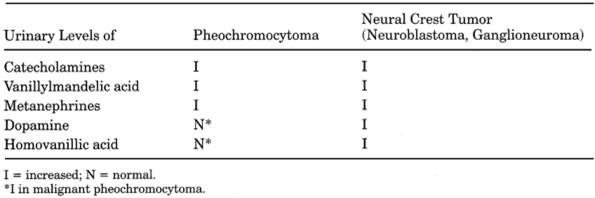
|
|
Table 13-21. Laboratory Tests in
Differential Diagnosis of Benign Pheochromocytoma and Neural Crest Tumors
(Neuroblastoma, Ganglioneuroma)
|
- Hyperkalemia,
hyponatremia, urinary sodium loss, hypovolemia corrected by administration
of mineralocorticoids
- Mild hyperchloremic
metabolic acidosis
- Decreased aldosterone and PRA that are not increased by combined diuretic and posture
establish the diagnosis.
- Normal adrenal
glucocorticoid response to ACTH stimulation test
- Laboratory findings of
associated diseases (e.g., diabetes mellitus, gout, pyelonephritis)
Neuroblastoma,
Ganglioneuroma, Ganglioblastoma
- See Table
13-21.
- Urinary concentrations of catecholamines (norepinephrine, normetanephrine,
dopamine, VMA, and HVA) are increased. Excretion of epinephrine is not
increased because of rapid catabolism. If only one of these substances is
measured, only ~75% of cases are diagnosed. If VMA and HVA or VMA and
total catecholamines are measured, 95-100% of cases are diagnosed.
- These tests are also useful for differentiating Ewing's tumor from metastatic
neuroblastoma of bone and to show response to therapy (surgery,
irradiation, or chemotherapy), which should bring return to normal in 1-4
mos. Continued increase indicates need for further treatment.
- Cystathionine in urine
suggests active disease but absence is not significant because it is not
normally present.
- Serum neuron-specific
enolase may be increased in neuroblastoma; high level is associated with
poor prognosis. Ratio of neuron-specific to nonneuronal enolase is
reported to improve specificity to >85% for neuroblastoma.
- Laboratory findings due
to metastases (e.g., tumor in biopsy of marrow, liver, or other sites,
anemia, etc.)
Pheochromocytoma
- (Tumor
of chromaffin cells of sympathetic nervous system; may secrete
epinephrine, norepinephrine, dopamine. Occurs in 0.1-0.2% of hypertensive
population in the United
States. Five percent of patients with
pheochromocytoma have normal blood pressure most or all of the time.
Sustained hypertension in 50% of cases.)
- See Figs.
13-19, , Tables 13-21
and .
- Diagnosis is based on increased blood or urine concentrations of catecholamines
(norepinephrine and, to a lesser extent, epinephrine) and their
metabolites (normetanephrine and metanephrine), which are usually
increased even when patient is asymptomatic and normotensive; rarely are
increases found only after a paroxysm. When other studies are negative, a
timed urine specimen or plasma concentration for catecholamines and
metabolites taken after a typical "spell" may be useful. However, repeated
testing may be necessary. Concentrations of >400 pg/dL (normal <100
pg/dL) for epinephrine or >2000 pg/dL (normal <500 pg/dL) for
norepinephrine
P.659
|
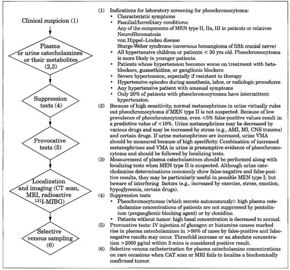
|
|
Fig. 13-19. Algorithm for diagnosis of
pheochromocytoma. (CAT = computerized axial tomography; CT = computed
tomography; I-MIBG =
metaiodobenzylguanidine labeled with iodine 131; MAO = monoamine oxidase; MEN
= multiple endocrine neoplasia; MRI = magnetic resonance imaging; VMA =
vanillylmandelic acid.)
|
are considered diagnostic. Concentrations are usually
5-100× normal, although considerable overlap and wide range of normal values
are seen. Intermediate values require further workup. Measurement of plasma
concentrations is particularly useful to compare paroxysm and basal
concentrations and to localize tumors by selective venous sampling. Blood
should be drawn in the unstressed supine patient without interfering conditions
or drugs. 24-hr urine free norepinephrine has reported sensitivity of 89-100%
and specificity of 98%; plasma norepinephrine has sensitivity of 82% and
specificity of 95%.19 In one study, plasma metanephrines
were more sensitive than plasma catecholamines or urine metanephrines; normal
plasma metanephrines excluded pheochromocytoma.
P.660
|
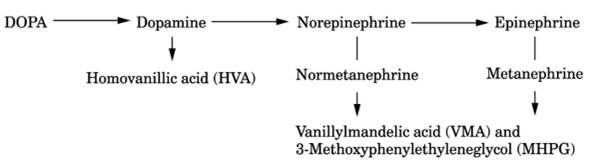
|
|
Fig. 13-20. Synthesis and breakdown of
catecholamines. Because the hormones are broken down before release,
metabolites are present in much larger amounts. When excretion of free
catecholamines is greater than that of metabolites, tumor is said to be
likely to be very small and difficult to locate.
|
Another report found 24-hr urine metanephrine values of
>0.9 mg to have sensitivity of 100% and positive predictive value of 83%.21 In a recent series plasma
normetanephrine or metanephrine had the highest sensitivity (97%) in patients
with familial predispositions; MEN type II patients had high plasma
metanephrine concentrations and von Hippel-Lindau disease patients had high
plasma concentrations of only normetanephrine.
Secretory Patterns
- Normally: epinephrine is
secreted primarily by adrenal medulla and norepinephrine is secreted
primarily at sympathetic nerve endings.
- Epinephrine is secreted
by tumors, usually of adrenal medulla, and causes characteristic symptoms.
- Norepinephrine is
secreted by almost all extra-adrenal tumors and many adrenal tumors; often
associated with sustained hypertension and hypermetabolism.
- Dopamine secretion is not
associated with hypertension.
- Malignancy: increased
dopamine and almost as much norepinephrine with very low epinephrine.
- Part of familial
syndrome: more likely to secrete both dopamine and epinephrine.
- Most common: increased
norepinephrine predominant with much less epinephrine and dopamine.
- Most patients show
increase of two or more catecholamines.
- Less common: equal
norepinephrine and epinephrine and some dopamine.
- Predominance of
epinephrine suggests tumor in adrenal or organ of Zuckerkandl; rarely
bladder or mediastinum.
- Isolated increase of
either adrenaline or dopamine is relatively common in normal persons but
uncommon in pheochromocytoma patients.
- Repeated urine pattern of
secretion is consistent in pheochromocytoma but some normal persons show
large changes.
- Presence of HVA is said
to suggest malignancy.
- Increased catecholamine
concentrations after surgical removal may indicate recurrence of tumor.
- When urine catecholamines
are fractionated, both epinephrine and norepinephrine must be measured
because some tumors produce only one of these hormones.
Catecholamines, Plasma/Urine
- Plasma
concentrations may not be increased when secretion is intermittent rather
than continuous; for these cases 24-hr urine values are more accurate.
Plasma concentrations are useful if 24-hr urine cannot be collected
P.661
|
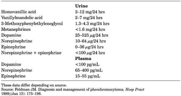
|
|
Table 13-22. Reference Range
for Catecholamines and Metabolites
|
Increased In
- Pheochromocytoma
- Neural crest tumors
(neuroblastoma, ganglioneuroma, ganglioblastoma)
- Adrenal medullary
hyperplasia
- Diabetic ketoacidosis
(markedly elevated)
- AMI (markedly elevated)
- Acute CNS disturbance (e.g.,
infarct, hemorrhage, encephalopathy, tumor)
- Progressive muscular
dystrophy and myasthenia gravis (some patients)
- May also be increased by
vigorous exercise before urine collection (<7×)
- Stress (emotional,
physical, postsurgery)
- Hypothyroidism
- Thyrotoxicosis
- Volume depletion (induced
by diuretics)
- Renal disease
- Heavy alcohol intake
- Hypoglycemia
- Has also been reported in
Guillain-Barré syndrome, acute intermittent porphyria, carcinoid syndrome,
acute psychosis
Interferences
- False increase may be due
to drugs that produce fluorescent urinary products (e.g., tetracyclines,
methyldopa (Aldomet), epinephrine and epinephrine-like drugs [nose drops,
cough and sinus remedies, bronchodilators, appetite suppressants], large
doses of vitamin B complex).
- Plasma
catecholamines decrease markedly after 5 mins if RBCs are not separated
from plasma
- Drugs that destroy
catecholamines in bladder urine, e.g., methenamine mandelate
- Not all methods include
dopamine in determination of urine total catecholamines.
- Urine norepinephrine/normetanephrine
may be less reliable than VMA or metanephrines in screening for
pheochromocytoma due to technical problems; best used to confirm diagnosis
(using HPLC) when other tests are equivocal.
- Avoid
medications for 1 wk before sampling
- Many drugs reported to
increase values of catecholamines or metabolites, including alpha -blockers,
aminophylline, amphetamines, ampicillin, beta-blockers, caffeine,
chlorpromazine, diazoxide, drug withdrawal (alcohol, clonidine),
epinephrine, ephedrine, imipramine, isoproterenol, labetalol, methyldopa,
monoamine oxydase inhibitors, nicotine, phenacetin, phenothiazine,
quinidine, theophylline, vasodilators (e.g., minoxidil, hydralazine
nitroglycerine, sodium nitroprusside), calcium channel blockers (acutely).
P.662
- Many drugs reported to
decrease values of catecholamines or metabolites, including anileridine,
aspirin, PAS, alpha agonists, bromocriptine,
sodium sulfobromophthalein (Bromsulphalein), calcium channel blockers
(long-term use), cimetidine, clofibrate, clonidine, chlorpromazine,
disulfiram, glyceryl guaiacolate, guanethidine, imipramine, isoproterenol,
L-dopa, monoamine oxidase inhibitors, propranolol, mephenesin,
methocarbamol, methyldopa, metyrosine, nalidixic acid, penicillin,
phenazopyridine, PSP, reserpine, sulfa drugs, thyroxine.
Urine VMA
- VMA is the urinary
metabolite of both epinephrine and norepinephrine. Excretion is
considerably increased in ~90% of patients. Because this analysis is
simpler than that for catecholamines, it has been more commonly used, but
it is less sensitive than other tests.
Increased In
- Pheochromocytoma
- Neuroblastoma,
ganglioneuroma, ganglioblastoma
Interferences
- Beware
of false-positive results due to ingestion of certain foods within 72 hrs
before the test (e.g., coffee, tea, chocolate, vanilla, some fruits and
vegetables, especially bananas) and drugs
- Beware of nonspecific
techniques for VMA assay that fail to detect 30% of cases of
pheochromocytoma.
Urine Metanephrines
- Is reliable screening
test as false-negatives = 4% and fewer interferences by drugs and diet are
seen than with VMA or catecholamines.
- Confirmation by urine
catecholamine fraction determinations has been considered an excellent
routine to identify pheochromocytoma patients.
- Plasma chromogranin A, a
marker for pheochromocytoma, has ~50% sensitivity.
- Hyperglycemia and
glycosuria are found in 50% of patients during an attack.
- GTT frequently shows a
diabetic type of curve; many patients develop clinical diabetes mellitus.
- Thyroid function tests
are normal.
- Urine changes are
secondary to sustained hypertension.
- PRA activity is
increased.
- Relative erythrocytosis
sometimes occurs.
- Increased incidence of
cholelithiasis
- Other hormones may be secreted
(e.g., serotonin, PTH, calcitonin, ACTH, gastrin, VIP, FSH, insulin).
Rarely, can cause Cushing's syndrome and hypercalcemia.
- 15% of
pheochromocytomas are extra-adrenal; 10% are multiple. 10% occur in children,
two-thirds of whom are male.
- 2-10%
of adrenal and 20-40% of extra-adrenal pheochromocytomas are malignant
- Familial inheritance in
10-20% of patients; 70% of these are bilateral. Associated with certain
neurocutaneous syndromes (e.g., von Hippel-Lindau disease [in ~20% of
cases], von Recklinghausen's disease, tuberous sclerosis).
- All
patients with pheochromocytoma should be screened for other components of
MEN type IIa and IIb present in ~4% of cases. Tumors associated with
familial syndromes are more likely to be asymptomatic, multiple, and extra-adrenal
Pseudo-Cushing's
Syndrome
Due To
- Major depressive
disorders: cortisol secretion is abnormal in 80% of these patients but
hypersecretion is usually minimal and transient; disappears with remission
of depression.
- . Evening nadir in plasma cortisol is preserved; level <5 µg/dL rules out
and >7.5 µg/dL indicates Cushing's syndrome.
- . Plasma cortisol
is low after administration of dexamethasone and remains low when CRH is
given soon after, whereas in Cushing's syndrome, plasma cortisol is not so
low after dexamethasone and increases after CRH.
P.663
- . Insulin-induced
hypoglycemia causes increased plasma cortisol but not in chronic Cushing's
syndrome.
- Chronic
alcoholism-abnormal liver function tests; resolves during abstinence as
liver function returns to normal.
Pseudoaldosteronism
Due to Ingestion of Licorice (Ammonium Glycyrrhizate)
- (Excessive
ingestion causes hypertension due to sodium retention)
- Decreased serum potassium
- Decreased aldosterone
excretion in urine
- Decreased PRA
- Urinary glycyrrhetinic acid can be measured by gas chromatography and mass spectrometry.
- Unstimulated
renin-aldosterone system may be suppressed for ≤ 4 mos after
cessation of long-term ingestion of licorice. Effect on electrolyte
balance may persist for ≤ 1 wk after cessation.
Pseudohyperaldosteronism
(Liddle's Syndrome)
- (Rare
familial nephropathic disorder [possibly at distal tubule] with clinical
manifestations closely resembling those due to aldosterone-producing
adrenal adenoma)
- Hypokalemia due to renal
potassium wasting
- Metabolic alkalosis
- Hypertension
- ○ All are corrected
by long-term administration of diuretics that act at distal tubule to
cause natriuresis and renal potassium retention (e.g., triamterene or
amiloride) and by restriction of sodium.
- Aldosterone secretion and excretion are greatly reduced and unresponsive to
stimulation by ACTH, angiotensin II, or low-sodium diet.
- Low plasma renin
- Sodium retention
Pseudohypoaldosteronism
Heterogeneous group
of disorders due to resistance to aldosterone action with signs and symptoms of
aldosterone deficiency, but aldosterone and PRA levels are markedly increased
and are resistant to mineralocorticoid therapy.
Tests
of Gonadal Function
Chromosome
Analysis
- Turner's syndrome
(gonadal dysgenesis): usually negative for Barr bodies
- Klinefelter's syndrome:
positive for Barr bodies
- Pseudohermaphroditism:
chromosomal sex corresponding to gonadal sex
Cytologic
Examination of Vaginal Smear (Papanicolaou Smear) for Evaluation of Ovarian
Function
- Maturation index is the
proportion of parabasal, intermediate, and superficial cells in each 100
cells counted.
- Lack of estrogen effect
shows predominance of parabasal cells (e.g., maturation index =100/0/0).
- Low estrogen effect
shows predominance of intermediate cells (e.g., maturation index =
10/90/0).
P.664
- Increased estrogen
effect shows predominance of superficial cells (e.g., maturation index =
0/0/100), as in hormone-producing tumors of ovary, persistent follicular
cysts.
Some
Patterns of Maturation Index in Different Conditions
|
|
Index
|
|
Childhood
|
|
Normal
|
|
|
Cortisone
therapy
|
|
|
Childbearing years
|
|
Preovulatory
(late follicular) phase
|
|
|
Premenstrual
(late luteal) phase
|
|
|
Pregnancy
(second month)
|
|
|
Cortisone
therapy
|
|
|
Amenorrhea
after ovarian irradiation
|
|
|
Surgical
oophorectomy
|
|
|
Bilateral
oophorectomy and adrenalectomy
|
|
|
Postmenopausal years, early (age 60)
|
|
|
Postmenopausal years, late (age 75)
|
|
Untreated
|
|
|
Moderate
estrogen treatment
|
|
|
High-dose
estrogen treatment
|
|
|
Years after
bilateral oophorectomy
|
|
|
Postadrenalectomy,
bilateral
|
|
|
- Karyopyknotic index is
the percentage of cells with pyknotic nuclei. Increased estrogen effect
(e.g., karyopyknotic index ≥ 85%) is seen, as in cystic glandular
hyperplasia of the endometrium.
- Eosinophilic index is the
percentage of cells showing eosinophilic cytoplasm; it may also be used as
a measure of estrogen effect.
- Combined
progesterone-estrogen effect: No quantitative cytologic criteria are
available. Endometrial biopsy should be used for this purpose.
- The
pattern may be obscured by cytolysis (e.g., infections, excess bacilli),
increased red or white blood cells, excessively thin or thick smears, or
drying of smears before fixation (artificial eosinophilic staining)
Estrogens
(Total), Serum
(Includes
estradiol produced by ovaries and placenta, and smaller amounts by testes and
adrenals; estrone and estriol)
Increased
In
- Granulosa cell tumor of
ovary
- Theca-cell tumor of ovary
- Luteoma of ovary
- Pregnancy
- Secondary to stimulation
by hCG-producing tumors (e.g., teratoma, teratocarcinoma)
- Gynecomastia
Decreased
In
- Primary hypofunction of
ovary
- Autoimmune oophoritis is
the most common cause. Usually associated with other autoimmune
endocrinopathies, e.g., Hashimoto's thyroiditis, Addison's disease,
insulin-dependent diabetes mellitus. May cause premature menopause.
- Resistant-ovary
syndrome.
- Toxic (e.g.,
irradiation, chemotherapy).
- Infection (e.g., mumps).
- Tumor (primary or
secondary).
- Mechanical (e.g.,
trauma, torsion, surgical excision).
- Genetic (e.g., Turner's
syndrome).
- Menopause.
- Secondary hypofunction of
ovary
- Disorders of
hypothalamic-pituitary axis
P.665
Follicle-Stimulating
Hormone (FSH) and Luteinizing Hormone (LH), Serum
(Pituitary
gonadotropins)
Use
- Differential diagnosis of
gonadal disorders
- Diagnosis and management
of infertility
Increased
In
- Primary hypogonadism
(anorchia, testicular failure, menopause)
- Gonadotropin-secreting
pituitary tumors
- Precocious puberty
(secondary to a CNS lesion or idiopathic)
- Complete testicular
feminization syndrome
- Luteal phase of menstrual
cycle
