 |
|
||||||||||||||
 |
|
||||||||||||||
ABFRACTION, ABRASION, ATTRITION, AND EROSION - James W.
Curtis Jr., DMD, Beverley A. Farley, DMD, Ronald E. Goldstein, DDS
INTRODUCTION
Modern dental practices frequently encounter patients who exhibit various forms
of wear to the dentition. The wear may present as abfraction, abrasion,
attrition, and/or erosion. During their lifetime, many people will experience the
effects of one or more of these conditions. The stresses of today's fast-paced
lifestyle may lead to various habits that can directly cause or contribute to
these problems. The etiologies of abfraction, abrasion, attrition, and erosion
may be interrelated. Therefore, multiple conditions may be seen in a single
patient (Figures 17-1A to
C

Figure 17-1A to C: These photographs illustrate the complex dental condition of a 71-year-old male: (A) The palatal cervical regions of the maxillary anterior teeth exhibit sharp, wedge-like lesions that are characteristic of abfraction. These areas would be difficult, if not impossible, to have resulted from toothbrush abrasion. (B) The buccal aspects of the maxillary posterior teeth show smooth, concave configurations that are consistent with toothbrush abrasion and/or erosion. (C) The buccal surfaces of the mandibular teeth have lesions that possess components of both abfraction (sharp margins in the occlusal regions) and abrasion (concave geometry and gingival recession in the cervical regions).
A review of the literature frequently reveals confusion, controversy, and
contradiction concerning the terminology and etiology related to the loss of
tooth structure due to noncarious processes. For example, erosion, as used in
the dental literature, indicates the loss of tooth structure due to chemical
dissolution. However, corrosion is the better term to denote the physical
deterioration of a material (including teeth) by a chemical or electrochemical
process. Erosion is actually the abrasive destruction of a material that occurs
as a result of movement of liquid or gas, with or without solid particles, over
the surface of the material. In this chapter, each term will be defined in an
effort to eliminate any confusion.
Often, the lines between the chemical and physical forces that cause noncarious
tooth structure loss are blurred. When the etiologic factors of more than one
of these conditions are simultaneously present, the resultant loss of tooth
structure will be accelerated or magnified. As an example, bulimics who brush
their teeth immediately after regurgitation may increase the rate of enamel
loss. This is due to the greater effect of abrasion on acid-etched enamel. In
assessing these various conditions, the possibility of multifactorial etiology
should always be kept in mind.93
In the new millennium, it is important to recognize that wear of the dentition
has been present since the origin of humankind. Young provides an extremely
interesting summary of the literature relating to dental wear in the aboriginal
populations of
In 1958, Barrett noted that teeth in modern populations rarely exhibit the
patterns of wear seen occurring in a natural fashion in aboriginal
civilizations.5 Herein lies part of the dilemma faced by the
profession today. Radical changes in our environment and diet over the past few
centuries have altered the extent and type of wear present in teeth.64
These same dietary alterations have increased the prevalence of plaque-related
dental diseases and the subsequent effects of such diseases.61
Further compounding the picture are cultural shifts that have led to heightened
awareness and the demand for esthetic dentistry.109 Ultimately, as a
profession, it is important that we recognize the anthropologic evidence
related to tooth wear and the consequences of basic stomatognathic function on
the longevity of teeth and restorations.
ABFRACTION
Abfraction is a wedge-shaped cervical lesion that results from repeated tooth
flexure caused by occlusal loading. Other terms have also been suggested for
this phenomenon, including noncarious cervical lesions and stress corrosion.
Although these lesions have been recognized for years, their etiology has been
debated. Numerous hypotheses were put forward over time to explain the cause of
these lesions. The most common theory was that of toothbrush abrasion occurring
independently or in conjunction with acid erosion.88 However, the
sharp angles and frequent subgingival location of these cervical lesions cannot
be adequately explained by any of the previous hypotheses. It was not until the
early to mid-1980s that the concept of tensile stress as the etiology of these
lesions came to the forefront.66,75,76 Sufficient experimental and
clinical evidence has now been garnered to establish the primary etiology of
these lesions as tensile stress of occlusal origin.4,19,26,34,42,44,46,47,65,68,87,98
However, even in light of strong scientific evidence, this topic remains highly
controversial.
As lateral occlusal forces are generated during mastication and parafunction,
flexure of the tooth occurs at the cervical fulcrum (Figure 17-2). This flexure concentrates tensile
stresses that disrupt the chemical bonds of the crystalline structure of enamel
and dentin. Small molecules then enter the microfractures and prevent the
reformation of the chemical bonds. Loss of tooth structure ultimately occurs in
the regions of concentrated stresses. After the initiation of these lesions,
they may be accelerated by acid erosion and/or abrasion.
When abfractions are restored, there tend to be relatively high failure rates
if the occlusal problems that initiated the lesions are not corrected. This is
true for both nonbonded and bonded restorations.23,36,45,50,51,73,77,82,85,87
Although not reported in the literature, some clinicians suspect that the
occasional case of facial porcelain debonding in ceramic-metal crowns used to
restore teeth with abfractions is caused by the same stresses that caused the
original lesions. This apparently occurs when the facial crown margin has been
placed at the same level as the apical aspect of the abfraction and the
occlusal disharmony has not been corrected. Again, the key to restorative
success for abfraction is control of the destructive occlusal forces that
initially caused the lesions.

Figure 17-2: Model of tensile stress etiology of abfraction. Lateral forces create tension and compression in the cervical region, as indicated by arrows. Magnified section shows disruption of hydroxyapatite crystals of enamel and microfractures of dentin. When small molecules enter microcracks, re-establishment of chemical bonds is prevented. These areas are more susceptible to destruction from factors such as abrasion and chemical dissolution. (Reproduced with permission from Lee WC, Eakle S. Stress-induced cervical lesions: review of advances in the past 10 years. J Prosthet Dent 1996;75:488.67)
Case Studies
A 34-year-old female presented for routine examination. She expressed no
complaints related to tooth sensitivity or occlusal dysfunction. Sharp angles
of the cervical lesions on the mandibular left first premolar and first molar
(mesial root) can be seen in Figure 17-3A. Although the lesions are darkly
stained, the dentin and cementum are not cariously involved. There is no
associated gingival recession on these teeth. Further clinical examination
revealed other abfractions. Occlusal analysis demonstrated centric relation
prematurities and a 2.5-mm left anterolateral shift from centric relation to
maximum intercuspation. During left lateral movement, the patient exhibited
group function, with the heaviest contacts occurring on the teeth with the
abfractions (Figure 17-3B). The patient was questioned in
detail after the clinical examination and eventually disclosed occasional
muscle symptoms that helped support the diagnosis of nocturnal bruxism.

Figure 17-3A: This 34-year-old woman does not show caries or gingival recession associated with the cervical lesions of the first premolar and mesial root of the first molar.

Figure 17-3B: During left lateral movement, the patient exhibited group function, with the heaviest contacts occurring on the teeth with the abfractions.
The case is a 42-year-old male with a complaint related to the space between
the maxillary left central and lateral incisors. He reported that the space had
been present as long as he could remember, but that it had increased over time.
Although he was aware of the notching defect on the central incisor, he could
not recall how long it had been present (Figure 17-4A). The abfraction extended from the
midfacial to the distolingual line angle. The discrepancy in the sizes of the
maxillary central incisors can be seen in the figure. There was significant
gingival recession, particularly on the distal aspect of the left central
incisor, that was accompanied by gingival inflammation and a probing defect of
4 to 5 mm. Further, there was a marked right shift of the dental midline and
the solid contact between the maxillary and mandibular left central incisors in
protrusion (Figure 17-4B

Figure 17-4A: This 42-year-old male has a space between the left lateral and central incisors.

Figure 17-4B: It is easy to see that traumatic occlusion could have played a role in the development of the cervical lesion on the distolabial area.
This
28-year-old man has an asymptomatic abfraction with gingival recession on the
maxillary right first premolar. The angle of the lesion is extremely acute and
extends approximately 2 mm into the facial surface of the tooth at its greatest
depth (Figure 17-5). Examination reveals
malpositioning of the mandibular canine and a hint of abfractions on the
mandibular premolars and shows a major mesiofacial wear facet on the affected
maxillary premolar.

Figure 17-5: This 28-year-old man has asymptomatic abfraction and gingival recession as well as malpositioning of the mandibular canine and a major wear facet on the affected maxillary premolar.
A 60-year-old woman presented with posterior bite collapse and poor health of
the dentition despite having received extensive dental care over the years.
Stained abfractions on the maxillary premolars and canine can be seen in Figure 17-6. Although the dentin was markedly
stained, it was hard and noncarious. The mandibular left lateral incisor also
exhibited an abfraction. Her mandibular partial denture (not shown) had been
fabricated approximately 12 years previously and accommodated the gross
occlusal plane discrepancy.

Figure 17-6: This 60-year-old woman exhibited stained abfractions on the maxillary cuspid and premolars that are noncarious but require restoration.
A 70-year-old male presented with an extreme loss of tooth structure owing to
abfraction. Figure 17-7 shows the severe nature of the
abfractions on the palatal surfaces of all posterior teeth on the right side.
Both molars had exposures of the pulp chambers due to the abfractions. The pulp
tissue in the second molar was clearly visible and vital, but the first molar
was necrotic. Neither tooth was symptomatic. The maxillary left region was
similarly involved. The mandibular arch also had generalized abfractions, but
they were not as severe as those in the maxilla.

Figure 17-7: This is an excellent example of how extreme loss of tooth structure due to abfraction can exist and invade the pulp chamber without causing any symptoms.
ABRASION
The loss of tooth structure due to repeated mechanical contact with objects
other than teeth is termed abrasion. This process is considered to be
pathologic. Any object placed against the teeth can cause abrasion. Evidence
exists of various forms of abrasion in prehistoric populations.2,24,39,40,110
A number of dental specimens recovered from the Sima de los Huesos Middle
Pleistocene cave site in Spain exhibited a particular type of interproximal
grooves between the posterior teeth. The grooves were found only in adults and
were apparently caused by the habitual probing of the interdental spaces with
rigid objects (ie, prehistoric toothpicks). Particles in the diet likely
enhanced this abrasive phenomenon.18 This same condition is seen in
present-day societies. Other articles present information on various forms of
tooth sharpening.80,108
Numerous oral habits cause abrasion; these are further discussed in Chapter 20. Examples of these habits include
the localized occlusal defects seen in some pipe smokers who clench the pipe stem
or individuals who chew on pens and pencils. Incisal notching is fairly common
among seamstresses who hold pins or needles between the anterior teeth. Unusual
cases of abrasion are reported in the dental literature and are discussed in Chapter 20.
Case Studies
The most conservative restoration of Class V defects is composite resin
bonding. It generally requires little or no tooth reduction, thereby retaining
as much tooth structure as possible to an already compromised tooth. A typical
procedure can be seen in Figures 17-8A
and B 17-8C 17-8D 17-8E 17-8F 17-8G 17-8H and I, which show a 45-year-old man with
evidence of gingival and incisal abrasion, erosion, and abfraction. It is
important to convey to patients that by treating these types of defects as
early as possible, less tooth structure is lost and more enamel is present to
enable a stronger bonded restoration.

Figure 17-8A and B: This 45-year-old man shows extreme tooth loss due to combination lesions both gingivally and incisally of abrasion, erosion, and abfraction.

Figure 17-8C: An air polisher is used to remove stain.

Figure 17-8D: Note how clean the teeth appear after a thorough prophylaxis with an air polisher.

Figure 17-8E: A rubber dam is placed, mylar strips are applied, and the teeth to be restored are etched.

Figure 17-8F: A dentin/enamel bonding agent is applied, then a dentin/enamel resin, and finally an appropriate tooth- colored microfilled composite resin is placed using a Goldstein #3 composite instrument (Hu-Friedy, Chicago, IL).

Figure 17-8G: Careful shade selection and attention to detail should produce an invisible margin.

Figure 17-8H and I: Although early intervention is the best approach, restoring the defects at any point is both functionally and esthetically beneficial. Bonding is especially effective in preventing further damage to the tooth surface it covers.
Figure 17-9A shows a 29-year-old female who has
abrasion and gingival recession confined to the anterior left segment,
involving the canine and two incisors on that side. Closer examination revealed
the smooth, rounded nature of the abraded areas (Figure 17-9B). Although she could not recall her
specific age at the time, she reported that she was told by a hygienist that
her brushing technique was improper when she was a teenager. She stated that
she was instructed in brushing and flossing by this hygienist and had noted no
progression of the recession since that time. For the past 8 years, she has
been a patient in the same dental practice, and the clinical charting indicates
that there has been no worsening of the problem. She has been informed about
gingival surgery to correct the defects but has declined since she does not
show her teeth when smiling.

Figure 17-9A: A 29-year-old female with abrasion confined to the maxillary left canine and lateral and central incisors.

Figure 17-9B: Close examination reveals smooth, rounded, abraded areas that are suspected to be the result of improper brushing.
ATTRITION
Attrition is the loss of tooth structure from tooth-to-tooth contact. According
to the classic definition, attrition was considered to be a physiologic process
occurring primarily from tooth contact during mastication. The wear from
attrition may be seen on the occlusal surfaces of posterior teeth, the incisal
edges of anterior teeth, the palatal surfaces of maxillary anterior teeth, and
the labial surfaces of mandibular anterior teeth. The affected surfaces are
usually hard, smooth, and shiny. However, the teeth may be sharp and jagged in
certain cases. The areas of attrition may exhibit a yellowish-brown
discoloration if the wear has penetrated the enamel. Wear may also occur
interproximally, causing mesial drifting and broadening of proximal contacts.
Young mouths typically do not exhibit severe attrition. However, wear may be
seen in the primary and mixed dentitions (Figure 17-10). Numerous articles have reported
on wear in children and adolescents.1,8,53,78,79,84,95,96 As
expected, increasing wear is seen with increasing age. This, as well as the
fact that men exhibit more wear as they age, was demonstrated in a study of 586
subjects aged 45 and older.35

Figure 17-10: This young girl demonstrated severe wear and attrition in her mixed dentition.
There is growing evidence that attrition is more likely the result of
pathologic conditions and environmental factors. Thus, the classic definition
of attrition as an entirely physiologic process is being challenged. It is well
established that teeth rarely contact during mastication, and there are many
other factors that more likely contribute to this type of wear. These factors
include bruxism, clenching, diet, malocclusion, and abrasive particles in the
environment.12,22,32,52,55,56,60,72,74,89,90,94,97 Dust in the tree
top canopy has been shown to contribute to wear in primates.107
Chronic exposure to dust and dirt can also cause increased wear in humans. This
can occur in agricultural settings49 or be associated with various
industrial settings, such as cement factories.102
Bruxism
Bruxism can lead to extreme loss of occlusal and incisal tooth structure (Figures 17-11A
to C 17-11D and E 17-11F to H 17-11I and J). For example, a young woman was
treated for her defective restorations in 1968 (see Figures 17-11D
and E).
Although she continued with routine maintenance appointments for a few years,
she never accepted the advice to have a bite appliance constructed to treat her
bruxism habit. Thirty-one years later, she returned with an extremely worn
dentition as seen in Figures 17-11F
to H. Crown
lengthening and full crowns were necessary to restore this patient's smile.

Figure 17-11A to C: This case illustrates the severe damage that can be caused by bruxism. The patient is a 56-year-old male who reports that his wife tells him he grinds his teeth while she is trying to sleep. He is also a farmer and is exposed to dust for extended periods of time for much of the year. The combined bruxism and environmental factors have likely contributed to the extreme wear present. As is most commonly seen with cases in which the wear progresses slowly, there has been no discernible loss of vertical dimension, as evidenced by lip position and speech patterns. Note the traumatic occlusal relationship when the patient is in complete intercuspation (A). Views of the severe wear of the maxillary and mandibular arches. Note the calcified, exposed pulp chambers and caries (B and C).

Figure 17-11D and E: This young woman's defective amalgam restorations were replaced in 1968 with tooth- colored restorations. At that time, and during the ensuing few years of maintenance recalls, she was advised to have a bite appliance constructed for her severe bruxism habit.

Figure 17-11F to H: She returned 31 years after her first appointment with an extremely worn dentition.

Figure 17-11I and J: Treatment consisted of crown lengthening and full crowns, which restored the patient's smile and her self-confidence.
Bruxism may
also produce abfractions in the cervical regions.62 Patients with
bruxism may experience symptoms of myofascial pain dysfunction syndrome or
related disorders.65,106 It is imperative to look closely at wear
patterns in patients suspected of bruxism and evaluate for other signs and
symptoms of occlusal dysfunction.
Due to the gradual loss of tooth structure that most commonly occurs with
bruxism, there is rarely loss of the vertical dimension of occlusion.
Attempting to increase the vertical dimension is often the first thought of
many dentists when planning the restoration of the severely worn dentition. This
approach, however, may be ill advised. Instead, the use of various occlusal
appliances may prevent or slow the loss of tooth structure and is highly
recommended (if not mandatory) following occlusal rehabilitation of a patient
with bruxism. Periodontal surgery to increase the clinical length of worn
crowns prior to restoration is often a useful adjunct.13 Nel and
colleagues described a variety of techniques that can be used in restoring wear
from bruxism.81
When the patient's main esthetic complaint is not showing enough tooth
structure when speaking or smiling, several treatment alternatives should be
considered. These include the following:
1. Orthodontics.36,63 Repositioning the teeth should be the
first treatment option when both functional and esthetic improvements can be
achieved. Although patient motivation may not be easily obtained, the slow
eruption of anterior teeth combined with functional orthodontic intervention
can many times result in the ideal solution to this problem. Therefore, it is wise
to seek an orthodontic consultation before providing the patient with
alternative treatment plans.
2. Prosthodontics.10,20,21,33,59,99 Depending on the
patient's intraoral condition, it may be possible to formulate a restorative
plan consisting of either reshaping mandibular anterior teeth to permit
lengthening of the maxillary anteriors or slightly opening the vertical
dimension. The best scenario is the patient who has worn the anterior teeth but
has maintained vertical dimension with the posterior teeth. If this patient is
treated with either direct composite resin bonding, porcelain laminates, or
full crowns, it will be essential to make and insist that the patient wear a
protective nightguard or bruxing appliance after the restorations are placed.
In most instances, it will not be easy to determine if the patient has actually
lost vertical dimension. If esthetics is the primary motivating factor and the
patient insists on a restorative solution, the best option is to determine if
it is possible to slightly open (or restore) the vertical dimension. This can
best be accomplished by slightly increasing the vertical dimension on a
properly articulated, mounted set of diagnostic casts. A wax-up or mock-up
using composite resin of the new occlusal scheme, at the increased vertical
dimension, can be completed. An acrylic or composite resin appliance is then
fabricated and cemented over the unprepared teeth. The patient should wear
these temporary restorations for approximately 3 months to ensure that the new
occlusal relationship is comfortable. If there is temporomandibular joint or
muscle discomfort, the occlusion can be adjusted until the patient is
comfortable. However, if it is necessary to adjust the occlusion to the
previous vertical dimension, it will be impossible to continue with this plan
of action. Instead, another option should be attempted: either orthodontics or
rearranging the incisal guidance of the anteriors if function permits (Figures 17-12A
and B 17-12C and D 17-12E 17-12F 17-12G 17-12H to J). This usually requires shortening
or beveling the mandibular incisors and lengthening the maxillary incisors.

Figure 17-12A and B: This 23-year-old female presented with advanced anterior incisal wear.

Figure 17-12C and D: The lower incisors were shortened and slightly beveled with a diamond stone.

Figure 17-12E: The maxillary right lateral and central incisors were etched and veneered with composite resin to add length.

Figure 17-12F: The final result shows a younger-appearing incisal plane.

Figure 17-12G: Note the improvement of the smile line by comparing this figure with Figure 17-12A.

Figure 17-12H to J: These diagrams show how this procedure was accomplished. A balance was achieved by shortening and beveling the mandibular anteriors (J) to compensate for the lengthening of the maxillary incisal edges.
3. Overlay denture. In elderly patients, it may be possible to create
the desired esthetics and functionally restore the lost occlusion with an
overlay denture. This serves as an economic alternative and an interim solution
(Figures 17-13A
and B 17-13C 17-13D 17-13E 17-13F 17-13G, and 17-13H), especially when there is loss of
vertical dimension.

Figure 17-13A and B: This 70-year-old man showed advanced incisal and occlusal wear on both the maxillary and mandibular teeth.

Figure 17-13C: The anterior view shows, in addition to advanced wear, that the maxillary anterior teeth are in crossbite.

Figure 17-13D: A removable all-acrylic overdenture that was made to fit over the patient's natural dentition was constructed to correct the crossbite, improve esthetics, and restore the vertical dimension.

Figure 17-13E: The extent of the patient's crossbite can easily be seen in this lateral view.

Figure 17-13F: The corrected crossbite seen in this lateral view also provided the patient with additional lip support.

Figure 17-13G: The patient complained of looking older because he showed no teeth when he smiled. This was partially due to tooth wear and collapsed lip support.

Figure 17-13H: Note the pleasing esthetics achieved with the removable overdenture, which provided increased support, correction of the crossbite, and additional tooth length.
Case Studies
This is a 24-year-old male who is a heavy bruxer. In addition to severe wear of
the dentition, he had buttressing bone (tori) throughout the mouth, as shown in
Figure 17-14. In the mandibular right second
molar region, a piece of this dense cortical bone had become necrotic and was
sequestrating.
The 29-year-old female shown in Figures 17-15A
and B 17-15C, and 17-15D is an admitted bruxer. The total
loss of the buccal cusp of the mandibular second premolar can be seen in Figure 17-15A and the wear of the lingual cusp of
the opposing maxillary second premolar in Figure 17-15B. The static occlusal relationship
is shown in Figure 17-15C. The working prematurities between
the maxillary first molar and mandibular first and second molars, shown in Figure 17-15D, may have precipitated the
grinding.

Figure 17-15A and B: Note the total loss of the buccal cusp of the mandibular second premolar (A) and the wear of the lingual cusp of the opposing maxillary second premolar (B) caused by bruxism.

Figure 17-15C: This shows the occlusal relationship.

Figure 17-15D: The grinding may have been precipitated by the working prematurities between the maxillary first molar and the mandibular first and second molars.
EROSION
Dental erosion is a perplexing and frustrating problem. It is defined as the
noncarious loss of tooth structure due to chemical dissolution not related to
acids produced by dental plaque. It can present as a solitary lesion or involve
a significant number of teeth. In certain medical conditions, such as
gastroesophageal reflux disease (GERD) and bulimia, the erosive lesions have a
characteristic pattern.9,11,25,27,37,83,86,91,92,112
There have been a number of theories regarding the etiology of erosion, and
there are numerous extrinsic causes of erosion.7,43,54,57,71,100
These include environmental, dietary, medication, and lifestyle factors.
Chronic contact with acidic fumes in factories that produce or use acids has
been cited as a notable cause of erosion.29,30,101,103,104 Another
environmental cause of erosion is prolonged swimming in pools with a low pH.
Dietary factors receive widespread attention and likely affect the greatest
number of people.76 Wine has been shown to lead to erosion in wine
makers,38 wine tasters,111 and wine merchants.28
Carbonated soft drinks and other acidic beverages play a major role in the
development of erosive lesions and dental caries.3,41,48,58,69,105
Whether the causes are acidic foods or beverages, the frequency and time of
consumption are major lifestyle factors that contribute to erosion. Certain
medications70 and oral hygiene products have also been implicated in
the development of dental erosion. It is well known that a drop in oral pH
below 5.5 initiates demineralization. Salivary flow rates and the buffering
capacity of saliva also affect demineralization. Additionally, it has been
postulated that extreme alkaline conditions promote chelation of calcium out of
teeth. Treatment of these types of lesions should only be done when the
causative problem is under control. Otherwise, restorations will have too short
a lifespan, ending in esthetic failure. It is acceptable, however, to use
provisional restorations during the corrective phase.
Case Studies
A 28-year-old woman had a severe bulimic condition over the course of many
years (Figures 17-16A 17-16B, and 17-16C). However, she underwent successful
treatment and desired to restore her smile. Since so much tooth structure had
been eroded, it was necessary to place provisional restorations followed by
crown lengthening (Figure 17-16D) and eventual replacement with the
final ceramic-metal restorations (Figures 17-16E 17-16F, and 17-16G

Figure 17-16A: This 28-year-old woman had a history of bulimia. After several years of therapy, she wanted to correct the damage caused by the bulimic condition. Note the severe occlusal erosion.

Figure 17-16B: The labial erosion seen here also contributed to the discoloration that bothered the patient.

Figure 17-16C: This before picture shows the severe labial erosion present on the posterior teeth.
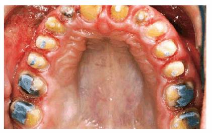
Figure 17-16D: Crown lengthening and build-ups with composite resin were necessary before making the impressions for the final restorations.
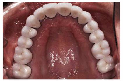
Figure 17-16E: The final splinted restorations were constructed using ceramic-metal.
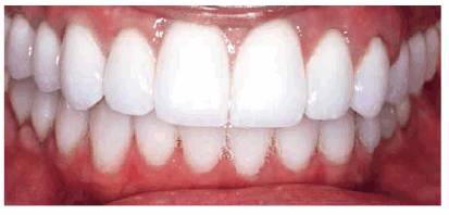
Figure 17-16F: The five splinted crowns restored this attractive lady's smile. Note how light a shade the patient selected.
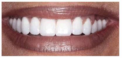
Figure 17-16G: The new, improved shapes and shade helped to accomplish the smile desired by the patient.
This is a 62-year-old man with angina pectoris. He developed the habit of
holding his nitroglycerine tablets between his teeth on the right side of the
mouth. As a result of this unusual behavior, erosive lesions affected the right
first molars. Figure 17-17 illustrates the defects on the
mandibular right first molar.
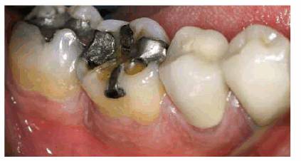
Figure 17-17: The habit of retaining nitroglycerine tablets between the teeth on the right side of the mouth resulted in erosive lesions as seen here on the mandibular right first molar.
DIFFERENTIAL DIAGNOSIS
Proper diagnosis is required to achieve successful treatment outcomes. As noted
at the beginning of this chapter, patients may simultaneously have more than
one of the conditions described. Thus, when occlusal or incisal changes are
noted, the cervical regions of the teeth should also be closely examined. Likewise,
the occlusal scheme should be fully evaluated if cervical notches or defects
are found.
When evaluating a patient who has any of these lesions, it is necessary to
correctly diagnose the condition and address the etiologic factors. Many
patients with these conditions may be asymptomatic and/or unaware of them. In
addition, they may have received "routine" dental care in the past
and be surprised when these conditions are brought to their attention. In the
case of bruxism, some patients are so surprised that they actually deny the
problem. If this is the case, the best way to demonstrate to them that they
have a bruxing problem is through visual images. A variety of means are
available to illustrate the problem, including intraoral photographs, surgical
microscopes, intraoral or extraoral video images, and diagnostic casts. With
the aid of even simple visual references, the patient can be shown the extent
of the damage that has been done and how he or she is causing it.31
Once the patient is convinced of the problem, the next step is attempting to
determine when it is occurring. If it occurs primarily during waking hours, the
patient may be able to control or correct the problem. If it occurs during
sleep, an appliance will be necessary to control the bruxing and/or prevent
further damage to the dentition.
A key criterion when examining a suspected abfraction is the presence of
lateral occlusal stresses during mastication or parafunctional movements. Thus,
signs of attrition in the form of notable wear facets and/or loss of anterior
guidance are highly probable when abfraction is present. The orientation of the
long axis of the tooth in relation to occlusal loading should also be
evaluated. The physical characteristics of abfraction are that of a sharp,
angular defect, and these lesions may be located completely beneath the
marginal gingiva. Abrasion in the cervical region can usually be distinguished
from abfraction by the smooth, rounded nature of the lesion. Minimal to extreme
gingival recession, with or without mucogingival defects, will likely accompany
abrasion. Gingival recession may also be seen with abfraction but is not a
hallmark of these defects.
Certain forms of abrasion are related to specific oral habits. The abraded
areas may be localized, such as with pen or pencil chewing or with pipe stem
clenching. Chapter 20 addresses these and the deleterious
effects of numerous other oral habits.
Dental practitioners should have a high level of suspicion when they see
generalized lingual erosion of the maxillary anterior teeth. Bulimia or GERD
will be the likely cause. It is important to carefully obtain a history that
will allow proper diagnosis. Individuals with GERD will more readily provide
information that will assist in the diagnosis. Patients who suffer from bulimia
may be reluctant to reveal their condition and are sometimes outwardly defensive
when questioned concerning the issues related to their eating disorder. Often,
however, a dentist may be the first medical professional to recognize signs of
bulimia and can be instrumental in initiating an appropriate referral to
address the overall condition.
Dentists must be diligent when they examine patients. They must look beyond the
routine of caries, periodontal diseases, and missing teeth and closely evaluate
patients for the loss of tooth structure due to noncarious processes. When
these conditions are found, dentists must take the time to assess potentially
interrelated etiologic factors by obtaining a proper history and performing a
thorough clinical examination.
Discovering and helping to identify destructive habits, such as bruxism, needs
to be a team effort. Frequently, the dental hygienist or assistant can be the
observant individual who calls attention to a potential problem before it
becomes an esthetic deformity. Team educational meetings are useful in teaching
staff exactly what signs to observe. Knowing the correct anatomy of anterior
and posterior teeth is of considerable value in being able to recognize even
minor cusp or incisal edge changes that are a result of bruxism. Thus, the
esthetics of the patient's smile not only depends on good oral hygiene but also
becomes a shared team responsibility to keep it looking as good as possible
throughout life.
REFERENCES
1. Abreu Tabarini HS. Dental attrition of Mayan Tzutujil children-a study based
on longitudinal materials. Bull Tokyo Med
Dent Univ 1995;42:31-50.
2. Alexandersen V, Noren JG, Hoyer I, et al. Aspects of teeth from
archeological sites in
3. al-Hiyasat AS, Saunders WP, Sharkey SW, Smith GM. The effect of a carbonated
beverage on the wear of human enamel and dental ceramics. J Prosthodont 1998;7:2-12.
4. Bader JD, McClure F, Scurria MS, et al. Case-control study of non-carious cervical
lesions. Community Dent
Oral Epidemiol 1996;24:286-91.
5. Barrett MJ. Dental observations on Australian aborigines. Continuously
changing functional occlusion. Aust Dent J 1958;58:39-52.
6. Barrett MJ. Functioning occlusion. Ann Aust Coll
Dent Surg 1969;2:68-80.
7.
8.
9.
10.
11.
12. Bauer W, van den Hoven F, Diedrich P. Wear in the upper and lower incisors
in relation to incisal and condylar guidance. J Orofac
Orthop 1997;58:306-19.
13. Becker W, Ochsenbein C, Becker BE. Crown lengthening: the
periodontal-restorative connection. Compend Cont
Educ Dent 1998;19:239-40, 242, 244-6.
14. Begg PR. Stone age man's dentition. Am J Orthod 1954;40:298-312.
15. Begg PR. Stone age man's dentition. Am J Orthod 1954;40:373-83.
16. Begg PR. Stone age man's dentition. Am J Orthod 1954;40:462-75.
17. Begg PR. Stone age man's dentition. Am J Orthod 1954;40:517-31.
18. Bermudez de Castro JM, Arsuaga JL, Perez PJ. Interproximal grooving in the
Atapuerca-SH hominid dentitions. Am J Phys
Anthropol 1997;102:369-76.
19. Bevenius J, L'Estrange P, Karlsson S, Carlsson GE. Idiopathic cervical
lesions: in vivo investigation by oral microendoscopy and scanning electron
microscopy. A pilot study. J Oral Rehabil
1993;20:1-9.
20. Bishop K, Bell M, Briggs P, Kelleher M. Restoration of a worn dentition
using a double-veneer technique. Br Dent J
1996;180:26-9.
21. Bishop K, Kelleher M, Briggs P, Joshi R. Wear now? An update on the
etiology of tooth wear. Quintessence
Int 1997;28:305-13.
22. Bishop KA, Briggs PF, Kelleher MG. Modern restorative management of
advanced tooth-surface loss. Prim Dent Care
1994;1:20-3.
23. Boghosian A. Clinical evaluation of a filled adhesive system in Class 5
restorations. Compend Cont
Educ Dent 1996;17:750-7.
24. Borrman H, Engstrom EU, Alexandersen V, et al. Dental conditions and
temporomandibular joints in an early Mesolithic bog man. Swed Dent J 1996;20:1-14.
25. Bouquot JE, Seime RJ. Bulimia nervosa: dental perspectives. Pract
Periodont Aesthet Dent 1997;9:655-63.
26. Braem M, Lambrechts P, Vanherle G. Stress-induced cervical lesions. J Prosthet
Dent 1992;67:718-22.
27. Brown S, Bonifazi DZ. An overview of anorexia and bulimia nervosa, and the
impact of eating disorders on the oral cavity. Compend Cont
Educ Dent 1993;14: 1594, 1596-1602, 1604-8.
28. Chaudhry SI, Harris JL, Challacombe SJ. Dental erosion in a wine merchant:
an occupational hazard? Br Dent J
1997;182:226-8.
29. Chikte UM, Josie-Perez AM, Cohen TL. Industrial dental erosion-a case
report. J Dent Assoc S
Afr 1996; 51:647-50.
30. Chikte UM, Josie-Perez AM, Cohen TL. A rapid epidemiological assessment of
dental erosion to assist in settling an industrial dispute. J Dent Assoc S
Afr 1998; 53:7-12.
31. Cook DA. Using crayons to educate patients about front-tooth wear patterns.
J Am Dent
Assoc 1998;129: 1149-50.
32. da Silva AM, Oakley DA, Hemmings KW, et al. Psychosocial factors and tooth
wear with a significant component of attrition. Eur J
Prosthodont Restor Dent 1997;5:51-5.
33. Darbar UR, Hemmings KW. Treatment of localized anterior toothwear with
composite restorations at an increased occlusal vertical dimension. Dent Update
1997;24:72-5 [published erratum appears in Dent Update 1997;24:157].
34. Dawid E, Meyer G, Schwartz P. The etiology of wedge-shaped defects: a
morphological and function-oriented investigation. J Gnathol 1991;10:49-56.
35. Donachie MA, Walls AW. The tooth wear index: a flawed epidemiological tool
in an aging population group. Community Dent
Oral Epidemiol 1996; 24:152-8.
36. Douglas WH. Form, function and strength in the restored dentition. Ann R Austra
Coll Dent Surg 1996; 13:35-46.
37. Evans RD, Briggs PF. Tooth-surface loss related to pregnancy-induced
vomiting. Prim Dent Care
1994; 1:24-6.
38.
39. Formicola V. Interproximal grooving: different appearances, different
etiologies. Am J Phys
Anthropol 1991;86:85-7.
40. Frayer DW. On the etiology of interproximal grooves. Am J Phys
Anthropol 1991;85:299-304.
41. Gedalia I, Dakuar A, Shapira L, et al. Enamel softening with Coca-Cola and
rehardening with milk or saliva. Am J Dent
1991;4:120-2.
42. Goel VK,
43. Grenby TH. Methods of assessing erosion and erosive potential. Eur J Oral Sci
1996;104:207-14.
44. Grippo JO. Abfractions: a new classification of hard tissue lesions of
teeth. J Esthet Dent
1991;3:14-9.
45. Grippo JO. Noncarious cervical lesions: the decision to ignore or restore. J Esthet Dent
1992;4(Suppl): 55-64.
46. Grippo JO. Bioengineering seeds of contemplation: a private practitioner's
perspective. Dent Mater
1996;12:198-202.
47. Grippo JO, Simring M. Dental "erosion" revisited. J Am Dent
Assoc 1991;122:41-7.
48. Harrison JL, Roeder LB. Dental erosion caused by cola beverages. Gen Dent
1991;39:23-4.
49. Healy WB. Soils and dental research. N Z Dent J
1998;94:114.
50. Heymann HO, Sturdevant JR, Bayne S, et al. Examining tooth flexure effects
on cervical restorations: a two year clinical study. J Am Dent
Assoc 1991; 122:41-7.
51. Horsted-Bindslev P, Knudsen J, Baelum V. 3-year clinical evaluation of
modified Gluma adhesive systems in cervical abrasion/erosion lesions. Am J Dent
1996;9: 22-6.
52. Hudson JD, Goldstein GR, Georgescu M. Enamel wear caused by three different
restorative materials. J Prosthet
Dent 1995;74:647-54.
53. Hugoson A, Ekfeldt A, Koch G, Hallonsten AL. Incisal and occlusal tooth
wear in children and adolescents in a Swedish population. Acta Odontol
Scand 1996;54:263-70.
54. Imfeld T. Dental erosion. Definition, classification and links. Eur J Oral Sci
1996;104:151-4.
55. Jagger DC, Harrison A. An in vitro investigation into the wear effects of
selected restorative materials on enamel. J Oral Rehabil
1995;22:275-81.
56. Jagger DC, Harrison A. An in vitro investigation into the wear effects of
selected restorative materials on dentine. J Oral Rehabil
1995;22:349-54.
57. Jarvinen VK, Rytomaa II, Heinonen OP. Risk factors in dental erosion. J Dent Res
1991;70:942-7.
58. Johansson AK, Johansson A, Birkhead D, et al. Dental erosion associated
with soft-drink consumption in young Saudi men. Acta Odontol
Scand 1997;55:390-7.
59. Josephson CA. Restoration of mandibular incisors with advanced wear. J Dent Assoc S
Afr 1992;47:419-20.
60. Kelleher M, Bishop K. The aetiology and clinical appearance of tooth wear. Eur J
Prosthodont Restor Dent 1997;5:157-60.
61. Kerr NW. Dental pain and suffering prior to the advent of modern dentistry.
Br Dent J
1998;184:388.
62. Khan F, Young WG, Daley TJ. Dental erosion and bruxism. A tooth wear
analysis from south east
63. Kokich VG. Esthetics and vertical tooth position: orthodontic
possibilities. Compend Cont
Educ Dent 1997;18:1225-31.
64. Langsjoen OM. Dental effects of diet and coca-leaf chewing on two
prehistoric cultures of northern
65. Lavigne GJ, Rompre PH, Montplaisir JY. Sleep bruxism: validity of clinical
research diagnostic criteria in a controlled polysomnographic study. J Dent Res
1996;75:546-52.
66. Lee WC, Eakle WS. Possible role of tensile stress in the etiology of
cervical erosive lesions of teeth. J Prosthet
Dent 1984;52:374-80.
67. Lee WC, Eakle WS. Stress-induced cervical lesions: review of advances in
the past 10 years. J Prosthet
Dent 1996;75:487-94.
68. Levitch LC, Bader JD, Shugars DA, Heymann HO. Non-carious cervical lesions.
J Dent
1994;22:195-207.
69. Lussi A, Jaeggi T, Jaeggi-Scharer S. Prediction of the erosive potential of
some beverages. Caries Res
1995;29:349-54.
70. Lussi A, Portmann P, Burhop B. Erosion on abraded dental hard tissues by
acid lozenges: an in situ study. Clin Oral
Investig 1997;1:191-4.
71. Lussi A, Schaffner M, Hotz P, Suter P. Dental erosion in a population of
Swiss adults. Community Dent
Oral Epidemiol 1991;19:286-90.
72. Mair LH, Stolarski TA, Vowles RW, Lloyd CH. Wear: mechanisms,
manifestations and measurement. Report of a workshop. J Dent 1996;24:141-8.
73. Matis BA, Cochran M, Carlson T. Longevity of glass-ionomer restorative
materials: results of a 10-year evaluation. Quintessence
Int 1996;27:373-82.
74. Mayhall JT, Kageyama I. A new, three-dimensional method for determining
tooth wear. Am J Phys
Anthropol 1997;103:463-9.
75. McCoy G. The etiology of gingival erosion. J Oral
Implantol 1982;10:361-2.
76. McCoy G. On the longevity of teeth. J Oral Implantol 1983;11:248-67.
77. Miller MB. Restoring class V lesions. Part 2: abfraction lesions. Pract Periodont Aesthet Dent 1997;9:505-6.
78. Millward A, Shaw L, Smith AJ, et al. The distribution and severity of tooth wear and
the relationship between erosion and dietary constituents in a group of
children. Int J Paediatr
Dent 1994;4:151-7.
79. Milosevic A, Lennon MA, Fear SC. Risk factors associated with tooth wear in
teenagers: a case control study. Community Dent
Health 1997;14:143-7.
80. Murray CG, Sanson GD. Thegosis-a critical review. Aust Dent J 1998;43:192-8.
81. Nel JC, Marais JT, van Vuuren PA. Various methods of achieving restoration of tooth structure loss due to
bruxism. J Esthet Dent
1996;8:183-8.
82. Neo J, Chew CL. Direct tooth-colored materials for noncarious lesions: a
3-year clinical report. Quintessence Int 1995;27:183-8.
83. Nunn JH. Prevalence of dental erosion and the implications for oral health.
Eur J Oral Sci
1996;104:156-61.
84. Nystrom M, Kononen M, Alaluusua S, et al. Development of horizontal tooth
wear in maxillary anterior teeth from five to 18 years of age. J Dent Res
1990; 69:1765-70.
85. Osborne-Smith KL, Burke FJ, Farlane TM, Wilson NH. Effect of restored and
unrestored non-carious cervical lesions on the fracture resistance of
previously restored maxillary premolar teeth. J Dent 1998;26:427-33.
86. O'Sullivan EA, Curzon ME, Roberts GJ, et al. Gastroesophageal reflux in children and its
relationship to erosion of primary and permanent teeth. Eur J Oral Sci
1998;106:765-9.
87. Powell
88. Radentz WH, Barnes GP,
89. Richards LC, Miller SL. Relationships between age and dental attrition in
Australian aboriginals. Am J Phys
Anthropol 1991;84:159-64.
90. Ritchard A, Welsh AH, Donnelly C. The association between occlusion and
attrition. Aust Orthod J
1992;12:138-42.
91. Ruffs JC,
92. Rytomaa I, Jarvinen V, Kanerva R, Heinonen OP. Bulimia and tooth erosion. Acta Odontol
Scand 1998;56:36-40.
93. Seow WK. Clinical diagnosis of enamel defects: pitfalls and practical
guidelines. Int Dent J
1997;47:173-82.
94. Shaw L. The epidemiology of tooth wear. Eur J
Prosthodont Restor Dent 1997;5:153-6.
95. Silness J, Berge M, Johannessen G. Longitudinal study of incisal tooth wear
in children and adolescents. Eur J Oral Sci
1995;103:90-4.
96. Silness J, Berge M, Johannessen G. Re-examination of incisal tooth wear in
children and adolescents. J Oral Rehabil
1997;24:405-9.
97. Smith BG, Robb ND. The prevalence of toothwear in 1007 dental patients. J Oral Rehabil
1996;23:232-9.
98. Spranger H. Investigation into the genesis of angular lesions at the
cervical region of teeth. Quintessence Int 1995;26:183-8.
99. Stewart B. Restoration of the severely worn dentition using a systematized
approach for a predictable prognosis. Int J
Periodont Rest Dent 1998;18:46-57.
100. ten Cate JM, Imfeld T. Dental erosion, summary. Eur J Oral Sci
1996;104:241-4.
101. Touminen M, Touminen R. Dental erosion and associated factors among
factory workers exposed to inorganic acid fumes. Proc Finn Dent
Soc 1991;87:359-64.
102. Touminen M, Touminen R. Tooth surface loss among people exposed to cement
and stone dust in the work environment in
103. Touminen M, Touminen R. Tooth surface loss and associated factors among
factory workers in
104. Touminen ML, Touminen RJ, Fubusa F, Mgalula N. Tooth surface loss and
exposure to organic and inorganic acid fumes in workplace air. Community Dent
Oral Epidemiol 1991;19:217-20.
105. Touyz LZ. The acidity (pH) and buffering capacity of Canadian fruit juice
and dental implications. J Can Dent
Assoc 1994;60:454-48.
106. Tsolka P, Walter JD,
107. Ungar PS, Teaford MF, Glander KE, Pastor RF. Dust accumulation in the
canopy: a potential cause of dental microwear in primates. Am J Phys
Anthropol 1995;97:93-9.
108. Ungar PS, Fennell KJ, Gordon K, Trinkaus E. Neanderthal incisor beveling. J Hum Evol
1997;32:407-21.
109.
110. Villa G, Giacobini G. Subvertical grooves of interproximal facets in
Neanderthal posterior teeth. Am J Phys
Anthropol 1995;96:51-62.
111. Wiktorsson AM, Zimmerman M, Angmar-Mansson B. Erosive tooth wear:
prevalence and severity in Swedish winetasters. Eur J Oral Sci
1997;105:544-50.
112. Yettram AL, Wright KWJ, Rickard HM. Finite element stress analysis of the
crowns of normal and restored teeth. J Dent Res
1976;55:1004-11.
113. Young WG. Anthropology, tooth wear, and occlusion ab origine. J Dent Res
1998;77:1860-3.
ADDITIONAL RESOURCES
Abdullah A, Sherfudhin H, Omar R, Johansson A. Prevalence of occlusal tooth
wear and its relationship to lateral and protrusive contact schemes in a young
adult Indian population. Acta Odontol
Scand 1994;52:191-7.
al-Hiyasat AS, Saunders WP, Sharkey SW, et al. Investigation of human enamel
wear against four dental ceramics and gold. J Dent
1998;26:487-95.
al-Hiyasat AS, Saunders WP, Sharkey SW, et al. The abrasive effect of glazed,
unglazed, and polished porcelain on the wear of human enamel, and the influence
of carbonated soft drinks on the rate of wear. Int J
Prosthodont 1997;10:269-82.
Altshuler BD. Eating disorder patients. Recognition and intervention. J Dent Hyg
1990;64:119-25.
Attin T, Koidl U, Buchalla W, et al. Correlations of microhardness and wear in
differently eroded bovine dental enamel. Arch Oral Biol
1997;42:243-50.
Attin T, Zirkel C, Hellwig E. Brushing abrasion of eroded dentin after
application of sodium fluoride solutions. Caries Res
1998;32:344-50.
Beckett H. Dental abrasion caused by a cobalt-chromium denture base. Eur J
Prosthodont Restor Dent 1995;3: 209-10.
Berge M, Johannessen G, Silness J. Relationship between alignment conditions of
teeth in anterior segments and incisal wear. J Oral Rehabil
1996;23:717-21.
Blair FM, Thomason JM, Smith DG. The traumatic anterior overbite. Dent Update 1997;24:144-52.
Bohmer CJ, Klinkenberg-Knol EC, Niezen-de Boer MC, et al. Dental erosions and
gastro-oesophageal reflux disease in institutionalized intellectually disabled
individuals. Oral Dis
1997;3:272-5.
Bowles WH, Wilkinson MR, Wagner MJ, Woody RD. Abrasive particles in tobacco
products: a possible factor in dental attrition. J Am Dent
Assoc 1995;126:327-31.
Brady JM, Woody RD. Scanning microscopy of cervical erosion. J Am Dent
Assoc 1977;94:726-9.
Briggs P, Bishop K. Fixed prostheses in the treatment of tooth wear. Eur J
Prosthodont Restor Dent 1997; 5:175-80.
Briggs PF, Bishop K, Djemal S. The clinical
evolution of the 'Dahl Principle.' Br Dent J 1997;183:171-6.
Burke FJ, Whitehead SA, McCaughey AD. Contemporary concepts in the pathogenesis
of the Class V non-carious lesion. Dent Update
1995;22:28-32.
Carlson-Mann LD. Recognition and management of occlusal disease from a
hygienist's perspective. Probe
1996;30:196-7.
Donachie MA, Walls AW. Assessment of tooth wear in an ageing population. J Dent
1995;23:157-64.
Douglas WH. Considerations for modeling. Dent Mater
1996;12:203-7.
Evans RD. Orthodontics and the creation of localised inter-occlusal space in
cases of anterior tooth wear. Eur J
Prosthodont Restor Dent 1997;5:69-73.
Goldstein RE. Esthetics in dentistry.
Goldstein RE. Current concepts in esthetic treatment. Proceedings of the Second
International Prosthodontic Congress; 1979;
Goldstein RE. Esthetics in dentistry. J Am Dent
Assoc 1982;104:301-2.
Goldstein RE. Diagnostic dilemma: to bond, laminate, or crown? Int J Periodont
Restor Dent 1987;87(5):9-30.
Goldstein RE. Finishing of composites and laminates. Dent Clin
North Am 1989;33:305-18.
Goldstein RE, Feinman RA, Garber DA. Esthetic considerations in the selection
and use of restorative materials. Dent Clin
North Am 1983;27:723-31.
Goldstein RE, Garber DA, Schwartz CG, Goldstein CE. Patient maintenance of
esthetic restorations. J Am Dent
Assoc 1992;123:61-6.
Goldstein RE, Garber DA, Goldstein CE, et al. The changing esthetic dental
practice. J Am Dent Assoc
1994;125:1447-57.
Gregory-Head B, Curtis DA. Erosion caused by gastroesophageal reflux:
diagnostic considerations. J Prosthodont
1997;6:278-85.
Hacker CH, Wagner WC, Razzoog ME. An in vitro investigation of the wear of
enamel on porcelain and gold in saliva. J Prosthet
Dent 1996;75:14-7.
Haines DJ, Berry DC,
Hazelton LR, Faine MP. Diagnosis and dental management of eating disorder
patients. Int J
Prosthodont 1996;9:65-73.
Imfeld T. Prevention of progression of dental erosion by professional and
individual prophylactic measures. Eur J Oral Sci
1996;104:215-20.
Irish JD, Turner CG 2nd. Brief communication: first evidence of LSAMAT in
non-native Americans: historic Senegalese from
Isacsson G, Bodin L, Selden A, Barregard L. Variability in the quantification
of abrasion on the Bruxcore device. J Orofac Pain
1996;10:362-8.
Johansson A. A cross-cultural study of occlusal tooth wear. Swed Dent J
Suppl 1992;86:1-59.
Kaidonis JA, Richards LC, Townsend GC, Tansley GD. Wear of human enamel: a
quantitative in vitro assessment. J Dent Res
1998;77:1983-90.
Kiliaridis S, Johansson A, Haraldson T, et al. Craniofacial morphology,
occlusal traits, and bite force in persons with advanced occlusal tooth wear. Am J Orthodont
Dentofac Orthop 1995;107:286-92.
Knight DJ, Leroux BG, Zhu C, et al. A longitudinal study of tooth wear in
orthodontically treated patients. Am J Orthod
Dentofac Orthop 1997;112:194-202.
Lambrechts P, van Meerbeek B, Perdigao J, et al. Restorative therapy for
erosive lesions. Eur J Oral Sci
1996;104:229-40.
Leinfelder KF, Yarnell G. Occlusion and restorative materials. Dent Clin
North Am 1995;39:355-61.
Lussi A. Dental erosion clinical diagnosis and case history taking. Eur J Oral Sci
1996;104:191-8.
Lyttle HA, Sidhu N, Smyth B. A study of the classification and treatment of
noncarious cervical lesions by general practitioners. J Prosthet
Dent 1998;79:342-6.
Magnusson T. Is snuff a potential risk factor in occlusal wear? Swed Dent J
1991;15:125-32.
McIntyre JM. Erosion. Aust
Prosthodont J 1992; 6:17-25.
Meurman JH, ten Cate JM. Pathogenesis and modifying factors of dental erosion. Eur J Oral Sci
1996;104: 199-206.
Millward A, Shaw L, Smith AJ. Dental erosion in four-year-old children from
differing socioeconomic backgrounds. ASDC J Dent
Child 1994;61:263-6.
Milosevic A. Tooth wear: an aetiological and diagnostic problem. Eur J
Prosthodont Restor Dent 1993;1:173-8.
Milosevic A. Toothwear: aetiology and presentation. Dent Update
1998;25:6-11.
Milosevic A, Brodie DA, Slade PD. Dental erosion, oral hygiene, and nutrition
in eating disorders. Int J Eat
Disord 1997;21:195-9.
Milosevic A, Lo MS. Tooth wear in three ethnic groups in Sabah (northern
Mixson JM, Spencer P, Moore DL, et al. Surface morphology and chemical
characterization of abrasion/erosion lesions. Am J Dent
1995;8:5-9.
Morley J. The esthetics of anterior tooth aging. Curr Opin
Cosmet Dent 1997;4:35-9.
Nemcovsky CE, Artzi Z. Erosion-abrasion lesions revisited. Compend Cont
Educ Dent 1996;17:416-8.
Neo J, Chew CL, Yap A, Sidhu S. Clinical evaluation of tooth-colored materials
in cervical lesions. Am J Dent
1996;9:15-18.
Nunn J, Shaw L, Smith A. Tooth wear-dental erosion. Br Dent J
1996;180:349-52.
Owens BM, Gallien GS. Noncarious dental "abfraction" lesions in an
aging population. Compend Cont
Educ Dent 1995;16:552-62.
Pintado MR, Anderson GC, DeLong R, Douglas WH. Variation in tooth wear in young
adults over a two-year period. J Prosthet
Dent 1997;77:313-20.
Plavcam JM, Kelley J. Evaluating the "dual selection" hypothesis of
canine reduction. Am J Phys
Anthropol 1996;99:379-87.
Ramp MH, Suzuki S, Cox CF, et al. Evaluation of wear: enamel opposing three
ceramic materials and a gold alloy. J Prosthet
Dent 1997;77:523-30.
Robertson PB, DeRouen TA, Ernster V, et al. Smokeless tobacco use: how it
affects the performance of major league baseball players. J Am Dent
Assoc 1995;126: 1115-21.
Sakaguchi RL, Brust EW, Cross M, et al. Independent movement of cusps during
occlusal loading. Dent Mater
1991;7:186-90.
Schmidt U, Treasure J. Eating disorders and the dental practitioner. Eur J
Prosthodont Restor Dent 1997;5: 161-7.
Seligman DA, Pullinger AG. The degree to which dental attrition in modern
society is a function of age and of canine contact. J Orofac Pain
1995;9:266-75.
Seligman DA, Pullinger AG. A multiple stepwise logistic regression analysis of
trauma history and 16 other history and dental cofactors in females with
temporomandibular disorders. J Orofac Pain
1996;10:351-61.
Silness J, Berge M, Johannessen G. A 2-year follow-up study of incisal tooth
wear in dental students. Acta Odontol
Scand 1995;53:331-3.
Smith BG, Bartlett DW,
Sognnaes RF, Wolcott RB, Xhonga FA.
Suzuki S, Cox CF, Leinfelder KF, et al. A new copolymerized composite resin
system: a multiphased evaluation. Int J
Periodont Restor Dent 1995;15:482-95.
Suzuki S, Suzuki SH, Cox CF. Evaluating the antagonistic wear of restorative
materials when placed against human enamel. J Am Dent
Assoc 1996;127:74-80.
Tay FR, Gwinnett AJ, Pang KM, Wei SH. Structural evidence of a sealed tissue
interface with a total-etch wet-bonding technique in vivo. J Dent Res
1994;73:629-36.
Teo C, Young WG, Daley TJ, Sauer H. Prior fluoridation in childhood affects
dental caries and tooth wear in a south east
Tyas MJ. The Class V lesion-aetilogy and restoration. Aust Dent J
1995;40:167-70.
Ungar PS, Teaford MF. Preliminary examination of non-occlusal dental microwear
in anthropoids: implications for the study of fossil primates. Am J Phys
Anthropol 1996;100:101-13.
van Foreest A, Roeters J. Restorative dental treatment of abraded canine teeth
in a Sumatran tiger (Panthera tigris sumatrae). J Vet Dent
1997;14:131-6.
Villa G, Giacobini G. Dental microwear. Morphological, functional and
phylogenetic correlations. Ital J Anat
Embryol 1998;103:53-84.
Yaacob HB, Park AW. Dental abrasion pattern in a selected group of Malaysians. J Nihon Univ
Sch Dent 1990;32:175-80.
Yap AU, Neo JC. Non-carious cervical tooth loss: part 1. Dent Update
1995;22:315-8.
Yap AU, Neo JC. Non-carious cervical tooth loss. Part 2: management. Dent Update
1995;22:364-8.
Zero DT. Etiology of dental erosion-extrinsic factors. Eur J Oral Sci 1996;104:162.

Politica de confidentialitate |