ESTHETIC PROBLEMS OF SPECIAL POPULATIONS
CHAPTER 27. ESTHETICS IN PEDIATRIC
DENTISTRY - Claudia Caprioglio, DDS, MS, Alberto Caprioglio, DDS, MS,
Damaso Caprioglio, MD, MS
INTRODUCTION
In the period ranging from the end of the primary dentition to the first phases
of the early mixed one, the esthetics and harmony of dental arches are
determined by the physiologic change of dental elements, the presence of
diastemas, the correct canine relationship, and the correct occlusive plane.
The occlusion of the primary dentition should be considered as a biological
unit, having special esthetic, functional, and skeletal characteristics. In
fact, the main duty of the pediatric dentist is the monitoring of growth
through adolescence.
The dual duty of the pediatric dentist is expressed not only by the application
of preventive and/or conservative dentistry but also by the space management
required to produce a morphic-functional recovery.
MATERIALS AND TECHNIQUES
In the primary dentition, it is necessary to consider a therapeutic strategy
evaluating the physiologic state of the deciduous element and the efficacy of
the treatment. A careful diagnosis must be carried out to define the relevant
prognosis. Insignificant therapeutic improvements can result from a poor
awareness of pulpal treatment options and lead to unnecessary treatment
procedures and materials. The introduction of light-cured composite resins has
changed clinical pediatric dentistry. In fact, these materials are welcome
treatment options that address both esthetic and functional issues. Their
advantages are represented by considerable hardness, high rigidity, and a high
level of resistance to compression. However, these materials are very sensitive
to technique and can show marginal infiltrations, a reduced resistance to wear,
polymerization contractions, surface roughness, and discoloration.
Composite resins are the material of choice to restore anterior teeth. The
composite resins recommended are microfilled hybrid composite resins. More
research into these materials has led to considerable improvements,
particularly in traumatology, thus making possible the tooth fragment
reattachment. This, in turn, has allowed dentists to proceed to a true
biological restoration to achieve a good anterior guide, improved resistance to
wear, and higher color stability in the follow-up years.2
Composite resins for posterior teeth can be used for Class I and II
restorations, where etching time is extremely important. Some authors
emphasized that few statistical differences were found for surface roughness in
the primary dentition.15,16 Further, the use of a glass ionomer
cement as a cavity base and the reconstruction of the tooth by applying the
incremental technique and using a rubber dam have reduced the wear index and
improved cavity adhesion. The kind of (direct or incremental) polymerization
influences the marginal adaptation.
Glass Ionomers and Modified Ionomer
Cements
These materials appeared for the first time in the early 1970s.20
They are composed of a powder, a calcium-fluoride-aluminium silicate glass, and
a liquid, generally a polyacrylic or polymaleic acid. Considering their link
with dentin, fluoride-leaching properties, and high resilience range, the use
of these materials has been advantageous in the treatment of caries lesions in
primary molar teeth.
Although the percentage of failure is higher in comparison with the amalgam
(33% versus 20% for amalgam), and although they lack resilience to abrasion and
have a low brightness, they have a great advantage: they result in minimal
destruction of sound tooth tissue and a reduced use of local anesthetic.
Berg described the resin-modified glass ionomer cements as materials that can
be polymerized and whose resin compound improves the resistance to fractures.6
They are suggested for Class I and II restorations in primary teeth, which
typically do not last beyond 3 years.
Compomers
These materials were introduced in the early 1990s. They are composed of a
mixed composite resin with an acid modification, which makes them more similar
to composite resins than to glass ionomers. They do not have the improved
characteristics of resins but are easy to handle, which reduces operative time
and makes them a good restorative solution.
A recent study by El-Kalla and Garcia-Godoy evaluated and measured the
resistance to compression, resistance to flexing, microhardness, and roughness
of the surface of three different compomers (Compoglass [Ivoclar Vivadent,
Amherst, NY], Dyract [DENTSPLY/Caulk, Milford, DE], and Hytac [3M ESPE, St.
Paul, MN]).12 Subsequently, these values have been compared to those
of a composite resin (Z 100 [3M ESPE]) and to a modified glass-ionomer
(Vitremer [3M ESPE]). The results demonstrated that the tested compomers had
flexing, compression, and microhardness qualities that were higher than cement
but lower than composite resin, whereas no significant differences in surface
roughness were reported.
The properties of compomers consist of
. A good adhesion to dental tissues (a dentinal adhesive is used instead of
acid etching);
. Easy handling, enhanced by the possibility of incremental polymerization;
. A reduced marginal fissure due to their property of absorbing water during
hardening;
. A good fluoride adsorption-release system; and
. An acceptable range of colors and brightness that produces good esthetics,
although not quite comparable to that of composite resin.
Because of the availability of these restorative materials, the pediatric
dentist can apply preventive measures and perform early conservative therapy
or, in the most severe cases, restore function and improve esthetics.
The increased predictive capabilities of the outcome of treatment along with
improvement in materials enable compliance with the relevant postulates for
successful pediatric dentistry:
. improvement of esthetic restoration
. elimination of infection, inflammation, and pain
. maintenance of the arch perimeter length
. stimulation of the alveolar growth
See Table 27-1 for a selection of materials for
clinical use.
The improved treatment techniques, better materials, and heightened awareness
of the benefits of preventive dentistry have led to better management and
predictable results. There are also additional means of cavity excavation,
representing alternative therapeutic solutions: Carisolv and air abrasion.
Carisolv
Carisolv (Mediteam Dental,
Obviously, to intervene on a closed cavity or one with a small opening in the
enamel, it will be necessary to use rotating instruments or excavators to reach
the dentin affected by the carious process.
The softening mechanism induced by Carisolv on the decayed dentin develops
mainly through the destruction of collagen fibrils already denatured by the
carious process. This is a very complex process called chloramination,
involving interaction between chlorine ions freed from hypochlorite and amine
groups of the three amino acids in a highly basic environment.
The Carisolv system can be used without anesthesia because it is not invasive
and is without the troublesome vibration and thermal dentinal stimulations
produced by rotating instruments. Therefore, it is suitable for phobic or
anxious persons and for young patients whenever there are contraindications to
conventional anesthesia and in all cases in which there is a risk of
accidentally reaching the pulp chamber. This last possibility is considerably
frequent in very deep cavities in very close proximity to the pulp. 18418c29s For all of
these reasons, Carisolv is a useful operative means. In fact, due to the system
selectivity (as already mentioned, this jelly acts exclusively on the decayed
dentin), it is able to detect even the thinnest amounts of healthy dentin as
opposed to the more aggressive rotating systems that easily reach into the pulp
after passing the thin dentin barrier.
Statistical surveys have shown that this system results in a high level of
satisfaction and compliance. It seldom requires anesthesia, and the remaining
dentinal substratum is receptive to current adhesives. Therefore, Carisolv represents
a valid alternative to conventional methods for removing decayed dentin.
Air Abrasion
Air abrasion is a caries excavation system that, as opposed to other
conventional or unconventional means, bombards the dental surface with small
particles of aluminium oxide projected by a high-pressure air jet. This method
was invented in 1954 by Robert Black. It was reintroduced about 20 years ago
and in the recent past has enjoyed wider acceptance by dentists and patients
alike.
At present, several air-abrasive instruments are available at a reasonable cost
for the cavity preparation. Although these systems provide valid assistance to
the daily practice of the pedodontist, they are still not positioned to replace
conventional instruments for cavity preparation. In fact, they are suitable for
the treatment of small carious processes in fissure sealing, amelogenes
imperfecta, and whenever an adhesive restoration technique is performed. The
various systems are all able to achieve rapid, effective removal of enamel and
of healthy dentin (the action on the decayed dentin is less invasive). Various
particle sizes may be used (those approved by the U.S. Food and Drug
Administration measure 27.5 microns); however, the abrasive effect is
conditioned by the particles' kinetic energy, particles' outlet nozzle size,
and the distance between the powder outlet hole and the surface to be treated.
The advantages of this technique are the absence of vibration, reduced or
elimination of anesthestics for small cavities, no need to change rotating
instruments as excavation continues, and absence of pulpal exposure. The
disadvantages are a lack of tactile sensitivity (which is present with usual
rotating instruments), reduced control in depth of dentinal tissue removal,
possible toxicity of aluminium oxide particles if inhaled (hence the need for a
rubber dam), the need for protection of the dentist and his or her assistants,
and the need for a very efficient air-aspiration system to avoid the dispersion
of particles into the environment. The system may be improved by using
particles of a different nature, transported not only by air but also by water,
thus inducing the particles to fall and be more easily removed by aspiration.
RESTORATION OF PRIMARY ANTERIOR TEETH
In the last 15 years, a great evolution has taken place in composite resins.
Their adhesion, polish, and esthetics have improved so much that they are now
the best restorative material for anterior teeth damaged by caries or by either
direct or indirect traumas. Among the undeniable merits of this kind of
restoration is the fact that this is a "reversible" treatment;
therefore, it can be redone when necessary.
Procedure
Step 1. Carefully evaluate the tooth shape and the place of the
contralateral tooth, thus predicting the reconstruction.
Step 2. Choose the restoration color, as the subsequent isolation with a
rubber dam prevents an accurate survey.
Step 3. Isolate the area and remove the carious lesion while aiming to
maintain as much sound tooth tissue as possible.
Step 4. Treat the dentin and enamel to obtain good adhesion (compliance
with instructions described for the selected material).
Step 5. Reconstruction. Ensure modeling is as precise as possible to
reduce chair time and improve the esthetic final result.
Step 6. Finishing. Use a flame-shaped diamond bur to reproduce
microanatomy of the rather irregular enamel surface. The finishing phase is
completed by using a needle-shaped bur.
Step 7. Remove the rubber dam and evaluate the results. Next, polish the
interproximal areas with pop-on disks and abrasive strips, taking care not to
remove the contact point. Then, polish the other areas of the buccal face using
rubber cups and polishing pastes with decreasing particle size.
Step 8. Color check. After initial dehydration, the tooth regains its
original color. Note that chromatic considerations should be postponed to the
subsequent visit.
CASE STUDIES
Caries Lesions in Anterior Teeth
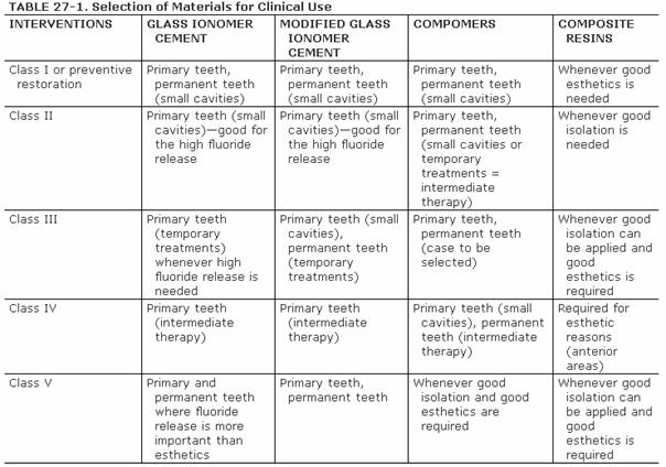
A male patient, 2 years and 8 months old, with interproximal caries of D, E, F,
and G (Figures 27-1A
and B
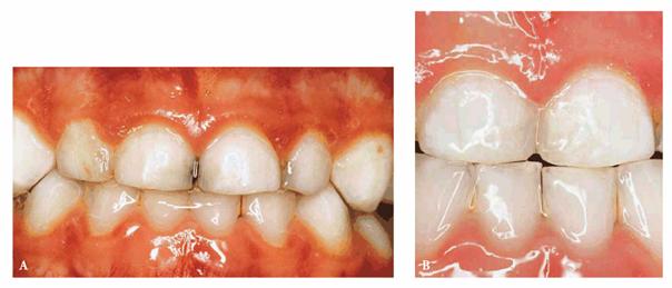
Figure 27-1A and B: Interproximal caries of frontal anterior teeth are removed, and a morphic-functional composite restoration is performed.
PROBLEM: The patient was not compliant, but an initial radiographic
examination was accomplished.
TREATMENT: Under conscious sedation, in only one visit, caries lesions
are removed, and the morphic-functional restoration with composite resin is
placed.
RESULT: The restoration achieved the esthetic goal and restored function
and anatomy. Furthermore, in a situation of tooth crowding, the arch length was
preserved. Both the patient and his parents were pleased.
Rampant Caries in Very Young Patients: Conservative
Approach
A male patient, 36 months old.
PROBLEM: He presented with rampant caries involving four maxillary
anterior teeth and caries in the lower arch. The parents hoped that the teeth
could be saved. Initial radiographs were taken (Figures 27-2A
and B
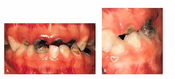
Figure 27-2A and B: Rampant caries involving the anterior teeth and the lower arch.
TREATMENT: Caries were removed, and endodontic treatment was performed.
Aluminum oxide posts were used. A rubber-base impression was made for
laboratory-processed full acrylic crowns (Figure 27-2C
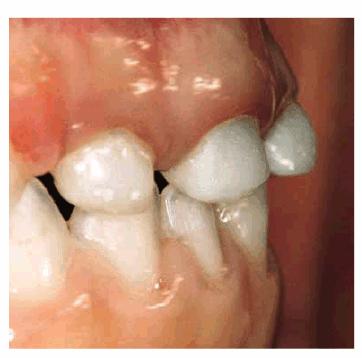
Figure 27-2C: Crowns are seated with an acceptable result.
RESULT: Crowns were seated with an acceptable result. Figures 27-2A and 27-2D show the sharp contrast between
before and after treatment. The child was able to resume his usual activities
without discomfort or fear of future embarrassment.
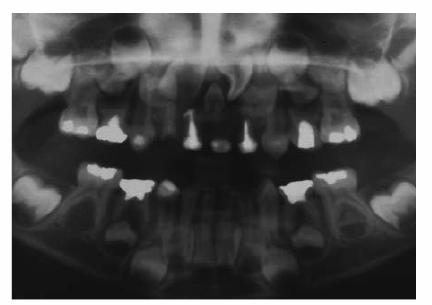
Figure 27-2D: The radiograph shows the endodontic treatment performed and the restoration of the lower caries.
Nursing Bottle Syndrome and/or Tooth
Loss due to Caries of the Anterior Teeth: The Pedodontic Prosthesis
The consequences of this pathology are serious because of the possible loss of
one or more anterior teeth due to serious caries lesions. The most critical
teeth are the maxillary incisors and, in relation to their eruptive succession,
the first primary molars.
When children present with chronic and recurrent fistulas and abscesses (Figures 27-3A
and B),
tooth function becomes limited. Radiographic investigation and clinical
evidence frequently show an infectious necrosis of the pulp in an advanced
phase, and the involved teeth (if an endodontic restorative therapy is not
possible) are extracted and a pediatric prosthetic appliance is constructed.
The correct space management and maintenance allow for the normal physiologic
evolution and eruption of the permanent teeth and improved esthetics and
speech.9,14,18
Figure 27-3A and B: When chronic and recurrent fistulas and abscesses are present and a conservative therapy cannot be performed, teeth are extracted.
Pedodontic prostheses (also used in cases of trauma and/or tooth agenesis) are
removable appliances that can offer a simple, safe, and efficient therapeutic
solution because they can lead to reduction of the orthodontic treatment time.
The decision to use these prostheses is guided by the child's and parents'
cooperation and by precise clinical conditions (tooth class, available space,
both general and oral health conditions).
Removable space maintenance appliances present considerable advantages: they
can determine orthodontic movements and can help prevent orofacial muscle
imbalance and/or harmful sucking habits, such as finger or thumb sucking or lip
sucking. Furthermore, they can be modified during the patient's growth, improve
esthetics, and reduce psychological problems. On the contrary, they can be
uncomfortable to the young patient because of their volume; they need periodic
checks and high patient and parental cooperation and can be more prone to
breakage than fixed appliances.
A male patient, 3 years, 9 months old.
PROBLEM: The patient presented with rampant caries, loss of the anterior
teeth, and advanced caries in the posterior teeth (Figure 27-4A
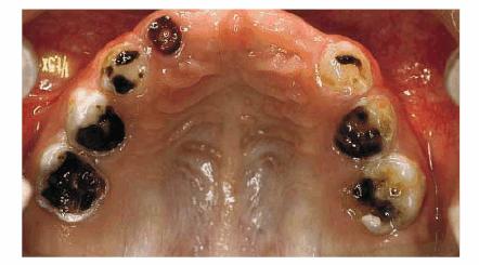
Figure 27-4A: A patient of 3 years, 9 months, with rampant caries and loss of an anterior tooth.
TREATMENT: The root of G is extracted, and the posterior teeth are
restored with composite resins. An attempt to maintain pulp vitality is made by
placing calcium hydroxide on the pulp. A pedodontic prosthesis has been used to
maintain the anterior space, preserve the vertical dimension, improve alveolar
growth, and avoid supereruption of the lower anterior teeth (Figures 27-4B to
D
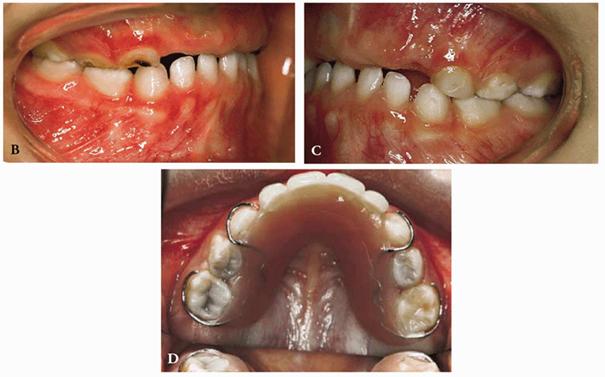
Figure 27-4B to D: The posterior teeth are restored with composite resin, and a pedodontic prosthesis is placed.
RESULT: Good function is restored, and the desired psychological result has been achieved, with lasting benefits. The patient has undergone periodic yearly visits: teeth #3 and #14 have erupted (Figures 27-4E and F
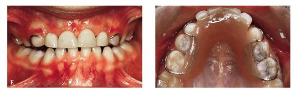
Figure 27-4E and F: Proper function is reached and the desired psychological result has been achieved in the long-term follow-up. Note that teeth #3 and #14 have erupted.
TRAUMA MANAGEMENT IN PRIMARY DENTITION
AND IN THE FIRST PHASE OF MIXED DENTITION
In pediatric dentistry, trauma is a very frequent event. Often it is very
difficult to make an accurate diagnosis as to the extent and severity of the
traumatic injury, manage the initial treatment of the acute aspect of the
injury, and determine the long-term follow-up. Dental traumas (as well as dental
caries) represent a true emergency and need an accurate diagnosis to provide
guidance in saving teeth, restoring the function of the dental arches,
improving esthetics, and avoiding complications.
As this is a high-incidence pathology, effective preventive measures need to be
taken to reduce the effects of trauma and ensuing complications that can occur
in young patients. It is extremely important to develop an effective prevention
and information plan for the public. The goal of this plan should be the
reduction of the functional and esthetic damage, the reduction of the
biological damage involving the orofacial area, and the awareness and
sensibility of both patients and practitioners to reduce sequelae, avoid
unnecessary treatment procedures, and provide the biological basis for healing
after injury.
Among the most effective preventive measures, we specify a timely orthodontic
correction to reduce the increased overjet; early correction of habits such as
finger sucking, thumb sucking, lip sucking, and abnormal swallowing; use of a
mouthguard to protect permanent teeth during sport activities; and correct
initial diagnosis and timely treatment, which are essential to produce a
correct initial treatment, avoid overtreatment, and avoid sequelae in the long-term
follow-up.
Trauma to the Primary Dentition
Andreasen and Andreasen's epidemiologic studies reported that one child in
three undergoes dental trauma. In fact, primary teeth, mainly because of their
anatomic characteristics, report more luxations than fractures, and 25% of them
suffer avulsions.1
Regarding trauma to hard tissues, young patients often present with crown
fractures (with or without pulp exposure). Therefore, the treatment plan
depends on the extent of the pulp exposure, the patient's and the family's
cooperation, the skill of the dentist, and the time interval between the trauma
and emergency care. If the pulp exposure is very small, the exposed area should
be cleaned and a pulp capping applied to the exposed pulp. For a larger pulp exposure,
pulp extirpation and root canal treatment should be performed. Examining and
diagnosing children's teeth can be especially challenging because accurate
radiographs may be difficult to obtain.
The treatment plan for primary teeth is usually different from one for
permanent teeth. There are several different reasons to be considered: the
healing mechanism of pulp and periodontal tissues in the primary dentition is
different than in permanent dentition. The healing mechanism characteristic in
permanent teeth may not occur in primary teeth, and sometimes extraction is
necessary to limit damage to permanent successors. Often treatment cannot be
performed because of the uncooperative behavior of children.
Reimplants of Primary Teeth
Traumatic avulsion is a frequent event in the primary dentition. It is
essential to conduct a differential diagnosis in the presence of a total
intrusive luxation, and in case of a multiple loss, it is necessary to verify
that the teeth have been neither swallowed nor inhaled. Actually, the debate is
open as to whether to reimplant only one tooth or even multiple primary teeth.
Avulsed primary teeth in which the roots have begun normal resorption are not
indicated for reimplantation. There is little value in reimplantation because
of the possibility of rapid root resorption or infection.
However, in young patients, the absence of teeth until the eruption of
permanent ones may cause esthetic and functional problems, as well as
psychological complications (such as anxiety), not only for the patients but
also for their parents. Therefore, attempting reimplantation is sometimes
worthwhile. Reimplantation cannot be performed when the tooth is not far from
normal resorption, radicular pathologic processes are present, or there is a
risk of infection and damage to the permanent tooth bud.
Recent clinical investigations carried out by Caprioglio et al.10
and Tsukiboshi19 have begun to define specific guidelines and
protocols. The tooth can be reimplanted only if these clinical situations are
present: (1) the child has acceptable occlusion, has no harmful habits, and is
in good health and (2) the avulsed tooth is far from root resorption, has been
avulsed not more than an hour, and has been stored hydrated. In the most
successful cases, the tooth will remain vital; otherwise, the root canal will
be treated with calcium hydroxide (for necrosis of the pulp). Unlike permanent
teeth, with reimplantation of primary teeth, healing of the pulp and
periodontal membrane should not be expected.
Sequelae after Trauma to Primary Teeth
One of the problems of trauma to the primary dentition is the possibility of
damaging the permanent successor tooth buds. The patient's age and the degree
and direction of the malposition of the primary teeth, as well as the type of
trauma, are some of the most important factors to be considered. The effect may
be either direct or indirect. An accurate diagnosis at the time of injury
ensures that appropriate care is prescribed. Combined with careful follow-up,
this care will, in many cases, prevent hypoplasia and hypomineralization
affecting the permanent successors.
The most serious deciduous tooth injuries in terms of damage to permanent
successors are intrusive luxation, avulsion, extrusive luxation, and subluxation.
The permanent teeth that are most often affected are the central incisors. The
effects on the successional tooth may be discoloration and hypoplasia of the
enamel, bending and malformation of the anatomic crown and root, hypoplasia of
the root, and retarded eruption. These problems may occur regardless of the
treatment of the traumatized primary teeth. It is very important to inform
parents about these possibilities; therefore, regular reviews are clearly
important to try to avoid or resolve the problems.
A male patient, 5 years, 8 months old.
PROBLEM: Occasionally, parents complain about discoloration of their
children's primary teeth, or the discoloration may go unnoticed. Discoloration
of primary teeth, as in this case, may be due to slight damage, such as
concussion or subluxation. If discoloration continues without pulp
obliteration, there is a possibility of pulp necrosis. Because of the original
trauma, a malformation of the anatomic crown of teeth #8 and #9 is observed.
TREATMENT: After the complete eruption of the two upper central
incisors, the pigmentation (hypoplasia) of the teeth is restored with composite
resins.
RESULT: Because enamel hypoplasia is superficial damage, it can be
easily and esthetically resolved (Figures 27-5A
and B).
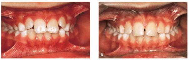
Figure 27-5A and B: Due to a trauma to primary teeth, a malformation of the anatomic crown of teeth #8 and #9 is observed (hypoplasia). Teeth are restored with composite resin.
Traumas in the Early Mixed Dentition
Although we acknowledge the importance of providing guidelines and promoting
informative and preventive protocols, for the purpose of brevity, we are not
addressing any reference to classifications, clinical examinations, medical
history, or special investigation, which are absolutely essential for
comprehensive treatment planning. We simply describe some clinical trauma cases
in which the cooperation between the pedodontist and the orthodontist has led
to a good esthetic and functional result.
Reattachment of the Tooth Fragment in
the Fracture of Anterior Teeth
As far back as 1961, Chosak and Aidelman proposed a technique to manage the
reattachment of the tooth fragment after trauma.11 Therefore, if the
fragment is available, this treatment procedure achieves excellent esthetic
results, particularly if the fragment is complete.
Another advantage is the ability to reconstruct the palatal face, which will
benefit the occlusal stability.
The fragment must be kept hydrated (water, milk, physiologic saline solution,
or other special storage media). Alternatively, dehydration will distort the
tooth color. The tooth must not have additional fractures, and the soft tissues
must not have lacerations, contusions, or bleeding.
Before reattachment, the fit of the tooth fragment to the remaining tooth
should be confirmed and checked for enamel defects. A fragment that is highly
damaged may be unsuitable for reattachment.
After the pretreatment preparation, local anesthesia is administered, and the
tooth is isolated with a rubber dam, the tooth fragment is cleaned and
prepared, and the pulp is dressed, if necessary. The remaining tooth is
beveled, the tooth fragment is tried in, the tooth is etched, and the fragment
is bonded with composite resin. Next, the tooth fragment is attached to the
remaining tooth, and the resin is reshaped and polished. The patient should be
examined after 1 week, 1 month, and 3 months and then checked annually for
discomfort and possible pulp necrosis and to evaluate esthetics.
A female patient, 9 years, 2 months old.
PROBLEM: This patient presented with an extended, noncomplicated enamel
and dentin fracture of teeth #8 and #9. Four days had passed since the trauma
occurred, and the fragments had not been hydrated (Figures 27-6A
and B
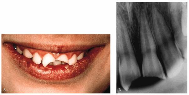
Figure 27-6A and B: An extended but not complicated enamel and dentin fracture of teeth #8 and #9.
TREATMENT: After performing routine clinical, instrumental, and
radiographic examinations, it was decided to try to rehydrate the tooth
fragments by putting them in a physiologic saline solution for 1 day. In the
meantime, the fit of the tooth fragments was confirmed (Figures 27-6C
and D). The
following day, the two rehydrated tooth fragments had reached their normal
color, and the reattachment of the two fragments proceeded as described above.
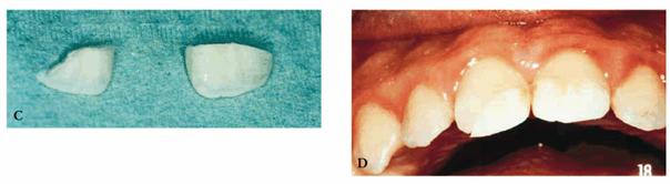
Figure 27-6C and D: The tooth fragments have not been hydrated for 4 days. They fit perfectly but are discolored.
RESULT: The result obtained was much better functionally and
esthetically than it would have been using only composite resin. This
one-appointment procedure (due to a perfect fitting of the fragments) has restored
the anterior guide and has reached a correct reproduction of the biting edge (Figures 27-6E
and F
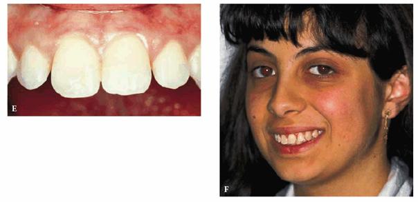
Figure 27-6E and F: The fragments have been rehydrated for 1 day and then reattached. The final result and the patient's smile are satisfying.
When
physiologic effects are more important than cosmetic ones; this technique
allows a "true biological restoration" with a true "restitutio
ad integrum" of the tooth crown. No additional treatment has been required
on the teeth other than periodic examinations.
A male patient, 8 years, 3 months old.
PROBLEM: The patient presented with a complicated enamel-dentin fracture
of the left central incisor with an immature apex. The patient had poor oral
hygiene and a class II molar relationship. The tooth fragment was available,
although it was not complete (Figures 27-7A
and B
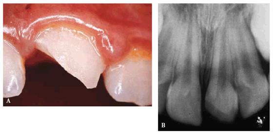
Figure 27-7A and B: A complicated enamel-dentin fracture of tooth 9 and the radiograph.
TREATMENT: After cleansing and stopping the flow of blood, the restoration was done in accordance with standard procedures. After pulp capping with calcium hydroxide, the tooth fragment was reattached. However, because the fragment was not complete, the missing parts were reconstructed with composite resin, and the entire periphery of the fractured surface of the remaining tooth was beveled to improve esthetics (Figure 27-7C
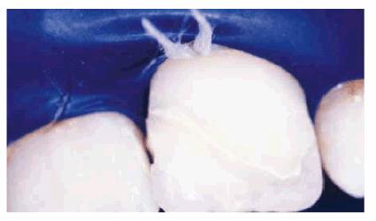
Figure 27-7C: The tooth fragment is reattached with a bevel to improve esthetics.
RESULT: Immediately after restorative treatment, the patient expressed
his satisfaction. After 18 months, the patient returned, reporting an extrusive
luxation of tooth 9 following a school incident. The tooth was immediately
repositioned and splinted orthodontically (Figure 27-7D). A. 018 nytinol wire was used for
15 days. It was subsequently removed after clinical and radiographic
confirmation of results.
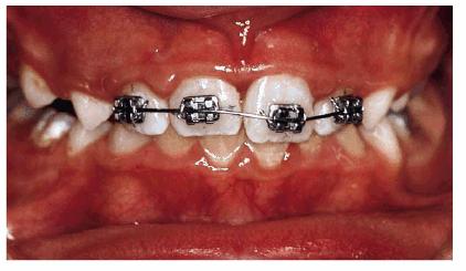
Figure 27-7D: After 18 months, the same tooth has an extrusive luxation and is orthodontically splinted.
The patient had periodic radiographic examinations and sensitivity tests to
monitor pulp and root healing, as well as tooth vitality (Figure 27-7E).
Figure 27-7E: The radiograph after the reposition.
From an orthodontic perspective, a serious skeletal Class II with deep bite is becoming more and more evident. Therefore, orthodontic treatment has been initiated in both arches to align the dentition over the basal bone in harmony with the surrounding hard and soft tissues, as well as to achieve good esthetics (Figures 27-7F and G
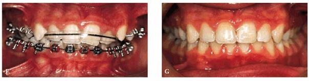
Figure 27-7F and G: The same patient during the orthodontic treatment and after. Good esthetics has been achieved.
Traumatic Avulsion
A traumatic avulsion is a serious clinical event. The traumatic loss of one or
more teeth, particularly when they are immature, represents a highly dramatic
event. Recommended therapy will vary according to the time elapsed from the
trauma to the first visit as follows:
. Immediate reimplantation (45 minutes or less) if the tooth is preserved in
milk or in a preservative solution and it is reimplanted within 24 hours
. Delayed reimplantation
There are three factors that affect the success of the reimplantation:
1. Time elapsed since the trauma occurred. If the tooth is preserved hydrated
or in saliva for a period of 20 to 120 minutes, it can be reimplanted as vital
and then followed up periodically to avoid pulp complications or ankylosis. If
2 hours have elapsed since the trauma or after 30 minutes of dryness, the tooth
must be treated endodontically with calcium hydroxide and then monitored
periodically.
2. Preservation of the avulsed tooth. The tooth must be kept in a liquid in
osmotic balance with the tissues. These include saliva, a physiologic saline
solution, milk, or special storage medium. It should be noted that teeth kept
in water and reimplanted have shown a high percentage of ankylosis.
3. Treatment plan. It is essential for the dentist to have a wide experience in
endodontics, pediatric dentistry, orthodontics, and oral surgery to perform the
treatment plan correctly to achieve an esthetic result and reduce sequelae.
RESTORATION OF POSTERIOR TEETH
When reference is made to the fundamental concepts governing pediatric dentistry,
the importance of the awareness of the correct evolution of arches, as well as
treating any irregular condition, must be a primary focus.
The first primary molars are the ones that, during eruption, determine the
first proprioceptive reflexes on the transversal plane. Their role, as well as
their maintenance, comes second when compared with the second primary molars.
The presence of the second primary molar is strategic to guide the eruption and
the articulation of the first permanent molars. The restoration and
preservation of the posterior primary teeth are vital to maintain the arch
length and eliminate mesial drift of the permanent molars.
Arch length and arch anatomy can be modified by
1. Tooth crowding: loss of space (unilateral or bilateral) due to a premature
extraction because of caries or loss from a traumatic event or ectopic
eruption, impaction, transposition, ankylosis, agenesis, or supernumerary
teeth.
2. Presence of habits such as oral breathing, sleep apnea syndromes, and thumb
sucking. In the presence of a harmful oral habit, these patients usually
present with a reduction of transversal (cross) diameters, as well as the loss
of one or more primary molars, which could lead to a further collapse of the
arch.
3. Presence of a malocclusion: Class II with an increased overjet and/or
overbite or Class III with an anterior crossbite and/or posterior crossbite
and/or open bite.
The pedodontist, often working with the orthodontist, must
perform a careful analysis of the dentition. The goal is arch harmony and good
balance among function, arch form, and oral tissue condition.
Optimum space maintenance therapy is the preservation of the primary molars
until natural exfoliation. Dental education and improved prevention have
reduced the number of children who develop malocclusion because of premature
loss of primary teeth. Therefore, it has become one of the most controllable
causes of malocclusion. When posterior teeth are damaged or lost, stainless
steel crowns for grossly broken-down teeth, space maintainers (fixed or
removable appliances), or esthetic posterior restoration techniques can be used
to maintain arch length.
Interproximal caries in primary teeth, due to the different thickness of enamel
and dentin, can more easily extend to the pulp, requiring endodontic therapy.
This aside, compomers and composite resins are the materials of choice for
their easy handling, reduced tooth preparation, reasonable wear properties,
good esthetics, and release of fluoride leaching.
Preventive Resin Restoration
Composite resin is the material of choice for the treatment of early occlusal
caries in the permanent dentition. The development and use of preventive resin
restoration has greatly changed the management of occlusal caries in very young
patients. The indications are an enamel-only lesion, incipient lesion just into
the dentin, and a small Class I lesion.
A male patient, 7 years, 2 months old.
PROBLEM: The patient was a high-caries-risk subject with poor oral
hygiene and an enamel-only lesion of tooth 3.
TREATMENT: Preventive resin restoration. After local anesthesia and
rubber dam isolation, a small high-speed diamond burr was applied to the
questionable fissure. It was essential to have adequate access to the
underlying dentin to be certain of complete caries removal. A glass ionomer
liner was placed over the dentin, extended to the amelodentinal junction, and
light-cured. An etching gel was placed on the enamel margins and on the
occlusal surface, washed, and dried. The bonding resin and the composite resin
were placed and polymerized, and, finally, a fissure sealant was placed over
the restoration and cured. After the rubber dam was removed, the occlusion was
checked (Figures 27-8A and B).
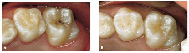
Figure 27-8A and B: An enamel caries is treated with a preventive resin restoration.
RESULT: The durability of preventive resin restoration has been proved
to be as good as amalgam, with less removal of sound tooth tissue and with
better esthetics.
ESTHETICS AND HARMONY OF DENTAL ARCHES:
SPACE MANAGEMENT IN PEDIATRIC DENTISTRY
Correct space management, starting from the emergence of primary dentition through
the late phase of mixed dentition, requires cooperation between the pedodontist
and the orthodontist. The preventive strategy not only simplifies the
subsequent orthodontic therapy by making it less complex and more reliable, it
also helps to improve esthetics and function.
Considering some fundamental concepts and new therapeutic trends focused on the
resetting of shape and the esthetics and harmony of the dental arches, it is
necessary to balance the arch symmetrically and to check the eruption of the
first permanent molar (tipping, uprighting) to prevent mesialization of the
first lower molars. Next, a correct dentoskeletal analysis, cephalometric
study, and careful evaluation of the means and materials must be done before
formulating a diagnosis and logical prognostic evaluation. Last, if orthodontic
brackets are applied to primary teeth, the advantages include reduced
demineralization risks; the possibility of good anchorage, which decreases the
reaction counterforce; and the reduction of acid-etching time and of problems
associated with the removal of orthodontic brackets (bonding and debonding).
The dentist can perform several different treatments:
1. Slicing of the primary cuspids and/or primary second molars
2. Lip bumper on the primary second molars
3. Mechanics of symmetrically balancing the arches following a premature loss
or extraction of the primary cuspid
4. Uprighting of the first permanent molars
A female patient, 6 years, 7 months old.
PROBLEM: The patient's frontal view showed a serious bimaxillary
crowding with deviation of the midline. When teeth #7 and #10 erupted, crowding
problems greatly increased. Also, tooth #10 was in crossbite (Figure 27-9A).
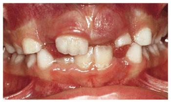
Figure 27-9A: A serious bimaxillary crowding with deviation of the midline in a 6-year, 7-month-old female.
TREATMENT: After a
short observation period, while the lateral incisors erupted, the pediatric
dentist and the orthodontist initiated therapy. M and R were sliced, and a lip
bumper was applied to K and T. The occlusal surface rose to resolve the
crossbite of the upper left lateral incisor. The maxillary arch was treated orthodontically
to correct the alignment (Figure 27-9B).
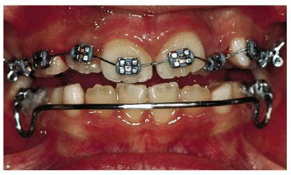
Figure 27-9B: After a short observation period, the early orthodontic therapy is started.
RESULT: The patient underwent an early orthodontic treatment for 1 year
that achieved arch balance, improvement of esthetics, and health of the oral
tissues. Furthermore, the improved smile contributed to greater self-confidence
and an improved sense of well-being (Figures 27-9C and D).
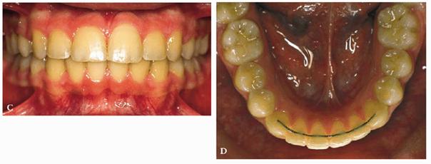
Figure 27-9C and D: The patient's smile and the lower arch after the orthodontic treatment: good balance and esthetics are achieved.
It is important to emphasize that the correct space management may require, in
addition to orthodontic treatment, consultation with and/or treatment by an
endodontist, prosthodontist, or oral surgeon. In fact, dental anomalies in
number (such as agenesis, supernumerary tooth and/or teeth) or in shape (micro-
or macrotooth) may require the cooperation of many different specialists to
improve esthetics and/or function.
A female patient, 9 years, 10 months old.
PROBLEM: The initial orthopantography showed agenesis of tooth #10 (with
deviation of the middle line), microdontia of tooth #7, and skeletal dental
Class III. These are often interrelated (Figure 27-10A).
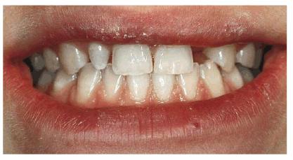
Figure 27-10A: After orthodontic treatment, the patient with microdontia of tooth 7 and agenesis of tooth 10 shows a good alignment.
TREATMENT: After applying a fixed orthodontic appliance, the dental
arches were aligned, and a correct transversal relationship was obtained with a
reduction in midline deviation. To improve esthetics in the anterior region,
tooth #7 was restored with a composite material, and tooth #10 was replaced
with a resin-bonded fixed Maryland bridge (Figures 27-10B and C). This bridge has esthetic appeal
because it does not require the use of full crowns on either side of the
missing tooth, and little or no tooth reduction is involved. The metal
framework was bonded to the tooth with resin cement. If the adjacent teeth to a
missing tooth are intact and in good condition, a resin-bonded bridge may be
the method of choice.
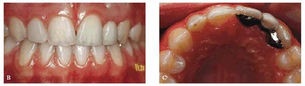
Figure
27-10B and C: A resin-bonded
RESULT: This intervention resulted in an acceptable masticatory and
esthetic function. Maximum oral hygiene was emphasized to the patient.
A male patient, 8 years, 3 months old.
PROBLEM: The patient presented with a supernumerary incisor and a double
tooth in place of the central upper left incisor (tooth #9) (Figures 27-11A and B). This anomaly manifests itself as a
structure resembling two teeth that have been joined together. In the anterior
region, the anomalous tooth usually has a groove on the buccal surface and a
notch in the incisal edge. Radiographs are necessary to determine if there is a
(fusion) union of the pulp chambers. Fusion exists when there is a joining of
two teeth by pulp and dentin. Two canals are usually present, as in this case.
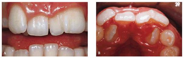
Figure 27-11A and B: A supernumerary incisor and a double tooth in place of the central upper left incisor. Frontal and occlusal views.
TREATMENT: Both the supernumerary tooth and fused tooth were extracted (Figure 27-11C); also, the fixed orthodontic appliance was
applied to the maxillary arch to close the anterior diastema. Subsequently, the
incisal margin and the interproximal area of tooth #9 were restored to improve
esthetics (Figure 27-11D).
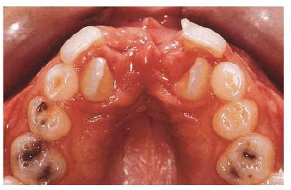
Figure 27-11C: Both the supernumerary tooth and fused tooth are extracted.
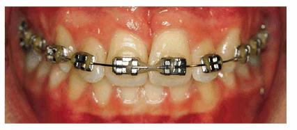
Figure 27-11D: An orthodontic appliance is applied to the maxillary arch to close the anterior diastema.
RESULT: The team work of several specialists created a good
morphic-functional recovery (Figures 27-11E and F) and an esthetic result that satisfied
the patient (Figures 27-11G and H).
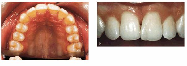
Figure 27-11E and F: The morphic-functional recovery at the end of the orthodontic treatment.
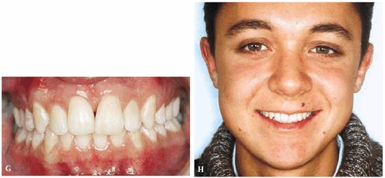
Figure 27-11G and H: The final result showing the patient's smile at the end of the treatment.
THE
FACE IN PEDIATRIC DENTISTRY: ESTHETIC KEYS
The concept of "beauty" has always been subjective. As to the
individual esthetic aspect, many attempts have been made during the centuries
with the purpose of extrapolating the golden cut or divine proportion. In spite
of all efforts of standardization, each age and every century has its own
esthetic canons, as each individual may have his or her own esthetic ideals.
However, our society is continuously creating new ideals of beauty, new trends
to which people would like to aspire. During the last few decades in the
Western world, an individual's appearance has assumed much more importance and
is essential in establishing self-image. Ultimately, it contributes to success
in all aspects of professional and social life. For this reason, many branches
of medicine that have an esthetic component continue to research and improve
their techniques. Among these entities is orthognathodontics, which is
enlarging its field of activity from the smile to the entire face of the
patient. As Goldstein says, "The way you see yourself and think others see
you has a great deal to do with the way you feel about yourself. A charming
smile can open doors; our own self-image is the key to our happiness."17
Several authors have shown that orthodontic treatment can improve facial
harmony, including ortho gnathic surgery. Traditional cephalometrics, based on
angular and linear measurements of the soft and hard tissues of the patient,
have proven to be less than reliable for correct diagnosis and a satisfactory
esthetic result. It suffices to say that there is no cephalometric analysis
that has universal appeal. Most cephalometric analyses use as a reference some
intracranial skeletal plans. Diagnosis and treatment planning are based on them
because of the assumption that the correction of definite dental and bone
parameters achieves facial esthetics, harmony, and facial balance. However,
many authors now agree that a careful analysis of the soft tissue is also
needed.
For this reason, cephalometrics now includes studies and measurements involving
soft tissues by using in general the usual teleradiographies of the skull in
norma lateralis or the photographic records of the patient in lateral view or,
more rarely, in frontal view.
At issue is the fact that a good functional occlusion with the usual skeletal
parameters does not always correspond with an esthetically pleasing facial
balance. This phenomenon is mainly due to the thickness of the soft tissues
covering the skeleton of the face, which can make the dentoskeletal analysis
unreliable in the evaluation of facial harmony. (In other words, if the lips
are not well balanced and closed at rest, facial dysmorphosis can be present in
the absence of dentoskeletal alterations.)
According to Blanchette et al., soft tissues have a tendency to mask
discrepancies of the bone base (maxilla and mandible); therefore, we would have
thinner soft tissues in low-angle subjects and thicker soft tissues in
high-angle ones.7 Perhaps it was for this reason that Ferrario et
al. found significant correlations between the skeletal class and the soft
tissues,13 and that Burnstone et al. argued that any dentoskeletal
standard can present unpredictable final esthetics of the face.8
At present, the purpose of an orthodontic treatment should be the achievement
of a good functional occlusion along with appealing dentofacial esthetics, thus
maintaining the integrity of the dentoperiodontal tissues. For these reasons,
several practitioners have started to focus their interest mainly on the study
of the patient's face rather than the skeleton. Therefore, the transition has
been from a diagnostic system, which can be defined as "centrifugal"
and which, starting from the skeleton, goes outward, to a
"centripetal" system, which begins instead with the analysis of soft
tissues to determine the corrections to be effected on hard tissues. Arnett and
Bergman's3,4 and Ayala's5 cephalometric analyses visually
evaluated the facial contour of the patient's soft tissue exclusively in a
natural position, both frontal and lateral views, to determine the diagnosis
and treatment plan.
Currently, all of the existing diagnostic systems based on the analysis of soft
tissues refer to adult subjects, especially those who must undergo orthognathic
surgery. The purpose of this chapter is to propose a method of analysis that
can determine esthetic reference parameters that are reliable for the child's
face (in the different ages of growth) and be useful to create a clinical
alternative to cephalometric analysis of the soft tissues. Moreover, this
method could help the clinician to integrate and complement the usual
cephalometric analysis to achieve not only esthetic facial harmony but also
good balance.
By analyzing the different methodologies used by authors to evaluate the
harmony of soft tissues in adults, reference parameters and data have been
selected that would be useful and reliable when evaluating growing patients.
These parameters and data have been subsequently modified after considering
craniofacial growth. At birth, in fact, the splanchnocranium is considerably
hypodeveloped if compared with the neurocranium (Figure 27-12). Furthermore, the mandible is the least
developed of the face's lower third and tends to grow more and for a longer
period of time when compared with the rest of the face. Moreover, a sequence
exists both in the maxilla and in the mandible. This has been defined as a
completion of growth in the three planes of space: first, growth completes in
width, then in length, and then in height. The transversal growth of both bones
(including width of the dental arches) tends to be complete before the pubertal
growth peak and is scarcely influenced by growth variations during adolescence.
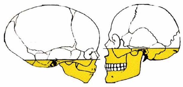
Figure 27-12: At birth, the splanchnocranium is considerably hypodeveloped if compared with the neurocranium.
Sagittal growth of the two maxillaries continues during puberty. In girls, it
stops almost immediately, on average between 14 and 15 years of age. In boys,
such growth usually does not stop before 18 years of age. The maxillaries' and
face's vertical growth continues longer in both sexes when compared with the
growth in length.
In light of these considerations, canons of esthetic evaluation have been
altered to adapt them to growing patients. The selected reference parameters do
not predict deliberate linear measures as the growing patient, unlike adults,
cannot have fixed values.
Frontal View Considerations
Symmetry among the Different Parts of the Face. Like in adults, the
child's face (Figure 27-13) must show perfect symmetry with the eyes, ears,
and mandibular angles placed at the same height.
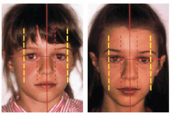
Figure 27-13: These children's faces appear to be in perfect symmetry.
Correct Distance between the Eyes, Nose, and Lips. The 1:1 ratio
between the width of the lips and the distance between the inside margins of
the iris (Figure 27-14) remains valid. However, the child's nose base
should be smaller than the intercantal distance as it will grow considerably.
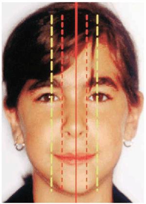
Figure 27-14: Correct distance between the eyes, nose, and lips.
Middle to Lower Facial Third Ratio. The reliable parameters for
adults cannot be the same as those for children (Figure 27-15). As previously stated, the neurocranium grows
earlier than the splanchnocranium; therefore, the middle third of the face
develops before the lower third. In fact, the lower third should be smaller
than the middle and upper thirds. Furthermore, when the lower third of the face
develops earlier, it is of special concern as it is indicative of excessive
growth in a vertical direction. Such considerations are inversely proportional
to the patient's age.
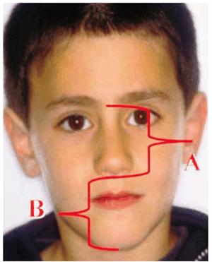
Figure 27-15: The middle third of the face develops before the lower third.
Ratio for Esthetic Balance. This is the division of the face by a
symmetry line passing through the glabella, nasal tip, midpoint of the upper
lip, midpoint of the chin, and the suborbital line. The Tr-Me/ZA-ZA ratio,
which in the adult is 1.35 for the male and 1.3 for the female, should have a
lower value for the adolescent, who will grow more vertically than in width (Figure 27-16). Therefore, the value shall start from about 1
in younger subjects to increase gradually during growth and ultimately to reach
normal (adult) reference values.
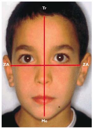
Figure 27-16: The ratio for esthetic balance.
Tr = trichion: the point of the hardline in the midline of the forehead. In
early childhood, identification of this landmark may be difficult because of an
irregular or indistinguishable hairline.
ZA = Zygion angle: the most lateral point of each zygomatic arch. It is
identical to the bony zygion of the malar bone.
ME = Menton (chin): the lowest median landmark on the lower border of the
mandible.
Sclera Exposure. An excessive exposure of sclera, the firm white
fibrous membrane that forms the outer covering of the eyeball, implies a
developmental deficit of the middle third of the face. If this is visible and
other symptoms are present, such as oral breathing with a narrow pointed nose,
reduced transversal diameters of the upper maxillary with crossbite, and,
dentally, upper arch crowding with a tendency to cuspal inclusion, a skeletal
Class III with maxillary hypoplasia is present.
Incisal Exposure. In children, when teeth can be exfoliating or
erupting, there are no reliable reference points. However, if, when smiling, a considerable
quantity of marginal gingiva is exposed, an excessive facial anterior vertical
growth or a maxillary excessive protrusion could exist.
Lip Closure without Tissue Strain. Over time, all soft tissues have
the tendency to relax or strain; therefore, it is acceptable for a young
subject to have the upper lip slightly shorter than that of an adult and,
hence, moderate lip incompetence. However, this should be present no longer
than 7 to 8 years of age.
Profile View Considerations
Skeletal Convexity from the Zygomatic Area to the Interlabial Gap. Considering
that, in children, the lower facial third develops ahead of the middle third,
it is normal that the cheek's profile is more convex than in adults (Figure 27-17). Also, a curve indicating a trend to high-angle
mandibular growth is alarming, even more so if it appears in children rather
than adults.
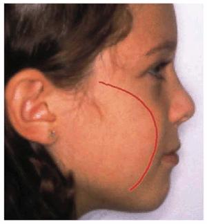
Figure 27-17: In children, the cheek's profile is more convex than in adults.
Nose Prominence. This is measured from the subnasal (the point at
which the columella merges with the upper lip in the midsagittal plane) to the
pronasal (the most prominent anterior point of the nose) parts of the nose.
Such distance, which ranges from 16 to 20 mm for normal values in adults, will
obviously have a lower value in children (Figure 27-18). It is important to note that a prominent nose
is generally a contraindication to an extractive treatment. (See also Chapter
9, Esthetics in Dentistry, Volume 1, 2nd edition.)
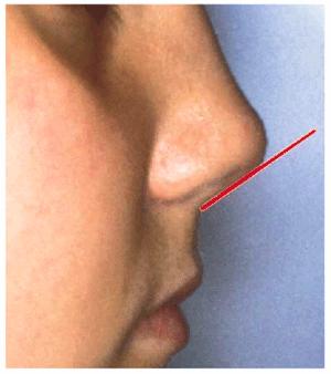
Figure 27-18: The nose prominence (subnasal - pronasal).
The shape of the nose must also be considered. In fact, with growth, the point
of the nose tends to move downward and forward. Therefore, it is evident that a
convex nose shape in a child worsens considerably with growth. On the contrary,
prognosis improves in young subjects with a concave or flat nose shape. In
these cases, it is also very useful to observe the child's parents. In fact,
the eyes and nose are the somatic features of the face, which present the
highest heredity level.
An increased nasolabial angle must not be an absolute contraindication to a
protocol of serial extractions but only one of the clinical factors evaluating
the case.
Lip Curvature. Both the upper and lower lips must present a slight
curvature, with concavity pushing forward.
A very marked labiomental sulcus in a child may indicate a sagittal mandibular
and maxillary vertical deficit, thus presupposing a low-angle facial typology (Figure 27-19). Alternatively, the total disappearance of this
sulcus can indicate a mandibular sagittal and vertical development involving
both planes and therefore a high-angle facial typology. High-angle subjects
camouflage the dentoskeletal Class III and the low-angle subjects the Class II,
improving the dental compensations that are present in such cases.
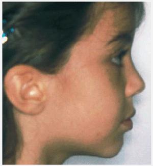
Figure 27-19: In a child, a very marked labiomental sulcus may indicate a sagittal mandibular and maxillary vertical deficit.
Nasolabial Angle. The nasolabial angle can be more open in the
child because the nose tip grows lower. Generally, in adults, all soft tissues
tend to relax and become less toned. In fact, for this reason, it is acceptable
for a young patient to have the upper lip slightly short or strained and a
gingival smile not more than 3 to 4 mm.
Correct Ratio between the Submental Area and the Lower Facial Third
Inferior: NTP-Gn/Sn-Gn*
This ratio, which in adults has a normal value of about 0.8, will be higher in
children even though the mandible will still develop in length, for two
reasons: the lower third of the face will continue to develop in height, and
the chin-neck contour is modest in children. Therefore, the usual value in the
young patient ranges between 1 and 1.2. Lower values indicate a hypomandible;
conversely, higher values indicate a hypermandible.
The skeletal type of the patient also has to be considered. For example, a
decrease in the normal value of this ratio in an obese child and an increase in
an athletic, long-limbed child are expected.
CONCLUSION
The esthetic measurements and treatments considered available and reliable for
adults cannot be considered for children. Research must eventually supply
reliable data and modify the well-worn cliche of the ideal face and proportion
for the Caucasian population during growth. The result should be to alter
existing esthetic analyses and adapt them to growing patients by following the
predictable craniofacial growth mechanism.
When we are familiar with the growth mechanism and the different factors
determining it, it will be possible to reduce the need for
orthopedic-orthodontic treatments. True esthetic orthodontics may be a protocol
to obtain true facial esthetics and balance when combined with effective
pediatric esthetic dentistry.
As Goldstein has proposed in Volume 1, 2nd Edition of Esthetics in Dentistry,
esthetics is the fourth dimension in dentistry, in addition to the biological,
physiologic, and mechanical dimensions. Esthetic balance is increasing in
importance because in the 21st century, our culture is more aware of the
essentials of attractiveness in the face and smile and in general physical
appeal.
Esthetic harmony is synonymous with skeletal, dental, and neuromuscular harmony
and temporal mandibular joint harmony. The concept of note is that esthetics in
pediatric dentistry is the basic guideline for esthetics in adults and will
become a subject of growing interest in the decades ahead. As a clinical issue,
esthetic considerations are increasing in frequency and importance in pediatric
dentistry. The pedodontist must work in close cooperation with an orthodontist
to apply correct preventive or early interceptive orthodontics and esthetic
principles. This close cooperation can reduce treatment times and costs and
increase long-term stability because space management will also reduce
extraction cases. It is imperative to understand that esthetic harmony can lead
to psychological health and higher self-assurance; it improves intrapersonal
relationships and strengthens self-confidence.
To quote Jean Cocteau, "A defect of our body, if corrected, can improve
our soul," or, to paraphrase an old Jewish saying, "He who gives a
smile to a child gives a smile to the world."
REFERENCES
1. Andreasen JO, Andreasen FM. Traumatic dental injuries-a manual. Copenhagen:
Munskgaard, 1999.
2. Andreasen JO, Borum MK, Jacobsen HL, Andreasen FM. Replantation of 400
avulsed permanent incisors: 1-4. Endod Dent Traumatol 1995;11:51-89.
3. Arnett WG, Bergman RT. Facial keys to orthodontic diagnosis and treatment
planning. Part I. Am J Orthod Dentofac Orthop 1993;103:299-312.
4. Arnett WG, Bergman RT. Facial keys to orthodontic diagnosis and treatment
planning. Part II. Am J Orthod Dentofac Orthop 1993;103:395-410.
5. Ayala M. Estetica facciale. Atti congresso SIDO Roma 2/12/99.
6. Berg JH. The continuum of restorative materials in pediatric dentistry-a
review for the clinician. Petriatr Dent 1998;20:93-100.
7. Blanchette ME, Nanda RS, Currier FG, et al. A longitudinal cephalometric
study of the soft tissue profile of short- and long-face syndromes from 7 to 17
years. Am J Orthod Dentofac Orthop 1996;109:116-31.
8. Burnstone CJ, James RB, Legan H. Cephalometrics for orthognathic surgery. J Oral Surg 1978;36:269-78.
9. Caprioglio C. Odontoiatria infantile. Cap. 2. In: Paglia L, ed. Progressi in
odontoiatria. Milano: Utet, 1999: 33-83.
10. Caprioglio C, Caprioglio D, Garcia-Godoy F. Traumatic dental injuries. In:
Garcia-Godoy F, ed. Clinical pediatric dentistry. Heidelberg: Springen (in
press).
11. Chosak A, Aidelman E. Rehabilitation of fractured incisor using the
patients natural crown-case report. J Dent Child 1961.
12. El-Kalla H, Garcia-Godoy F. Bond strength and interfacial micromorphology
of four adhesive systems in primary and permanent molars. J Dent Child 1998;65:
169-76.
13. Ferrario VF, Sforza C, Serrao G, et al. Reliability of soft tissue
references for anteroposterior measurement of dental bases. Int J Adult Orthodont Orthognath Surg 1998;13:210-6.
14. Fortier JP, Demars-Freemault CH. Pedodonzia. Milano: Masson, 1988.
15. Garcia-Godoy F, Flaits CM, Hicks MJ. Secondary caries adjacent to amalgam
restoration lined with fluoridated dentin desensitizer. Am J Dent 1998;11:254-8.
16. Garcia-Godoy F, Hosoya Y. Bonding mechanism of compo-glass to dentin in
primary teeth. J Clin Pediatr Dent 1998;22:217-20.
17. Goldstein RE. Change your smile. 2nd rev. edn. Carol Stream, IL: Quintessence,
1988:22.
18. Pinkam JR, Casamassimo PS, Fields M, et al. Pediatric dentistry: infancy
through adolescence. Philadelpha: WB Saunders, 1994.
19. Tsukiboshi M. Treatment planning for traumatized teeth. Tokyo:
Quintessence, 2000.
20. Wilson AD, Kent BE. A new translucent cement for dentistry: a glass
isonomer cement. Br Dent J 1972; 32:133-5.
ADDITIONAL RESOURCES
Andreasen JO. Atlas of replantation and transplantation of teeth. Fribourg:
Mediglobe, 1992.
Andreasen JO, Andreasen FM. Text book and colour atlas of traumatic injuries to
the tooth. 3rd edn. Copenhagen: Munskgaard, 1994.
Bass NM. The esthetic analysis of the face. Eur J Orthod 1991;13:343-50.
Berkman MD, Goldsmith D, Rothschild D. Evaluation-diagnosis-planning. The
challenge in the correction of dentofacial deformities. J Clin Orthod
1979;13:526-38.
Black RB. Application and re-evaluation of air abrasive technique. J Am Dent
Assoc 1955;50:408-14.
Cameron AC, Widmer RP, et al. Handbook of pediatric dentistry. London: Mosby,
Wolfe, 1997.
Caprioglio D, Falconi P. Odontoiatria infantile pratica. Milano: Libraria
internazionale, 1992.
Caprioglio D, Manna A, Paglia L, et al. Manuale di traumatologia
dento-alveolare. Milano: Ciba, 1996.
Ericson D, et al. Clinical evaluation of efficacy and safety of a new method
for chemo-mechanical removal of caries. Caries Res 1999;33:171-7.
Goldstein RE. Esthetics in dentistry. Philadelphia: JB Lippincott, 1976.
Goldstein RE, Parkins FM. Air-abrasive technology: its new role in restorative
dentistry. J Am Dent Assoc 1994; 125:151.
Holdaway RA. A soft-tissue cephalometric analysis and its use in orthodontic
treatment planning. Part I. Am J Orthod 1983;84:279-93.
Jacobson A. Planning for orthognathic surgery-art or science? Int J Adult Orthodont Orthognath Surg 1990;5:217-24.
Loevy HT. Dental management of the child patient. Chicago: Quintessence, 1987.
McDonald RE. Dentistry for the child and adolescent. Mosby, 1982.
Park YC, Burstone CJ. Soft-tissue profile-fallacies of hard-tissue standards in
treatment planning. Am J Orthod Dentofac Orthop 1986;90:52-62.
Staele HJ, Koch MJ. Kinder und Jugendzahn heilkunde. Koln: Arzte-Verlag Grubtl,
1996.
Steiner CC. Cephalometrics in clinical practice. Angle Orthod 1959;29:8-29.
Tweed CH. Indications for extraction of teeth in orthodontic procedure. Am J
Orthod Oral Surg 1944;30: 405-28.
Worms FW, Spiedel TM, Bevis RR, Waite DE. Post-treatment stability and
esthetics of orthognathic surgery. Angle Orthod 1980;50:251-73.
Wylie GA, Fish LC, Epker BN. Cephalometrics: a comparison of five analyses
currently used in the diagnosis of dentofacial deformities. Int J Adult Orthodont Orthognath Surg 1987;2:15-36.
|
