Respiratory Diseases
Laboratory
Tests for Respiratory System Disease
Bronchoscopy
And Bronchoalveolar Lavage (BAL)1
(Saline lavage
of lung subsegment via fiberoptic bronchoscope)
Use
- For biopsy of
endobronchial tumor in which obstruction may cause secondary pneumonia
with effusion but still a resectable tumor
- To obtain bronchial
washings for
- Diagnosis of
nonresectable tumors that may be treated with radiation (e.g., oat cell
carcinoma, Hodgkin's disease), metastatic tumors, peripheral lesions that
cannot be reached by bronchoscope.
- Diagnosis of pulmonary
infection, especially when sputum examination is not diagnostic.
Quantitative bacterial culture and cytocentrifugation for staining slides
provides overall diagnostic accuracy of 79% for pulmonary infection.
Negative predictive value = 94%.
- Giemsa stain
- Healthy persons show
<3% neutrophils, 818% lymphocytes, 8089% alveolar macrophages.
- >10% neutrophils:
indicates acute inflammation (e.g., bacterial infection, including
Legionella, acute respiratory distress syndrome [ARDS], drug reaction).
- >1% squamous
epithelial cells: indicates that a positive culture may reflect saliva
contamination.
- >80% macrophages:
common in pulmonary hemorrhage. Aspergillosis is the only infection
associated with significant alveolar hemorrhage, which may also be found
in >10% of patients with hematologic malignancies.
- >30% lymphocytes: may
indicate hypersensitivity pneumonitis (often up to 5060% with more
cytoplasm and large irregular nucleus).
- >10% neutrophils and
>3% eosinophils: characteristic of idiopathic pulmonary fibrosis;
alveolar macrophages predominate. Lymphocyte percentage may be increased.
- >10 colony-forming bacteria/mL indicates bacterial infection if
<1% squamous epithelial cells are present on Giemsa stain.
- Gram stain
- Many bacteria suggests
bacterial infection if there are <1% squamous epithelial cells,
especially if culture shows >10 bacteria/mL.
- No bacteria suggests
that bacterial infection is unlikely but Legionella
should be ruled out with direct fluorescent antibody (DFA) test if Giemsa
stain shows increased neutrophils.
- Combined with
methenamine silver or Pap stain, 94% sensitivity for diagnosis of Pneumocystis infection; increased to 100% when BAL is
combined with transbronchial biopsy.
- Acid-fast stain: positive
result may indicate Mycobacterium tuberculosis
or Mycobacterium avium-intracellulare infection.
P.134
- Toluidine blue stain: may
show Pneumocystis carinii cysts in Pneumocystis pneumonia or Aspergillus
hyphae in immunocompromised host with invasive aspergillosis.
- Prussian bluenuclear red
stain: strongly positive result indicates severe alveolar hemorrhage;
moderately positive indicates some hemorrhage; absent indicates no
evidence of alveolar hemorrhage.
- DFA stain for Legionella, herpes simplex virus (HSV) I and II
(stains bronchial epithelial cells and macrophages), and CMV (stains
mononuclear cells) may indicate infection with corresponding organism.
- Pap stain: atypical
cytology may be due to cytotoxic drugs, radiation therapy, viral infection
(intranuclear inclusions of herpesvirus or CMV), tumor.
- Oil red O stain: shows
many large intracellular fat droplets in one-third to two-thirds of cells
in some patients with fat embolism due to bone fractures but in <3% of
patients without embolism.
Gases,
Blood
See Chapter 12.
Decreased
pO (Anoxemia)
- Hypoventilation (e.g.,
chronic airflow obstruction): due to increased alveolar CO ,
which displaces O
- Alveolar hypoxia (e.g.,
high altitude, gaseous inhalation)
- Pulmonary diffusion
abnormalities (e.g., interstitial lung disease): supplemental O
usually improves pO
- Right-to-left shunt:
supplemental O has no effect; requires
positive end-expiratory pressure
- Congenital anomalies of
heart and great vessels
- Acquired (e.g., ARDS)
- Ventilation-perfusion
mismatch: supplemental O usually improves pO
- Airflow obstruction
(e.g., chronic obstructive pulmonary disease [COPD], asthma)
- Interstitial
inflammation (e.g., pneumonia, sarcoidosis)
- Vascular obstruction
(e.g., pulmonary embolism)
- Decreased venous
oxygenation (e.g., anemia)
Increased
pCO (Hypercapnia)
- Decreased ventilation
- Airway obstruction
- Drug overdose
- Metabolic disorders
(e.g., myxedema, hypokalemia)
- Neurologic disorders
(e.g., Guillain-Barré syndrome, multiple sclerosis)
- Muscle disorders (e.g.,
muscular dystrophy, polymyositis)
- Chest wall abnormalities
(e.g., scoliosis)
- Increased dead space in
lungs (perfusion decreased more than ventilation decreased)
- Lung diseases (e.g.,
COPD, asthma, pulmonary fibrosis, mucoviscidosis)
- Chest wall changes
affecting lung parenchyma (e.g., scoliosis)
- Increased production
(e.g., sepsis, fever, seizures, excess carbohydrate loads)
Lymph
Node (Scalene) Biopsy
- (Biopsy
of scalene fat pad even without palpable lymph nodes)
- Positive in 15% of
bronchogenic carcinoma cases. May also be positive in various granulomatous
diseases (e.g., TB, sarcoidosis, pneumoconiosis).
Pleural
Needle Biopsy (Closed Chest)
- (Whenever
cannot diagnose otherwise)
- Positive for tumor in ~6%
of malignant mesothelioma cases and ~60% of other cases of malignancy.
- Positive for tubercles in
two-thirds of cases on first biopsy with increased yield on second and
third biopsies; therefore repeat biopsy if suspicious clinically. Can also
culture biopsy material for TB. Fluid culture alone establishes diagnosis
of TB in 25% of cases.
P.135
Sputum
- Color in various
conditions
|
Rusty
|
Lobar pneumonia
|
|
Anchovy
paste (dark brown)
|
Amebic liver abscess rupture into
bronchus
|
|
Red currant
jelly
|
Klebsiella pneumoniae
|
|
Red
(pigment, not blood)
|
Serratia marcescens; rifampin
overdose
|
|
Black
|
Bacteroides melaninogenicus pneumonia;
anthracosilicosis
|
|
Green (with
WBCs, sweet odor)
|
Pseudomonas infection
|
|
Milky
|
Bronchioalveolar carcinoma
|
|
Yellow
(without WBCs)
|
Jaundice
|
|
- Smears and cultures for
infections (e.g., pneumonias, TB, fungi) must be adequate samples of
sputum showing ciliated cells, macrophages; neutrophils (usually
>25/LPF in good specimen) if acute inflammation is present unless
patient is neutropenic; monobacterial population if due to bacterial
infection; acute infl 515d37f ammation without a definite bacterial pattern may be
due to Legionella or RSV or influenza viruses. Must be promptly refrigerated Saliva contamination may
show squamous epithelial cells (>19/LPF = poor specimen; 1119/LPF =
fair specimen; <10/LPF = good specimen), extracellular strands of
streptococci, clumps of anaerobic Actinomyces,
candidal budding yeasts with pseudohyphae. For possible anaerobic
aspiration, fine needle aspiration (FNA) or alveolar lavage is needed.
- Cytology for carcinoma
- Positive in 40% on first
sample
- Positive in 70% with
three samples
- Positive in 85% with
five samples
- False-positive in <1%
- Cytology in bronchogenic
carcinoma
- Positive in 6785% of
squamous cell carcinoma
- Positive in 6470% of
small-cell undifferentiated carcinoma
- Positive in 55% of
adenocarcinoma
Thoracoscopy/Open
Lung Biopsy
Use
- Diagnosis of pleural
malignancy
- Accuracy = 96%;
sensitivity = 91%, specificity = 100% negative predictive value = 93%2
- Diagnosis of pulmonary
infection or neoplasm when BAL is not diagnostic
Respiratory
Diseases
Abscess,
Lung
- Sputum: marked increase; abundant, foul, purulent;
may be bloody;
contains elastic fibers.
- Gram stain is
diagnosticsheets of PMNs with a bewildering variety of organisms.
- Bacterial cultures
(including tubercle bacilli)anaerobic as well as aerobic; rule out
amebas, parasites.
- Cytologic examination
for malignant cells.
- Blood culture: may be
positive in acute stage.
- Increased WBC in acute
stages (15,00030,000/cu mm)
- Increased ESR
- Normochromic normocytic
anemia in chronic stage
- Albuminuria is frequent.
- Findings of underlying
diseaseespecially bronchogenic carcinoma; also drug addiction,
postabortion state, coccidioidomycosis, amebic abscess, TB, alcoholism
P.136
Adult
Respiratory Distress Syndrome (ARDS)
Defined
As3
- Ratio of pO
(partial pressure arterial O )/FiO
(fraction inspired O concentration) ≤
200 regardless of positive end-expiratory pressure. This ratio correlates
with patient's outcome. In acute lung injury (change in lung function)
this ratio is ≤ 300.
- Bilateral pulmonary
infiltrates on frontal radiography
- Pulmonary wedge pressure
≤ 18 mm Hg or no evidence of increased left atrial pressure
- Preceding or associated
event (e.g., sepsis [most common], aspiration, infection, pneumonia,
pancreatitis, shock, fat emboli, trauma, DIC, etc.; more than one cause is
often present). Infection is more likely due to gram-negative than
gram-positive organisms. Occurs in 23% of cases of gram-negative
bacteremia.
- Static pulmonary
compliance <50 mL/cm H O that markedly reduces
vital capacity, total lung capacity, functional residual capacity.
- Initially there is
respiratory alkalosis and varying degrees of hypoxemia resistant to
supplementary O ; then profound anoxemia
with pO <50 mm Hg on room
air.
- BAL shows increased PMNs
(≤ 80%). Eosinophilia occurs occasionally. Opportunistic organisms
may be found if presents as ARDS.
Asthma,
Bronchial
- Earliest change is
decreased pCO with respiratory
alkalosis with normal pO . Then pO
decreases before pCO increases.
- With severe episode
- Hyperventilation causes
decreased pCO in early stages (may be
<35 mm Hg).
- Rapid deterioration of
patient's condition may be associated with precipitous fall in pO2 and
rise in pCO (>40 mm Hg).
- pO <60 mm Hg may indicate severe attack or presence of
complication.
- Normal pCO suggests that the patient is tiring.
- Acidemia and increased
pCO suggest impending respiratory failure.
- Mixed metabolic and
respiratory acidosis occurs.
- When patient requires
hospitalization, arterial blood gases should be measured frequently to
assess status.
- Eosinophilia may be
present.
- Sputum is white and
mucoid without blood or pus (unless infection is present).
- Eosinophils, crystals
(Curschmann's spirals), and mucus casts of bronchioles may be found.
- Laboratory findings due
to underlying diseases that may be primary and that should be ruled out,
especially polyarteritis nodosa, parasitic infestation, bronchial
carcinoid, drug reaction (especially to aspirin), poisoning (especially by
cholinergic drugs and pesticides), hypogammaglobulinemia.
Bronchiectasis
- WBC usually normal unless
pneumonitis is present.
- Mild to moderate
normocytic normochromic anemia with chronic severe infection
- Sputum abundant and
mucopurulent (often contains blood); sweetish smell
- Sputum bacterial smears
and cultures
- Laboratory findings due
to complications (pneumonia, pulmonary hemorrhage, brain abscess, sepsis,
cor pulmonale)
- Rule
out cystic fibrosis of the pancreas and hypogammaglobulinemia or
agammaglobulinemia
Bronchitis,
Acute
Due To
- Viruses (e.g.,
rhinovirus, coronavirus, adenovirus, influenza) cause most cases.
- Mycoplasma pneumoniae, Chlamydia pneumoniae,
Bordetella pertussis, Legionella
spp.
- WBC and ESR may be
increased.
P.137
Bronchitis,
Chronic
- WBC and ESR normal or
increased
- Eosinophil count
increased if there is allergic basis or component
- Smears and cultures of
sputum and bronchoscopic secretions
- Laboratory findings due
to associated or coexisting diseases (e.g., emphysema, bronchiectasis)
- Acute exacerbations are
most commonly due to
- Viruses
- M.
pneumoniae
- Haemophilus
influenzae
- S.
pneumoniae
- Moraxella
(Branhamella) catarrhalis
Carcinoma,
Bronchogenic
- Cytologic examination of sputum for malignant cellspositive in 40% of patients on first
sample, in 70% with three samples, in 85% with five samples.
False-positive tests are <1%.
- Sputum cytology gives highest positive yield with squamous cell carcinoma (6785%),
intermediate with small cell undifferentiated carcinoma (6470%), lowest
with adenocarcinoma (55%).
- Biopsy of scalene lymph nodes for metastases to indicate inoperable
statuspositive in 15% of patients
- Biopsy of bronchus, pleura, lung, metastatic sites in appropriate cases
- Cytology of pleural effusion
- Needle biopsy of pleura is positive in 58% of cases with malignant effusion;
indicates inoperable status.
- Transthoracic needle aspiration provides definitive cytologic diagnosis of cancer in
8090% of cases; useful when other methods (e.g., sputum cytology,
bronchoscopy) fail to provide a microscopic diagnosis.
- Cancer cells in bone marrow and rarely in peripheral blood
- Biochemical tumor markers
- Serum CEA is increased
in one-third to two-thirds of patients with all four types of lung
cancer. Principal uses are to monitor response to therapy and to
correlate with staging. Values <5 ng/mL correlate with survival over 3
yrs compared to values >5 ng/mL. Values >10 ng/mL correlate with
higher incidence of extensive disease and extrathoracic metastases. A
fall to normal suggests complete tumor removal. A fall to still elevated
values may indicate residual tumor. An elevated unchanged value suggests
residual progressive disease. A value that falls and then rises during
chemotherapy suggests that resistance to drugs has occurred.
- Serum neuron-specific
enolase may be increased in 7987% of patients with small cell cancer and
in 10% of those with nonsmall cell cancer and nonmalignant lung
diseases. Pretreatment level correlates with stage of small cell cancer.
May be used to monitor disease progression; falls in response to therapy
and becomes normal in complete remission but not useful for initial
screening or detecting early recurrence.
- Paraneoplastic syndromes
- Endocrine and metabolic
(primarily due to small cell cancer)
- ACTH (Cushing's
syndrome) is most commonly produced ectopic hormone (50% of patients
with small cell cancer)
- Hypercalcemia occurs in
>12% of patients (mostly in epidermoid carcinoma); correlates with
large tumor mass that is often incurable and quickly fatal. (See Humoral Hypercalcemia of Malignancy.)
- Serotonin production by
carcinoid of bronchus.
- SIADH occurs in 11% of
patients with small cell cancer.
- Prolactin usually due
to anaplastic tumors.
- Gonadotropin production
predominantly with large cell carcinoma
- Renal tubular
dysfunction with glycosuria and aminoaciduria
- Hyponatremia due to
massive bronchorrhea in bronchoalveolar cell carcinoma
- Others (e.g.,
melanocyte-stimulating hormone, vasoactive intestinal peptides)
- Coagulopathies, e.g.,
P.138
- Migratory
thrombophlebitis
- Chronic hemorrhagic
diathesis
- Neuromuscular syndromes
(most commonly with small cell cancer), e.g.,
- Myasthenia
- Encephalomyelitisantineuronal
antibodies and small cell cancer associated with limbic encephalitis
- Cutaneous, e.g.,
- Dermatomyositis
- Acanthosis nigricans
- Syndromes due to metastases
(e.g., liver metastases with functional hepatic changes, Addison's
disease, diabetes insipidus)
- Findings of complicating
conditions (e.g., pneumonitis, atelectasis, lung abscess)
- Normochromic, normocytic
anemia in <10% of patients
Croup
(Epiglottitis, Laryngotracheitis)
- Group B H. influenzae causes >90% of cases of epiglottitis;
other bacteria include beta-hemolytic streptococci and pneumococci.
- Cultures, smears, and
tests for specific causative agents
- Blood cultures should be
taken at the same time as throat cultures.
- Neutrophilic leukocytosis
is present.
- Clinical picture in
infectious mononucleosis or diphtheria may resemble epiglottitis.
- Laryngotracheitis is
usually viral (especially parainfluenza) but rarely bacterial in origin.
Dysplasia,
Bronchopulmonary
- Usually seen in infants
recovering from respiratory distress syndrome (RDS) in whom endotracheal
tube and intermittent positive pressure ventilation have been used for
>24 hrs.
- Stage I
(first days of life)severe RDS is present.
- Stage II (late in first
week)clinical improvement but not asymptomatic
- Stage III (second week of
life)clinical deterioration, increasing hypoxemia, hypercapnia, acidosis,
diffuse radiographic changes in lungs
- Stage IV (after 1 mo of
age)chronic healing phase with further radiographic changes. 25% die,
usually due to pneumonia. Symptoms usually resolve by 2 yrs but abnormal
pulmonary function tests and right ventricular hypertrophy may persist for
several years.
Emphysema,
Obstructive
- Laboratory findings of
underlying disease that may be primary (e.g., pneumoconiosis, TB,
sarcoidosis, kyphoscoliosis, marked obesity, fibrocystic disease of
pancreas, alpha-1-antitrypsin deficiency)
- Laboratory findings of
associated conditions, especially duodenal ulcer
- Laboratory findings due
to decreased lung ventilation
- pO decreased and pCO increased
- Ultimate development of
respiratory acidosis
- Secondary polycythemia
- Cor pulmonale
Goodpasture's
Syndrome
- (Alveolar
hemorrhage and GN [usually rapidly progressive] associated with antibody against
pulmonary alveolar and glomerular basement membranes)
- Proteinuria and RBCs and
RBC casts in urine
- Renal function may
deteriorate rapidly or renal manifestations may be mild.
- Renal biopsy may show characteristic linear immunofluorescent deposits of IgG and
often complement and focal or diffuse proliferative GN.
- Serum may show antiglomerular basement membrane IgG antibodies by enzyme immunoassay
(EIA). Titer may not correlate with severity of pulmonary or renal
disease.
- Eosinophilia absent and
iron-deficiency anemia more marked than in idiopathic pulmonary
hemosiderosis
- Sputum or BAL showing
hemosiderin-laden macrophages may be a clue to occult pulmonary
hemorrhage.
P.139
- Other causes of combined
pulmonary hemorrhage and GN are
- Wegener's granulomatosis
- Hypersensitivity
vasculitis
- SLE
- Polyarteritis nodosa
- Endocarditis
- Mixed cryoglobulinemia
- Allergic angiitis and
granulomatosis (Churg-Strauss syndrome)
- Beh#231;et's syndrome
- Henoch-Schönlein purpura
- Pulmonary-renal
reactions due to drugs (e.g., penicillamine)
Hantavirus
Pulmonary Syndrome
Hernia,
Diaphragmatic
- Microcytic anemia (due to
blood loss) may be present.
- Stool may be positive for
blood.
Histiocytosis
X
- Diagnosis is established by open lung biopsy
- Pulmonary disorder is the
major manifestation of this disease; bone involvement in minority of cases
with lung disease. Pleural effusion is rare.
- BAL shows increase in
total number of cells; 220% Langerhans' cells, small numbers of
eosinophils, neutrophils, and lymphocytes, and 70% macrophages.
- Most adults do not have
positive gallium citrate 67 ( Ga) scans.
- Mild decrease in pO ,
which falls with exercise
Interstitial
Pneumonitis, Diffuse
Serum LD is increased.
Larynx
Diseases
- Culture and smears for specific organisms (e.g., tubercle bacilli, fungi)
- Biopsy for diagnosis of visible lesions (e.g., leukoplakia, carcinoma)
- May be due to any
respiratory viruses.
Legionnaires' Disease
See Chapter 15.
Nasopharyngitis,
Acute
Due To
- Bacteria (e.g., Group A
beta-hemolytic streptococci [causes 1030% of
cases seen by doctors], H. influenzae, M. pneumoniae,
etc.). (Mere presence of staphylococci, pneumococci,
alpha- and beta-hemolytic streptococci [other than groups A, C, and G] in
throat culture does not establish them as cause of pharyngitis and does
not warrant antibiotic treatment.)
- Virus (e.g., EBV, CMV,
adenovirus, RSV, HSV, coxsackievirus)
- M.
pneumoniae
- C.
pneumoniae (formerly TWAR agent)
- Fungus, allergy, foreign
body, trauma, neoplasm
- Idiopathic (no cause is
identified in ~50% of cases)
Microscopic
Examination of Stained Nasal Smear
- Large numbers of eosinophils suggest
allergy. Does not correlate with blood eosinophilia.
- Eosinophils and neutrophils suggest chronic allergy with superimposed
infection.
- Large numbers of neutrophils suggest
infection.
- Gram stain and culture of pharyngeal exudate may show significant
pathogen.
P.140
Neonatal
Respiratory Distress Syndrome (RDS)
- Hypoxemia
- Hypercapnia and acidosis
in severe cases
- pO is maintained between 5070 mm Hg to minimize retinal damage.
- Laboratory findings due
to complications (e.g., hypoglycemia, hypocalcemia, acidosis, anemia)
Pleural
Effusion
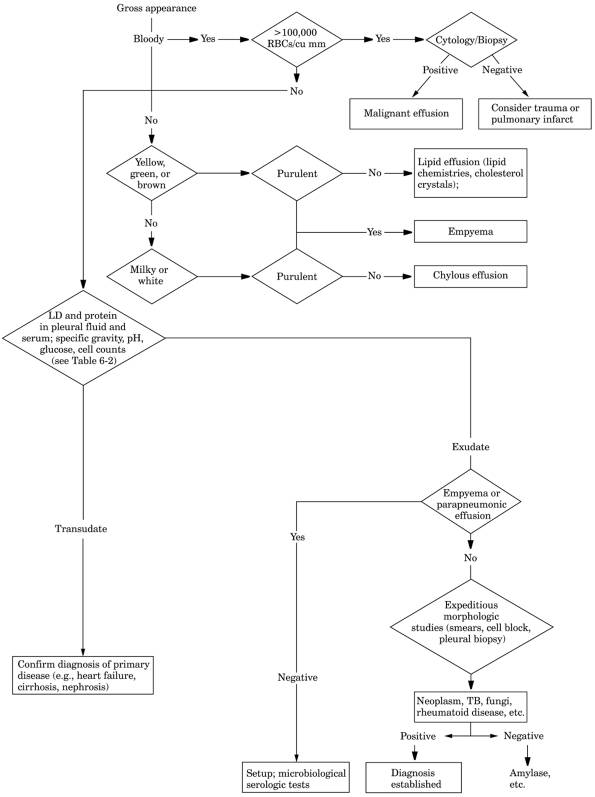
See Fig. 6-1 and Tables 6-1, and .
Normal
Values
|
Specific gravity
|
|
|
Total protein
|
|
|
Albumin
|
0.34.1 gm/dL
|
|
Globulin
|
|
|
Fibrinogen
|
|
|
pH
|
|
|
The underlying cause of an effusion is
usually determined by first classifying the fluid as an exudate or a
transudate. A transudate does not usually require additional testing but exudates always do.
Transudate
- Congestive heart failure
(causes 15% of cases)acute diuresis can result in pseudoexudate
- Cirrhosis with ascites
(pleural effusion in ~5% of these cases)rare without ascites
- Nephrotic syndrome
- Early (acute) atelectasis
- Pulmonary embolism (some
cases)
- Superior vena cava
obstruction
- Hypoalbuminemia
- Peritoneal
dialysisoccurs within 48 hrs of initiating dialysis
- Early mediastinal
malignancy
- Misplaced subclavian
catheter
- Myxedema (rare cause)
- Constrictive
pericarditiseffusion is bilateral
- Urinothoraxdue to
ipsilateral GU tract obstruction
Exudate
- Pneumonia, malignancy,
pulmonary embolism, and GI conditions (especially pancreatitis and
abdominal surgery, which cause 90% of all exudates)
- Infection (causes 25% of
cases)
- Bacterial pneumonia
- Parapneumonic effusion
(empyema)
- TB
- Abscess (subphrenic,
liver, spleen)
- Viral, mycoplasmal,
rickettsial
- Parasitic (ameba,
hydatid cyst, filaria)
- Fungal effusion (Coccidioides, Cryptococcus, Histoplasma, Blastomyces,
Aspergillus; in immunocompromised host, Aspergillus,
Candida, Mucor)
- Pulmonary
embolism/infarction
- Neoplasms (metastatic
carcinoma, especially breast, ovary, lung; lymphoma, leukemia,
mesothelioma, pleural endometriosis) (causes 42% of cases)
- Trauma (penetrating or
blunt)
- Hemothorax, chylothorax,
empyema, associated with rupture of diaphragm
- Immunologic mechanisms
- Rheumatoid pleurisy (5%
of cases)
- SLE
- Other collagen vascular
diseases occasionally cause effusions (e.g., Wegener's granulomatosis,
Sjögren's syndrome, familial Mediterranean fever, Churg-Strauss syndrome,
mixed connective tissue disease)
P.141
|
|
|
Fig. 6-1. Algorithm for pleural effusion.
|
- After myocardial
infarction or cardiac surgery
- Vasculitis
- Hepatitis
- Sarcoidosis (rare cause;
may also be transudate)
- Familial recurrent polyserositis
- Drug reaction (e.g.,
nitrofurantoin hypersensitivity, methysergide)
- Chemical mechanisms
P.142
|
|
|
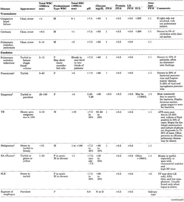
Table 6-1. Pleural Fluid Findings in
Various Clinical Conditions
|
P.143
P.144
|
|
|
Table 6-1. (continued)
|
P.145
|
|
|
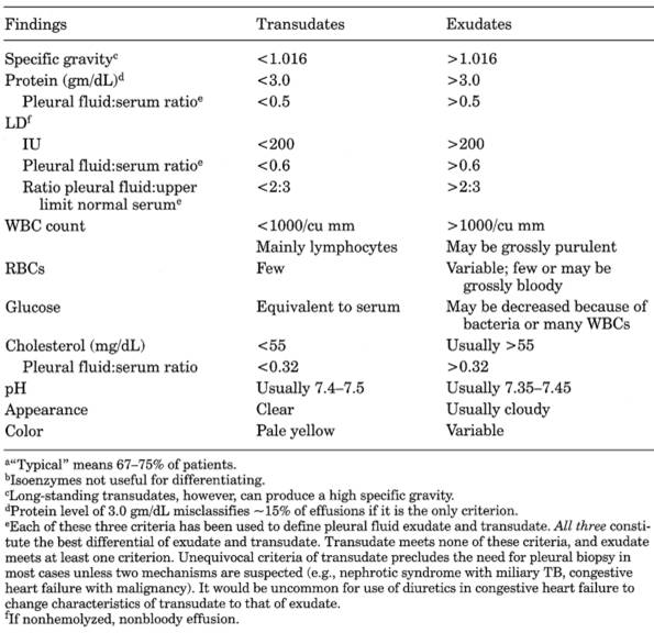
Table 6-2. Comparison of Typicala Findings in Transudates and Exudatesb
|
- Pancreatic (pleural
effusion occurs in ~10% of these cases)
- Esophageal rupture (high
salivary amylase and pH <7.30 that
approaches 6.00 in 4872 hrs)
- Subphrenic abscess
- Lymphatic abnormality
- Irradiation
- Milroy's disease
- Yellow nail syndrome
(rare condition of generalized hypoplasia of lymphatic vessels)
- Injury
- Altered pleural mechanics
- Late (chronic)
atelectasis
- Trapped lung
- Endocrine
- Movement of fluid from
abdomen to pleural space
- Meigs' syndrome (protein
and specific gravity are often at transudate-exudate border but usually not
transudate)
- Urinothorax
- Cancer
- Pancreatitis, pancreatic
pseudocyst
P.146
- Unknown (~15% of all
exudates)
- Cirrhosis,
pulmonary infarct, trauma, and connective tissue diseases comprise ~9% of
all cases
|
|
|
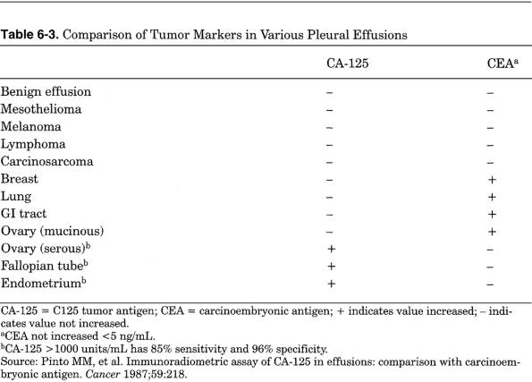
Table 6-3. Comparison of Tumor Markers in
Various Pleural Effusions
|
Exudates
That Can Present as Transudates
- Pulmonary embolism
(>20% of cases)due to atelectasis
- Hypothyroidismdue to
myxedema heart disease
- Malignancydue to
complications (e.g., atelectasis, lymphatic obstruction)
- Sarcoidosisstage II and
III
- Pleural fluid analysis
results in definitive diagnosis in ~25% and a probable diagnosis in
another 50% of patients; may help to rule out a suspected diagnosis in
30%.
Location
- Typically
left-sidedruptured esophagus, acute pancreatitis, RA. Pericardial disease
is left-sided or bilateral; rarely exclusively right-sided.
- Typically right-sided or
bilateralcongestive heart failure (if only on left, consider that right
pleural space may be obliterated or patient has another process, e.g.,
pulmonary infarction).
- Typically right-sidedrupture
of amebic liver abscess.
Gross
Appearance
- Clear, straw-colored
fluid is typical of transudate.
- Cloudy, opaque appearance
indicates more cell components.
- Bloody fluid suggests
malignancy, pulmonary infarct, trauma, postcardiotomy syndrome; also
uremia, asbestosis, pleural endometriosis. Bloody fluid from traumatic
thoracentesis should clot within several minutes, but blood present more
than several hours has become defibrinated and does not form a good clot.
Nonuniform color during aspiration and absence of hemosiderin-laden
macrophages and some crenated RBCs also suggest traumatic aspiration.
- Chylous (milky) fluid is
usually due to trauma (e.g., auto accident, postoperative) but may be
obstruction of duct (e.g., especially lymphoma; metastatic carcinoma,
granulomas). Pleural fluid triglyceride >110 mg/dL or triglyceride
pleural fluid to serum
P.147
ratio >2 occurs only in chylous effusion (seen
especially within a few hours after eating). After centrifugation, supernatant
is white due to chylomicrons, which also stain with Sudan III. Equivocal
triglyceride levels (60110 mg/dL) may require a lipoprotein electrophoresis of
fluid to demonstrate chylomicrons diagnostic of chylothorax. Triglyceride
<50 mg/dL excludes chylothorax.
- Pseudochylous in
chronic inflammatory conditions (e.g., rheumatoid pleurisy, TB, chronic
pneumothorax therapy for TB) due to either cholesterol crystals (rhomboid
shaped) in sediment or lipid-containing inclusions in leukocytes.
Distinguish from chylous effusions by microscopy. Fluid may have lustrous
sheen.
- White fluid suggests
chylothorax, cholesterol effusion, or empyema.
- Black fluid suggests Aspergillus niger
infection.
- Greenish fluid suggests
biliopleural fistula.
- Purulent fluid indicates
infection.
- Anchovy (dark red-brown)
color is seen in amebiasis, old blood.
- Anchovy paste in ruptured
amebic liver abscess; amebas found in <10%.
- Turbid and greenish
yellow fluid is classical for rheumatoid effusion.
- Turbidity may be due to
lipids or increased WBCs; after centrifugation, a clear supernatant
indicates WBCs as cause; white supernatant is due to chylomicrons.
- Very viscous (clear or
bloody) fluid is characteristic of mesothelioma.
- Debris in fluid suggests
rheumatoid pleurisy; food particles indicate esophageal rupture.
- Color of enteral tube
food or central venous line infusion due to tube or catheter entering
pleural space.
Odor
- Putrid due to anaerobic
empyema
- Ammonia due to
urinothroax
Protein,
Albumin, Lactate Dehydrogenase
- See Table
6-2.
- When exudate criteria are
met by LD but not by protein, consider malignancy and parapneumonic
effusions.
- Very high pleural fluid
LD (>1000 U/L) occurs in empyema, rheumatoid pleurisy, paragonimiasis;
sometimes with malignancy; rarely with TB.
Glucose
- Same concentration as
serum in transudate
- Usually normal but 3055
mg/dL or pleural fluid to serum ratio <0.5 and pH <7.30 may be found
in TB, malignancy, SLE; also esophageal rupture; lowest levels may occur
in empyema and RA. Therefore, only helpful if very low level (e.g.,
<30). 010 mg/dL highly suspicious for RA (see Rheumatoid
Effusion).
pH
Low pH (<7.30) always means exudate,
especially empyema, malignancy, rheumatoid pleurisy, SLE, TB, esophageal
rupture. Esophageal rupture is only cause of pH close to 6.0; collagen vascular
disease is only other cause of pH <7.0. pH <7.10
in parapneumonic effusion indicates need for tube drainage. In malignant
effusion, pH <7.30 is associated with short survival time, poorer prognosis,
and increased positive yield with cytology and pleural biopsy; tends to correlate
with pleural fluid glucose <60 mg/dL.
Amylase
- Increased (pleural fluid
to serum ratio >1.0 and may be >5 or pleural fluid greater than
upper limit of normal for serum)
- Acute pancreatitismay
be normal early with increase over time.
- Pancreatic pseudocystalways
increased, may be >100,000 U/L.
- Also perforated peptic
ulcer, necrosis of small intestine (e.g., mesenteric vascular occlusion);
10% of cases of metastatic cancer and esophageal rupture.
- Isoenzyme studies
- Pancreatic type amylase is found
in acute pancreatitis and pancreatic pseudocyst.
P.148
- Salivary type amylase is found
in esophageal rupture and occasionally in carcinoma of ovary or lung or
salivary gland tumor. Should be determined in undiagnosed left pleural
effusions.
Other
Chemical Determinations
- Cholesterol <55 mg/dL is said to be found in transudates and >55 mg/dL
in exudates.
- CEA
>10 ng/mL has specificity of >95% and sensitivity of 54100% for
lung cancer, 83% for breast cancer, 100% for GI tract cancers. May also be
increased in empyema and parapneumonic effusions.
- C125 tumor antigen (CA-125;) has
sensitivity of 71% and specificity of 99% for non-mucinous epithelial ovarian carcinoma.
- Combined CEA and CA-125
have sensitivity for detection of malignant effusions due to carcinomas of
lung, breast, GI tract, and ovary of 75100% and specificity of 98%. May
indicate primary site when the source is unknown or cytology is negative (Table 6-3).
- Other tumor markers have
been suggested for diagnosis of cancer, but value not established (e.g.,
acid phosphatase in prostatic cancer, hyaluronic acid in mesothelioma,
beta 2-microglobulin, etc.)
- Acid mucopolysaccharides
(especially hyaluronic acid) may be increased (>120 µg/mL) in
mesotheliomas.
- Immune complexes
(measured by Raji cell, C1q component of C, RIA, etc.) are often found in
exudates due to collagen vascular diseases (SLE, RA). RA latex
agglutination tests show frequent false-positives and should not be
ordered.
- Occasionally latex
agglutination for bacterial antigens is useful. Gas-liquid chromatography
has been reported to show butyric, isobutyric, propionic, and isovaleric
acids in anaerobic acute bacterial infection and increased lactic and
acetic acid levels in aerobic infections.
Cell
Count
- Total WBC count is almost
never diagnostic.
- >10,000/cu mm
indicates inflammation, most commonly with pneumonia, pulmonary infarct,
pancreatitis, postcardiotomy syndrome.
- >50,000/cu mm is
typical only in parapneumonic effusions, usually empyema.
- Chronic exudates (e.g.,
malignancy and TB) are usually <5000/cu mm.
- Transudates are usually
<1000/cu mm.
- 50006000 RBCs/cu mm
needed to give red appearance to pleural fluid
- Can be caused by needle
trauma producing 2 mL of blood in 1000 mL of pleural fluid.
- >100,000 RBCs/cu mm is grossly hemorrhagic and suggests malignancy,
pulmonary infarct, or trauma but occasionally seen in congestive heart
failure alone.
- Hemothorax (pleural fluid
to venous Hct ratio >2) suggests trauma, bleeding from a vessel,
bleeding disorder, or malignancy but may be seen in same conditions as
above.
Smears
- Wright's stain
differentiates PMNs from mononuclear cells; cannot differentiate
lymphocytes from monocytes.
- Mononuclear cells
predominate in transudates and chronic exudates (lymphoma, carcinoma, TB,
rheumatoid conditions, uremia). >50% is seen in two-thirds of cases due
to cancer. >8590% suggests TB, lymphoma, sarcoidosis, rheumatoid
causes.
- PMNs predominate in early
inflammatory effusions (e.g., pneumonia, pulmonary infarct, pancreatitis,
subphrenic abscess).
- After several days,
mesothelial cells, macrophages, lymphocytes may predominate.
- Large mesothelial cells
>5% are said to rule out TB (must differentiate from macrophages).
- Lymphocytes
- >85% suggests TB,
lymphoma, sarcoidosis, chronic rheumatoid pleurisy, yellow nail syndrome,
chylothorax.
- 5075% in >50% of
cases of carcinoma.
- Eosinophils in pleural
fluid (>10% of total WBCs) is not diagnostically significant.
- May mean blood or air in
pleural space (e.g., pneumothorax [most common], repeated thoracenteses,
traumatic hemothorax).
P.149
- It also is said to be
associated with asbestosis, pulmonary infarction, polyarteritis nodosa.
- Parasitic disease (e.g.,
paragonimiasis, hydatid disease, amebiasis, ascariasis).
- Fungal disease (e.g.,
histoplasmosis, coccidioidomycosis).
- Drug-related (e.g.,
nitrofurantoin, bromocriptine, dantrolene).
- Idiopathic effusion (in
approximately one-third of cases; may be due to occult pulmonary embolism
or asbestosis).
- Uncommon with malignant
effusions.
- Rare with TB.
- Not usually accompanied
by striking blood eosinophilia. Many diseases associated with blood
eosinophilia infrequently cause pleural effusion eosinophilia.
- Basophils >10% only in
leukemic involvement of pleura.
- Occasionally lupus
erythematosus (LE) cells make the diagnosis of SLE.
- Gram stain for early diagnosis
of bacterial infection.
- Acid-fast smears are
positive in 20% of tuberculous pleurisy.
- Culture is often positive in
empyema but not in parapneumonic effusions.
- Bacterial
antigens may detect H. influenzae type b,
Streptococcus pneumoniae, several
types of Neisseria meningitidis. Useful when
viable organisms cannot be recovered (e.g., due to prior antibiotic
therapy).
Cytology
- Positive in 60% of
malignancies on first tap, 80% by third tap. Therefore should repeat taps
with cytologic examinations if cancer is suspected. Is more sensitive than
needle biopsy. Combined with needle biopsy, increases sensitivity by
<10%.4 (See Carcinoma,
Bronchogenic.) High yield with adenocarcinoma, low yield with
Hodgkin's disease.
- Rheumatoid effusions:
cytologic triad of slender elongated and round giant multinucleated
macrophages and necrotic background material with characteristically low
glucose is said to be pathognomonic. Mesothelial cells are nearly always
absent.
- Flow cytometry assay for
DNA aneuploidy and staining with monoclonal antibodies (e.g., CEA,
cytokeratin) to distinguish malignant mesothelioma, metastatic tumor, and
reactive mesothelial cells can be performed (note: some malignant cells
may be diploid).
Pleural
Fluid Findings in Various Clinical Conditions
- See Fig.
6-1.
- Tuberculosis
- High protein
contentalmost always >4.0 gm/dL
- Increased lymphocytes
- Acid-fast smears are positive in <20%, and culture is positive in ~67% of
cases; culture combined with histologic examination establishes the
diagnosis in 95% of cases.
- Needle biopsy can be performed without hesitation
- Large mesothelial cells
>5% are said to rule out TB (must differentiate from macrophages).
- TB often presents as
effusion, especially in youth; pulmonary disease may be absent; risk of
active pulmonary TB within 5 yrs is 60%.
- Malignancy
- Can cause effusion by
metastasis to pleura, causing exudate-type fluid, or by metastasis to
lymph nodes, obstructing lymph drainage and giving transudate-type fluid.
Low pH and glucose indicate a poor prognosis with short survival time.
- Characteristic effusion
is moderate to massive, frequently hemorrhagic, with moderate WBC count
with predominance of mononuclear cells; however, only half of malignant
effusions have RBC >10,000/cu mm.
- Cytology establishes the diagnosis in ~50% of patients
- Combined cytology and pleural biopsy give positive results in
- In some instances of suspected lymphoma
with negative
conventional test results, flow cytometric analysis of pleural fluid
showing a monoclonal lymphocyte population can establish the diagnosis.
- Mucopolysaccharide level
may be increased (normal <17 mg/dL) in mesothelioma.
P.150
- Lung and breast cancer
and lymphoma cause 75% of malignant effusions; in 6%, no primary tumor is
found. Pleural or ascitic effusion occurs in 2030% of patients with
malignant lymphoma.
- CEA, CA-125see Table 6-3.
- Pulmonary
Infarct
- Effusion occurs in 50% of
patients with pulmonary infarct; is bloody in one-third to two-thirds of
patients; often no characteristic diagnostic findings occur.
- Small volume, serous or
bloody, predominance of PMNs, may show many mesothelial cells; this typical
pattern is seen in only 25% of cases.
- Congestive
Heart Failure
- Is predominantly
right-sided or bilateral. If unilateral or left-sided in patients with
congestive heart failure, rule out pulmonary infarct.
- Pneumonias
- Parapneumonic effusions
(exudate type of effusion associated with lung abscess, bronchiectasis;
~5% of bacterial pneumonias).
- Aerobic gram-negative
organisms (Klebsiella, Escherichia coli, Pseudomonas) are associated with
a high incidence of exudates (with 500040,000/cu mm, high protein, normal
glucose, normal pH) and resolve with antibiotic
therapy. Nonpurulent fluid with positive Gram stain or positive blood
culture or low pH suggests that effusion will become or behave like
empyema.
- S.
pneumoniae causes parapneumonic effusions in 50% of cases, especially with
positive blood culture.
- Staphylococcus
aureus
has effusion in 90% of infants, 50% of adults; usually widespread
bronchopneumonia.
- Streptococcus
pyogenes has effusion in 90% of cases; massive effusion, greenish color.
- Haemophilus
influenzae has effusion in 5075% of cases.
- Viral or Mycoplasma pneumoniapleural effusions develop in 20%
of cases.
- Legionnaires'
diseasepleural effusion occurs in up to 50% of patients; may be
bilateral.
- P.
carinii
pneumonia cases often have pleural effusion to serum LD ratio >1.0 and
pleural effusion to serum protein ratio <0.5.
- pH <7.0 and glucose
<40 mg/dL indicate need for closed chest tube drainage even without
grossly purulent fluid
- pH of 7.07.2 is
questionable indication and should be repeated in 24 hrs, but tube
drainage is favored if pleural fluid LD >1000 U/L. Tube drainage is
also indicated if fluid is grossly purulent or Gram stain or culture is
positive.
- Normal pH is alkaline and
may approach 7.6.
- Empyema
- Usually WBCs
>50,000/cu mm, low glucose, and low pH. Suspect clinically when
effusion develops during adequate antibiotic therapy.
- In Proteus mirabilis
empyema, high ammonia level may cause a pH ~8.0.
- Rheumatoid
Effusion
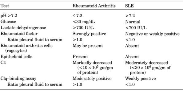
- See Table
6-4.
- Found in ~70% of RA
patients at autopsy.
- Exudate is frequently turbid and may be milky. Classic picture is cloudy greenish fluid
with 0 glucose level. Level is <50 mg/dL in 80% and <25 mg/dL in 66%
of patients; is the most useful finding clinically. Failure of level to
increase during IV glucose infusion distinguishes RA from other causes. Nonpurulent, nonmalignant effusions not due to TB or RA
almost always have glucose level >60 mg/dL.
- RF may be present but may
also be found in other effusions (e.g., TB, cancer, bacterial pneumonia).
RF titer ≥1:320 or equal to or greater than serum level suggests
rheumatoid pleurisy.
- RA cells may be found
(see Cytology).
- Cytologic examination for
malignant cells and smears and cultures for bacteria, tubercle bacilli,
and fungi are negative.
- Needle biopsy usually shows nonspecific chronic inflammation but may show characteristic
rheumatoid nodule microscopically. One-third of cases have parenchymal
lung disease (e.g., interstitial fibrosis).
- Other laboratory findings
of RA are found.
- Protein level is >3
gm/dL.
P.151
|
|
|
Table 6-4. Comparison of Pleural Fluid in
Rheumatoid Arthritis and Systemic Lupus Erythematosus (SLE)
|
- Increased LD (usually
higher than in serum) is commonly found in other chronic pleural effusions
and is not useful in differential diagnosis.
- Systemic
Lupus Erythematosus
- LE cells are specific for SLE but test has poor sensitivity.
- ANA titer ≥160 or pleural fluid to serum ratio >1.0 is
suggestive but not diagnostic.
Pneumoconiosis
- Biopsy of lung, scalene lymph nodehistologic, chemical, spectrographic, and
radiographic diffraction studies, electron microscopy (e.g., silicosis,
berylliosis; also metastatic tumor, sarcoidosis, TB, fungus infection)
- Bacterial smears and
cultures of sputum (especially for tubercle bacilli) should be done.
- Cytologic examination of
sputum and bronchoscopic secretions for malignant cells, especially
squamous cell carcinoma of bronchus and mesothelioma of pleura
- Asbestos bodies sometimes
occur in sputum after exposure to asbestos dust even without clinical
disease.
- Acute beryllium disease
may show occasional transient hypergammaglobulinemia.
- Chronic beryllium disease
- Secondary polycythemia
- Increased serum gamma
globulin
- Increased urine calcium
- Increased beryllium in
urine long after beryllium exposure has ended
- Increased WBC if
associated infection
- Secondary polycythemia or
anemia
- Silicosis
- Associated conditions
- ≤25% have mycobacterial infections, half of which are
nontuberculous.
- Increased incidence of
nocardiosis, cryptococcosis, sporotrichosis.
- 10% have connective
tissue diseases (e.g., progressive systemic sclerosis, RA, SLE).
- Increased incidence of
ANA, RF, hypergammaglobulinemia. ACE increased
in one-third of patients.
Pneumonia
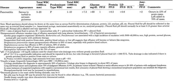
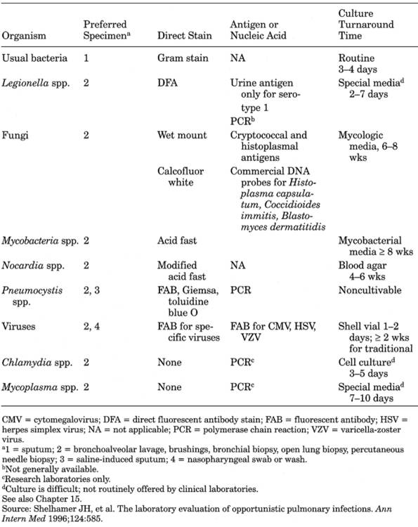
See Table 6-5.
P.152
|
|
|
Table 6-5. Opportunistic Pulmonary
Infections
|
Due To
- Bacteria
- S.
pneumoniae causes 6070% of bacterial pneumonia in patients requiring
hospitalization. May cause ~25% of hospital-acquired cases of pneumonia.
Blood culture positive in 25% of untreated cases during first 34 days.
- Staphylococcus causes <1% of all
acute bacterial pneumonia with onset outside the hospital but more
frequent after outbreaks of influenza; may be secondary to measles, mucoviscidosis,
prolonged antibiotic therapy, debilitating diseases (e.g., leukemia,
collagen
P.153
diseases). Frequent cause of
nosocomial pneumonia. Bacteremia in <20% of
patients.
- H.
influenzae is important in 6- to 24-mo age group; rare in adults except for
middle-aged men with chronic lung disease and/or alcoholism and patients
with immunodeficiency (HIV, multiple myeloma, chronic lymphocytic leukemia
[CLL]). Can mimic pneumococcal pneumonia; may be isolated with S. pneumoniae.
- Gram-negative bacilli (e.g.,
K. pneumoniae, enterobacteria, E. coli, P. mirabilis, Pseudomonas
aeruginosa) are common causes of hospital-acquired pneumonia but unlikely
outside the hospital. K. pneumoniae causes 1% of primary bacterial
pneumonias, especially in alcoholic patients and patients with upper lobe
pneumonia; tenacious red-brown sputum is typical.
- Tubercle bacilli
- Legionella
pneumophila
- M.
pneumoniaemost common in young adult male population (e.g., armed forces
camps)
- C.
pneumoniae, Chlamydia psittaci
- Others (e.g., streptococcosis,
tularemia, plague)
- See Table
6-5.
- Viruses
- Influenza, parainfluenza,
adenoviruses, RSV, echovirus, coxsackievirus, reovirus, CMV, viruses of
exanthems, herpes simplex, hantavirus
- Rickettsiae
- Q fever is most common in
endemic areas; typhus.
- Fungi
- P.
carinii, Histoplasma, and Coccidioides in
particular; Blastomyces, Aspergillus.
- Protozoans
- Toxoplasma
|
Underlying Condition
|
Organism
|
|
Obstructive cancer
|
S. pneumoniae, H. influenzae,
M. catarrhalis, anaerobes
|
|
Alcoholism
|
S. pneumoniae, H. influenzae,
Klebsiella spp., Legionella spp., anaerobes, M. tuberculosis
|
|
HIV infection
|
S. pneumoniae, H. influenzae,
S. aureus, gram-negative bacilli, P. carinii, M.
tuberculosis and MAI (mycobacterium avium-intracellulare),
Toxoplasma gondii, Cryptococcus, Nocardia, CMV, histoplasmosis, Coccidioides
immitis, Legionella, M. catarrhalis, Rhodococcus equi
|
|
Atypical pneumonia
|
M. pneumoniae, C. psittaci, C. pneumoniae, Coxiella bur-netii,
Francisella tularensis, many viruses
|
|
Laboratory
Findings
- WBC is frequently normal
or slightly increased in nonbacterial pneumonias; considerable increase in
WBC is more common in bacterial pneumonia. In severe
bacterial pneumonia, WBC may be very high or low or normal. Because
individual variation is considerable, it has limited value in
distinguishing bacterial and nonbacterial pneumonia.
- Urine protein, WBCs,
hyaline and granular casts in small amounts are common. Ketones may occur
with severe infection. Check for glucose to rule out
underlying diabetes mellitus.
- Sputum reveals abundant WBCs in bacterial pneumonias. Gram stain shows abundant organisms
in bacterial pneumonias (e.g., Pneumococcus,
Staphylococcus). Culture sputum for appropriate bacteria. Sputum that contains many organisms and WBCs on smear but no
pathogens on aerobic culture may indicate aspiration pneumonia. Sputum is
not appropriate for anaerobic culture.
- In all cases of pneumonia, blood culture
and sputum culture and smear for Gram stain should be performed before antibiotic therapy
is started. Optimum specimen of sputum shows >25 PMNs and ≤5
squamous epithelial cells/LPF (10× magnification), but >10 PMNs and
<25 epithelial cells may be considered acceptable sputum specimen.
>25 epithelial cells indicate unsatisfactory specimen from oropharynx
which should not be submitted for culture. If good sputum specimen is
obtained, further diagnostic microbiological tests are usually not
performed.
- Nasopharyngeal aspirate
may identify S. pneumoniae with few false
positives but S. aureus and gram-negative bacilli often represent
false-positive findings.
P.154
- In H.
influenzae pneumonia, sputum culture is negative in >50% of
patients with positive cultures from blood, pleural fluid, or lung tissue,
and may be present in the sputum in the absence of disease.
- Transtracheal aspiration (puncture of cricothyroid membrane) generally yields a
faster, more accurate diagnosis.
- Protected brush bronchoscopy and BAL have high sensitivity
- Diagnostic lung puncture to determine specific causative agent as a guide to antibiotic
therapy may be indicated in critically ill children.
- Open lung biopsy is gold standard with 97% accuracy but 10% complication rate.
- For pleural effusions
that are aspirated, Gram stain and culture should also be performed.
- Respiratory pathogens isolated from blood, pleural fluid, or transtracheal aspirate
(except in patients with chronic bronchitis) or identified by bacterial
polysaccharide antigen in urine may be considered the definite causal
agent.
- Urine testing for capsular antigen from S.
pneumoniae or type B H. influenzae by
latex agglutination may be helpful. Positive in ~90% of bacteremic
pneumococcal pneumonias and 40% of nonbacteremic pneumonias. May be
particularly useful when antibiotic therapy has already begun.
- Acute phase serum should
be stored at onset. If causal diagnosis is not established, a convalescent
phase serum should be taken. A 4× increase in antibody titer establishes
the causal diagnosis (e.g., L. pneumophila, Chlamydia
spp., respiratory viruses [including influenza and RSV]), M. pneumoniae. Serologic tests to determine whether
pneumonia is due to Histoplasma, Coccidioides,
etc.
Pneumonia,
Lipid
Sputum shows fat-containing macrophages that stain with Sudan. They may be present only intermittently; therefore, examine sputum more
than once
Pulmonary
Alveolar Proteinosis
- (Rare
disease characterized by amorphous, lipid-rich, proteinaceous material in
alveoli)
- PASpositive material appears in sputum.
- PSP dye injected intravenously is excreted in sputum for long periods of
time.
- BAL fluid contains
increased total protein, albumin, phospholipids, and CEA.
- Recently antibodies to surfactant protein
A (ELISA assay) in sputum and BAL have been reported to be highly specific.
- Serum CEA is increased and correlates with BAL findings. Reflects
severity of disease and decreases with response to treatment.
- Routine laboratory test findings are nonspecific.
- Serum LD increases when
protein accumulates in lungs and becomes normal when infiltrate resolves;
correlates with serum CEA.
- Decreased arterial O2.
- Secondary polycythemia
may occur.
- Diagnosis usually requires open lung biopsy. Electron microscopy shows many lamellar
bodies.
- Laboratory findings due
to superinfection.
Pulmonary
Embolism and Infarction
- No laboratory test is
diagnostic.
- <10% of emboli lead to
infarction
- Measurement of arterial
blood gases (obtained when patient is breathing room air) is the most
sensitive and specific laboratory test.
- pO <80 mm Hg in 88% of
cases but normal pO does not rule out
pulmonary embolus. In appropriate clinical setting, pO
<88 mm Hg (even with a normal chest radiograph) is indication for lung
scans and search for deep vein thromboses. pO
>90 mm Hg with a normal chest radiograph suggests a different
diagnosis. Normal complete lung scans exclude the diagnosis.
- Hypocapnia and
slightly elevated pH.
- Increased WBC in 50% of
patients but is rarely >15,000/cu mm (whereas in acute bacterial
pneumonia is often >20,000/cu mm).
- Increased ESR
P.155
- Triad of increased LD and
bilirubin with normal AST is found in only 15% of cases.
- Serum enzymes differ from
those in acute myocardial infarction.
- Increased serum LD (due
to isoenzymes LD-2 and LD-3) in 80% of patients
rises on first day, peaks on second, normal by tenth day.
- Serum AST is usually
normal or only slightly increased.
- cTn not increased.
- Serum indirect bilirubin
is increased (as early as fourth day) to ~5 mg/dL in 20% of cases.
- Pleural effusion may
occur.
Plasma D-dimer (ELISA or Latex Agglutination Kits)
- Use
- Detects lysis of fibrin
clot only, whereas fibrinogen degradation products test detects lysis of
both fibrin clot and fibrinogen (see Chapter 11).
At appropriate cutoff level, has >80% sensitivity but only ~30%
specificity. Negative predictive value >90%; normal test useful in
excluding pulmonary embolism in patients with low pretest probability.
Value less than cutoff level (which varies with assay kit) obviates need
for pulmonary angiography.
- Increased
In
- Deep venous thrombosis
- DIC with fibrinolysis
- Renal, liver, or cardiac
failure
- Major injury or surgery
- Inflammation (e.g.,
arthritis, cellulitis), infection (e.g., pneumonia)
- Thrombolytic therapy
- Measurements of serum CK,
LD, and fibrin products are not indicated routinely as they do not have
sufficient sensitivity or specificity to be of diagnostic value.
- Increased serum ALP
Sinusitis,
Acute
Due To
- Often precipitated by
obstruction due to viral URI, allergy, foreign body.
- S.
pneumoniae and H. influenzae cause >50% of
cases; also anaerobes, S. aureus, S. pyogenes
(group A).
- M.
catarrhalis causes ~20% of cases in children
- Viruses cause ~1020% of
cases
- P.
aeruginosa and H. influenzae are predominant
organisms in cystic fibrosis patients.
- Mucor spp., Aspergillus spp. should be ruled out in patients with
diabetes or acute leukemia and in renal transplant recipients.
- Anaerobes (e.g.,
streptococci, Bacteroides spp.) occur in ~50% of
cases of chronic sinusitis.
- Needle aspiration of
sinus is required for determination of organism. Culture of nose, throat,
and nasopharynx specimens do not correlate well.
- Mucosal biopsy may be
indicated if aspirate is not diagnostic in unresponsive patient with acute
infection.
REFERENCES
1. Kahn FW, Jones JM. Bronchoalveolar lavage in the rapid diagnosis of lung disease.
Lab Manage June 1986:31.
2. Menzies R, Charbonneau
M. Thoracoscopy for the diagnosis of pleural disease. Ann Intern Med .
3. Bernard GR, Artigas A,
Brigham KL, et al. The American-European Consensus Conference on ARDS:
definitions, mechanisms, relevant outcomes and clinical trial coordination. Am J Respir Crit Care Med .
4. Prakesh UBS, Reiman HM.
Comparison of needle biopsy with cytologic analysis for the evaluation of
pleural effusion: Analysis of 414 cases. Mayo Clin
Proc .
Wallach, Jacques
Interpretation of Diagnostic Tests, 7th
Edition
Copyright ©2000
Lippincott Williams & Wilkins
> Table of Contents > SECTION III - Diseases of
Organ Systems > Chapter 8 - Hepatobiliary Diseases and Diseases of the
Pancreas
Chapter 8
Hepatobiliary Diseases and Diseases of the
Pancreas
Liver
Function Tests
Common
Test Patterns
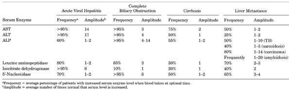
- See Table
8-1.
- See Figs.
8-1, , and .
- Patterns of abnormalities
rather than changes in single test results are particularly useful despite
sensitivities of only 65% in some cases.
- Test results may be
abnormal in many conditions that are not primarily hepatic (e.g., heart
failure, sepsis, infections such as brucellosis, SBE), and individual test
results may be positive in conditions other than liver disease. Results on
individual tests are normal in a high proportion of patients with proven
specific liver diseases, and normal values may not rule out liver disease.
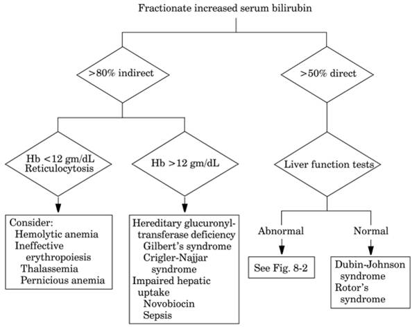
- Serum bilirubin
(direct/total ratio)
- <20% direct.
- Constitutional (e.g.,
Gilbert's disease, Crigler-Najjar syndrome).
- Hemolytic states.
- 2040% direct.
- Favors hepatocellular
disease rather than extrahepatic obstruction.
- Disorders of bilirubin
metabolism (e.g., Dubin-Johnson, Rotor's syndromes).
- 4060% direct: Occurs in
either hepatocellular or extrahepatic type.
- >50% direct: Favors
extrahepatic obstruction rather than hepatocellular disease.
- Serum total bilirubin
- Not a sensitive
indicator of hepatic dysfunction; may not reflect degree of liver damage.
- Must be >2.5 mg/dL to
produce clinical jaundice.
- >5 mg/dL seldom
occurs in uncomplicated hemolysis unless hepatobiliary disease is also
present.
- Is generally less
markedly increased in hepatocellular jaundice (<10 mg/dL) than in
neoplastic obstructions (≤20 mg/dL) or intrahepatic cholestasis.
- In extrahepatic biliary
obstruction, bilirubin may rise progressively to a plateau of 3040 mg/dL
(due in part to balance between renal excretion and diversion of
bilirubin to other metabolites). Such a plateau tends not to occur in
hepatocellular jaundice, and bilirubin may exceed 50 mg/dL (partly due to
concomitant renal insufficiency and hemolysis).
- Concentrations are
generally higher in obstruction due to carcinoma than that due to stones.
- In viral hepatitis, higher
serum bilirubin suggests more liver damage and longer clinical course.
- In acute alcoholic
hepatitis, >5 mg/dL suggests a poor prognosis.
- Increased serum
bilirubin with normal ALP suggests constitutional hyperbilirubinemias or
hemolytic states.
- Normal serum bilirubin,
AST, and ALT with increased ALP (of liver origin) and LD suggests
obstruction of one hepatic duct or metastatic or infiltrative disease of
liver. Metastatic and granulomatous lesions of liver cause 1.53.0×
increase of serum ALP and LD.
P.200
|
|
|
Table 8-1. Increased Serum Enzyme Levels
in Liver Diseases
|
P.201
|
|
|
Fig. 8-1. Algorithm illustrating workup
for jaundice.
|
- Due to renal excretion,
maximum bilirubin = 1035 mg/dL; if renal disease is present, level may
reach 75 mg/dL.
- Direct bilirubin >1.0
mg/dL in an infant always indicates disease.
- AST and ALT
- Most sensitive tests for
acute hepatocellular injury (e.g., viral, drug related). >500 U/L
suggests such a diagnosis. Seldom >500 U/L in obstructive jaundice,
cirrhosis, viral hepatitis in AIDS, alcoholic liver disease.
- Most marked increase
(1002000 U/L) occurs in viral hepatitis, drug injury, carbon
tetrachloride poisoning.
- >4000 indicates toxic
injury, e.g., from acetaminophen.
- Patient is rarely
asymptomatic with level >1000 U/L.
- AST >10× normal
indicates acute hepatocellular injury but lesser increases are
nonspecific and may occur with virtually any other form of liver injury.
- Usually <200 U/L in
posthepatic jaundice and intrahepatic cholestasis.
- <200 U/L in 20% of
patients with acute viral hepatitis.
- Usually <50 U/L in
fatty liver.
- <100 U/L in alcoholic
cirrhosis; ALT is normal in 50% and AST is normal in 25% of these cases.
- <150 U/L in alcoholic
hepatitis (may be higher if patient has delirium tremens).
- <200 U/L in 65% of
patients with cirrhosis.
- <200 U/L in 50% of
patients with metastatic liver disease, lymphoma, and leukemia.
- Normal values may not
rule out liver disease: ALT is normal in 50% of cases of alcoholic
cirrhosis and AST is normal in 25% of cases.
- AST soaring to peak of
10009000 U/L and declining by 50% within 3 days and to <100 U/L
within a week suggests shock liver with centrolobular necrosis (e.g., due
to congestive heart failure, arrhythmia, sepsis, GI hemorrhage); serum
bilirubin and ALP reflect underlying disease.
P.202
|
|
|
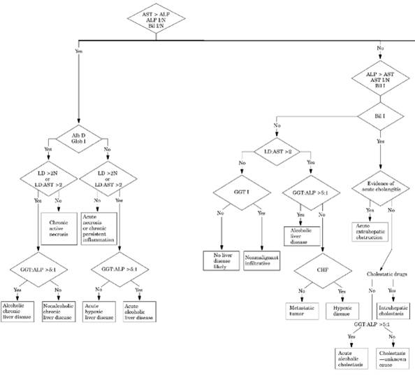
Fig. 8-2. Algorithm illustrating
sequential abnormal liver function test interpretation. (Alb = albumin; Bil =
bilirubin; CHF = congestive heart failure; Glob = globulin; I = increased; N
= normal. Enzymes all in same U/L.) (Adapted from
Henry JB. Clinical diagnosis and management by laboratory methods,
16th ed. Philadelphia:
WB Saunders, 1979.
|
P.203
P.204
|
|
|
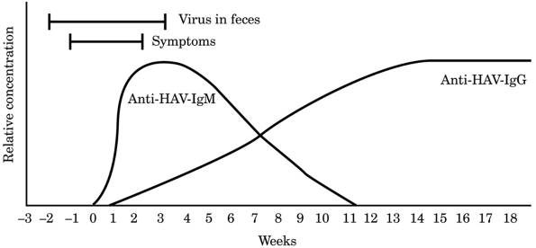
Fig. 8-3. Antibody markers in hepatitis A
virus infection. (IgG = immunoglobulin G; IgM = immunoglobulin M.)
(Reproduced with permission of Abbott Laboratories, Pasadena, CA.)
|
- Rapid rise of AST and
ALT to very high levels (e.g., >600 U/L and often >2000 U/L)
followed by a sharp fall in 1272 hrs is said to be typical of acute
biliary duct obstruction.
- Abrupt AST rise may also
be seen in acute fulminant viral hepatitis (rarely >4000 U and
declines more slowly; positive serologic tests) and acute chemical
injury.
- Degree of increase has
low prognostic value.
- Serial determinations
reflect clinical activity of liver disease.
- Mild increase of AST and
ALT (usually <500 U/L) with ALP increased >3× normal indicates
cholestatic jaundice, but more marked increase of AST and ALT (especially
>1000 U/L) with ALP increased <3× normal indicates hepatocellular
jaundice.
- Increased concentration
has poor correlation with extent of liver cell necrosis and has little
prognostic value.
- AST/ALT ratio >2 with
ALT <300 U/L is suggestive of alcoholic hepatitis, and ratio >3 is
highly suggestive, in cases of liver disease. Greater increase in AST than
in ALT also occurs in cirrhosis and metastatic liver disease. In patients
with cirrhosis or portal hypertension, AST/ALT ratio ≥3 suggests
primary biliary cirrhosis. Greater increase in AST than in ALT favors
viral hepatitis, posthepatic jaundice, intrahepatic
cholestasis. AST is increased in AMI and in muscle
diseases, but ALT is normal. ALT is more specific for liver disease than
AST.
- Serum ALP
- Is the best indicator of
biliary obstruction but does not differentiate intrahepatic cholestasis
from extrahepatic obstruction. Is increased out of proportion to other
liver function tests.
- Increases before
jaundice occurs.
- High values (>5×
normal) favor obstruction and normal levels virtually exclude this
diagnosis.
- Markedly increased in
infants with congenital intrahepatic bile duct atresia but is much lower
in extrahepatic atresia.
- Increase (310× normal)
with only slightly increased transaminases may be seen in biliary
obstruction and the converse in liver parenchymal disease (e.g.,
cirrhosis, hepatitis).
- Increased (210× normal)
in early infiltrative (e.g., amyloid) and space-occupying diseases of the
liver (e.g., tumor, granuloma, abscess).
- Increased >3× normal
in ≤5% of acute hepatitis.
- <3× normal is
nonspecific and may occur in all types of liver diseases (e.g.,
infiltrative liver diseases, cirrhosis, chronic hepatitis, viral
hepatitis) and in diseases affecting the liver (e.g., congestive heart
failure).
P.205
- GGT/ALP ratio >5
favors alcoholic liver disease.
- Isolated increase of GGT
is a sensitive screening and monitoring test for alcoholism. Increased GGT
due to alcohol or anticonvulsant drugs is not accompanied by increased
ALP.
- Serum 5′-NT and LAP
levels parallel the increase in ALP in obstructive type of hepatobiliary
disease, but the 5′-NT is increased only in the latter and is normal
in pregnancy and bone disease, whereas the LAP is increased in pregnancy
but usually normal in bone disease. GGT is normal in bone disease and
pregnancy. Therefore, these enzymes are useful in determining the source
of increased serum ALP. Although serum 5′-NT usually parallels ALP
in liver disease, it may not increase proportionately in individual patients.
|
Serum Enzyme
|
Biliary Obstruction
|
Pregnancy
|
Childhood; Bone Disease
|
|
ALP
|
I
|
I
|
I
|
|
5′-NT
|
I
|
N
|
N
|
|
LAP
|
I
|
I
|
N
|
|
GGT
|
I
|
N
|
N
|
|
I = increased; N = normal.
|
|
- Test for
antimitochondrial antibodies to rule out primary biliary cirrhosis in
females (present in >90% of cases;) and
radiologic studies to rule out primary sclerosing cholangitis.
- Bilirubin (bile) in
urine implies increased serum direct bilirubin and excludes hemolysis as
the cause. Often precedes clinical icterus. May occur without jaundice in
anicteric or early hepatitis, early obstruction, or liver metastases.
(Tablets detect 0.050.1 mg/dL; dipsticks are less sensitive; test is
negative in normal persons.)
- Complete absence of urine
urobilinogen strongly suggests complete bile duct obstruction; level is normal
in incomplete obstruction. Decreased in some phases of hepatic jaundice.
Increased in hemolytic jaundice and subsiding hepatitis. Increase may
indicate hepatic damage even without clinical jaundice (e.g., some
patients with cirrhosis, metastatic liver disease, congestive heart
failure). Presence in viral hepatitis depends on phase of disease. (Normal is <1 mg
or 1 Ehrlich unit per 2-hr specimen.)
- Serum cholesterol
- May be normal or
slightly decreased in hepatitis.
- Markedly decreased in
severe hepatitis or cirrhosis.
- Increased in
posthepatitic jaundice or intrahepatic cholestasis.
- Markedly increased in
primary biliary cirrhosis.
- PT
- May be prolonged due to
lack of vitamin K absorption in obstruction or lack of synthesis in
hepatocellular disease. Not useful when only slightly prolonged.
- Corrected within 2448
hrs by parenteral administration of vitamin K (10 mg/day for 3 days) in
obstructive but not in hepatocellular disease. Failure to correct
suggests poor prognosis; extensive hepatic necrosis should be considered.
- Markedly prolonged PT is
a good index of severe liver cell damage in hepatitis and cirrhosis and
may herald onset of fulminant hepatic necrosis.
- Serum gamma globulin
- Tends to increase with
most forms of chronic liver disease.
- Increases are not specific;
found in other chronic inflammatory and neoplastic diseases.
- Moderate increases
(e.g., >3 gm/dL) are suggestive of chronic active hepatitis; marked
increases are suggestive of autoimmune chronic hepatitis.
- Polyclonal increases in
IgG and IgM are found in most cases of cirrhosis.
- Increased IgM alone may
suggest primary biliary cirrhosis.
- Increased IgA may occur
in alcoholic cirrhosis.
- Immunoglobulins are
usually normal in obstructive jaundice.
- Serum albumin is slow to
reflect liver damage.
- Is usually normal in
hepatitis and cholestasis.
- Increase toward normal
by 23 gm/dL in treatment of cirrhosis implies improvement and more
favorable prognosis than if no increase with therapy.
- Some patients do not
present the usual pattern.
- Liver function test abnormalities
may occur in systemic diseases, e.g., SLE, sarcoidosis, TB, SBE,
brucellosis, sickle cell disease.
- A confusing pattern may
occur in mixed forms of jaundice (e.g., sickle cell disease producing
hemolysis and complicated by pigment stones causing duct obstruction).
P.206
Disorders
of the Liver, Gallbladder, Biliary Tree, and Pancreas
Abscess
Of Liver, Pyogenic
Due To
- Biliary tract infection,
33%
- Direct extension, 25%
- Trauma, 15%
- Bacteremia, 10%
- Pyelophlebitis, 6%
- Unknown, 10%
- Gram stain and culture
- Gram-negative bacilli
(e.g., Escherichia coli, Klebsiella spp.)
- Anaerobes (e.g., Bacteroides fragilis)
- Staphylococcus
aureus
or streptococci are found in children with bacteremia.
- Abnormalities of liver function tests
- Decreased serum albumin
in 50% of cases; increased serum globulin
- Increased serum ALP in
75% of cases
- Increased serum
bilirubin in 2025% of cases; >10 mg/dL usually indicates pyogenic
rather than amebic origin and suggests poorer prognosis because of more
tissue destruction
- See Space-Occupying
Lesions
- Increase in WBC due to
increase in granulocytes in 70% of cases
- Anemia in 60% of cases
- Ascites is unusual
compared to other causes of space-occupying lesions.
- Laboratory findings due
to complications (e.g., right pleural effusion in 20% of cases, subphrenic
abscess, pneumonia, empyema, bronchopleural fistula)
- Patients with amebic abscess of liver due to Entamoeba
histolytica also show positive serologic tests for ameba.
- Stools may be negative
for cysts and trophozoites.
- Needle aspiration of
abscess may show E. histolytica in 50% of
patients.
- Characteristic
brown or anchovy-sauce color may be absent; secondary bacterial
infection may be superimposed
- See Echinococcus
granulosus cyst.
Biliary
Atresia, Extrahepatic, Congenital
- Direct serum bilirubin is increased in
early days of life in some infants but not until second week in others.
Level is often <12 mg/dL during first months, with subsequent rise later
in life.
- Laboratory findings as in Biliary Obstruction, Complete (see next
section).
- Liver biopsy to differentiate from neonatal hepatitis
- Laboratory findings due
to sequelae (e.g., biliary cirrhosis, portal hypertension, frequent
infections, rickets, hepatic failure)
- I-rose bengal excretion test (see Neonatal Hepatitis)
- Most important to
differentiate this condition from neonatal hepatitis, for which surgery
may be harmful.
- >90% of cases of
extrahepatic biliary obstruction in newborns are due to biliary atresia;
occasional cases may be due to choledochal cyst (causes intermittent
jaundice in infancy), bile plug syndrome, or bile ascites (associated with
spontaneous perforation of the common bile duct).
Biliary
Obstruction, Complete (Intrahepatic Or Extrahepatic)
- Typical pattern of extrahepatic obstruction includes increased
serum ALP (>23× normal), AST <300 U/L, increased direct serum
bilirubin.
- In extrahepatic type, the
increased ALP is related to the completeness of obstruction. Normal ALP is
extremely rare in extrahepatic obstruction. Very high levels may also
occur in cases of intrahepatic cholestasis.
- Serum LAP parallels ALP.
P.207
- AST is increased (≤
300 U) and ALT is increased ≤ 200 U); levels usually return to
normal in 1 wk after relief of obstruction. In acute
biliary duct obstruction (e.g., due to common bile duct stones or acute
pancreatitis), AST and ALT are increased >300 U (and often >2000 U)
and decline 5876% in 72 hrs without treatment; simultaneous serum total
bilirubin shows less marked elevation and decline, and ALP changes are
inconsistent and unpredictable.
- Direct serum bilirubin is
increased; indirect serum bilirubin is normal or slightly increased.
- Serum cholesterol is
increased (acute, 300400 mg/dL; chronic, ≤ 1000 mg/dL).
- Serum phospholipids are
increased.
- PT is prolonged, with
response to parenteral vitamin K more frequent than in hepatic parenchymal
cell disease.
- Urine bilirubin is
increased; urine urobilinogen is decreased.
- Stool bilirubin and
urobilinogen are decreased (clay-colored stools).
- Laboratory findings due
to underlying causative disease are noted (e.g., stone, carcinoma of duct,
metastatic carcinoma to periductal lymph nodes).
Bile
Duct Obstruction (One)
- Characteristic pattern is serum bilirubin that remains normal in
the presence of markedly increased serum ALP.
Breast-Milk
Jaundice
- (Due
to the presence in mother's milk of
5-β-pregnane-3-α-20-β-diol, which inhibits glucuronyl
transferase activity)
- Severe unconjugated hyperbilirubinemia.
Develops in 1% of breast-fed infants by fourth to seventh day. Reaches
peak of 1525 mg/dL by second to third week, then
gradually disappears in 310 wks in all cases. If nursing is interrupted, serum
bilirubin falls rapidly by 26 mg/dL in 26 days and may rise again if
breast-feeding is resumed; if interrupted for 69 days, serum bilirubin
becomes normal.
- No other abnormalities
are present.
- Kernicterus does not
occur.
Cholangitis,
Acute
- Marked increase in WBC
(≤ 30,000/cu mm) with increase in granulocytes
- Blood culture positive in ~30% of cases; 25% of these are
polymicrobial.
- Laboratory findings of incomplete duct obstruction due to
inflammation or of preceding complete duct obstruction (e.g., stone, tumor,
scar). See Choledocholithiasis.
- Laboratory findings of parenchymal cell necrosis and malfunction
Increased serum AST, ALT, etc.
Increased urine urobilinogen
Cholangitis,
Primary Sclerosing
- (Chronic
fibrosing inflammation of intra- and extrahepatic bile ducts predominantly
in men younger than age 45 years; rare in pediatric patients; ≤ 75%
of cases are associated with inflammatory bowel disease, especially
ulcerative colitis; slow, relentless, progressive course of chronic cholestasis
to death [usually from liver failure]. 25% of patients are asymptomatic at
time of diagnosis.)
- Diagnosis should not be made if there is previous bile duct surgery, gallstones,
suppurative cholangitis, bile duct tumor, or damage due to floxuridine, AIDS,
congenital duct anomalies.
- Characteristic cholangiogram is required for diagnosis; distinguishes it from
primary biliary cirrhosis.
- Cholestatic biochemical profile for >6 mos
- Serum ALP may fluctuate
but is always increased >1.5× upper limit of normal (usually ≥3×
upper limit of normal).
- Serum GGT is increased.
- Serum AST is mildly
increased in >90%. ALT is greater than AST in three-fourths of cases.
P.208
- Serum bilirubin is
increased in one-half of patients; occasionally is very high; may
fluctuate markedly; gradually increases as disease progresses. Persistent
value >1.5 mg/dL is poor prognostic sign that may indicate
irreversible, medically untreatable disease.
- Increased gamma globulin
in 30% of cases and increased IgM in 4050% of cases
- ANCAs in ~65% of cases
and ANAs in <35% are present at higher levels than in other liver
diseases, but diagnostic significance is not yet known.
- In contrast to primary
biliary cirrhosis, antimitochondrial antibody, smooth-muscle antibody and
RF are negative in >90% of patients.
- HBsAg is negative.
- Liver biopsy provides only confirmatory evidence in patients with
compatible history, laboratory, and radiographic findings. Liver copper is
usually increased but serum ceruloplasmin is also increased.
- Laboratory findings due
to sequelae
- Cholangiocarcinoma in
1015% of patients may cause increased serum CA 19-9.
- Portal hypertension,
biliary cirrhosis, secondary bacterial cholangitis, steatorrhea and
malabsorption, cholelithiasis, liver failure.
- Laboratory findings due
to underlying disease, e.g.,
- ≤ 7.5% of
ulcerative colitis patients have this disease; many fewer patients with
Crohn's disease. Associated with syndrome of retroperitoneal and
mediastinal fibrosis.
Cholecystitis,
Acute
- Increased ESR, WBC
(average 12,000/cu mm; if >15,000 suspect empyema or perforation), and
other evidence of acute inflammatory process
- Serum
AST is increased in 75% of patients
- Increased serum bilirubin
in 20% of patients (usually <4 mg/dL; if higher, suspect associated
choledocholithiasis)
- Increased serum ALP (some
patients) even if serum bilirubin is normal
- Increased serum amylase
and lipase in some patients
- Laboratory findings of
associated biliary obstruction if such obstruction is present
- Laboratory findings of
preexisting cholelithiasis (some patients)
- Laboratory findings of
complications (e.g., empyema of gallbladder, perforation, cholangitis,
liver abscess, pyelophlebitis, pancreatitis, gallstone ileus)
Cholecystitis,
Chronic
- May be mild laboratory
findings of acute cholecystitis or no abnormal laboratory findings.
- May be laboratory
findings of associated cholelithiasis.
Choledocholithiasis
- During or soon after an
attack of biliary colic
- Increased WBC
- Increased serum
bilirubin in approximately one-third of patients
- Increased urine
bilirubin in approximately one-third of patients
- Increased serum and
urine amylase
- Increased serum ALP
- Laboratory evidence of fluctuating or transient cholestasis.
Persistent increase of WBC, AST, ALT suggests
cholangitis.
- Laboratory findings due
to secondary cholangitis, acute pancreatitis, obstructive jaundice,
stricture formation, etc.
- In duodenal drainage,
crystals of both calcium bilirubinate and cholesterol (some patients); 50%
accurate (only useful for nonicteric patients)
Cholelithiasis
- Laboratory findings of
underlying conditions causing hypercholesterolemia (e.g., diabetes
mellitus, malabsorption) may be present.
- Laboratory findings of
causative chronic hemolytic disease (e.g., hereditary spherocytosis)
- Laboratory findings due
to complications (e.g., cholecystitis, choledocholithiasis, gallstone
ileus)
P.209
|
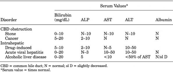
|
|
Table 8-2. Comparison of Various Types of
Cholestatic Disease
|
Cholestasis
- See Table
8-2.
- Increased serum ALP
- Increased GGT, 5′-NT, and LAP parallel ALP and confirm the
hepatic source of ALP.
- Increased serum cholesterol and phospholipids but not
triglycerides
- Increased fasting serum bile acid (>1.5 µg/mL) with ratio of cholic acid to
chenodeoxycholic acid >1 in primary biliary cirrhosis and many
intrahepatic cholestatic conditions but <1 in most chronic
hepatocellular conditions (e.g., Laënnec's cirrhosis, chronic active
hepatitis). (Relatively little experience exists with
this test.)
- Cholestasis
may occur without hyperbilirubinemia
Due To
- Canalicular
- Drugs (e.g., estrogens,
anabolic steroids)most common cause (see Table 8-3)
- Normal pregnancy
- Alcoholic hepatitis
- Infections, e.g.,
- Acute viral hepatitis
- Gram-negative sepsis
- Toxic shock syndrome
- AIDS
- Parasitic, fungal
infection
- Sickle cell crisis
- Postoperative state after
long procedure and multiple transfusions
- Benign recurrent familial
intrahepatic cholestasis (rare)
- Non-Hodgkin's lymphoma
more often than Hodgkin's disease
- Amyloidosis
- Sarcoidosis
- Interlobular
Bile Ducts
- Sclerosing
pericholangitis (associated with inflammatory bowel disease)
- Primary biliary cirrhosis
- Postnecrotic cirrhosis
(20% of cases)
- Congenital intrahepatic
biliary atresia
- Interlobular
and Larger Intrahepatic Bile Ducts
- Multifocal lesions (e.g.,
metastases, lymphomas, granulomas)
- Larger
Intrahepatic Bile Ducts
- Sclerosing cholangitis
- Intraductal stones
- Intraductal
papillomatosis
- Cholangiocarcinoma
- Caroli's disease
(congenital biliary ectasia)
P.210
|
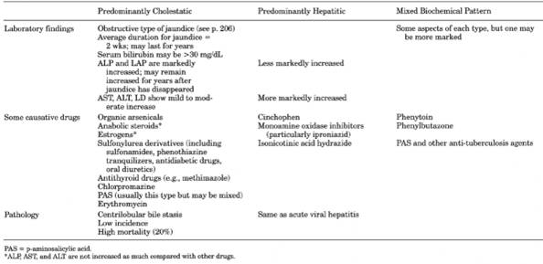
|
|
Table 8-3. Comparison of Three Main Types
of Liver Disease Due to Drugs
|
P.211
- Extrahepatic
Ducts (Surgical or Extrahepatic Jaundice)
- Carcinoma (e.g.,
pancreas, ampulla, bile ducts, gallbladder)
- Stricture, stone, cyst,
etc., of ducts
- Pancreatitis (acute,
chronic), pseudocysts
- Increased
risk of cholangiocarcinoma in progressive cholestatic diseases
Cholestasis,
Benign Recurrent Intrahepatic
- (Familial
condition; attacks begin after age 8 yrs, last weeks to months, complete
resolution between episodes, may recur after months or years; exacerbated
by estrogens.)
- Increased serum ALP
- Transaminase usually
<100 U.
- Serum bilirubin may be
normal or ≤ 10 mg/dL.
- Liver biopsy shows
centrolobular cholestasis without inflammation.
Cholestasis,
Neonatal
Due To
Idiopathic neonatal
hepatitis 5060%
Extrahepatic biliary
atresia 20%
Metabolic disease
Alpha -antitrypsin
deficiency 15%
Cystic fibrosis
Tyrosinemia
Galactosemia
Niemann-Pick disease
Defective bile acid synthesis
Infection (e.g., CMV infection, syphilis,
sepsis, GU tract infection)
Toxic causes (e.g., drugs, parenteral
nutrition)
Other conditions
Paucity of bile ducts (Alagille syndrome)
Indian childhood cirrhosis
Hypoperfusion/shock
Cirrhosis, Primary Biliary (Cholangiolitic Cirrhosis,
Hanot's Hypertrophic Cirrhosis, Chronic Nonsuppurative Destructive Cholangitis,
etc.)
- (Multisystem
autoimmune disease; chronic nonsuppurative inflammation and destruction of
small intrahepatic bile ducts producing chronic cholestasis and cirrhosis)
Diagnostic Criteria
- Laboratory findings of
- Cholestatic pattern
(increased ALP) of long duration (may last for
years) not due to known cause (e.g., drugs).
- Antimitochondrial
autoantibodies present.
- Confirmed patency of
bile ducts (e.g., with ultrasonography or computed tomographic [CT]
scan).
- Compatible liver biopsy
is highly desirable.
- Serum ALP is markedly increased; is of liver origin. Reaches a plateau early in the
course and then fluctuates within 20% thereafter; changes in serum level
have no prognostic value. 5′-NT and GGT parallel ALP. This is one of the few conditions that elevates
both serum ALP and GGT to striking levels.
- Serum mitochondrial antibody titer is strongly positive (1:401:80) in ~95% of
patients and is hallmark of disease (98% specificity); titer >1:160 is
highly predictive of primary biliary cirrhosis (PBC) even in absence of
other findings. Does not correlate with severity or rate of progression.
Titers differ greatly in patients. Similar titers occur in 5% of patients
with chronic hepatitis; low titers occur in 10% of
P.212
patients with other liver disease; rarely found in
normal persons. Titer usually decreases after liver transplantation but
generally remains detectable.
- Serum
bilirubin is normal in early phase but increases in 60% of patients with
progression of disease and is a reliable prognostic indicator; an elevated
level is a poor prognostic sign. Direct serum bilirubin is increased in
80% of patients; levels >5 mg/dL in only 20% of patients; levels >10
mg/dL in only 6% of patients. Indirect bilirubin is normal or slightly
increased.
- Laboratory findings show relatively little evidence of parenchymal damage.
- AST and ALT may be
normal or slightly increased (up to 15× normal), may fluctuate within a
narrow range, and have no prognostic significance.
- Serum albumin, globulin,
and PT normal early; abnormal values indicate advanced disease and poor
prognosis; not corrected by therapy.
- Marked increase in total cholesterol and phospholipids with
normal triglycerides; serum is not lipemic; serum triglycerides become
elevated in late stages. Associated with xanthomas and xanthelasmas. In
early stages, LDL and VLDL are mildly elevated and HDL is markedly
elevated (thus atherosclerosis is rare). In advanced stage, LDL is
markedly elevated with decreased HDL and presence of lipoprotein X
(nonspecific abnormal lipoprotein seen in other cholestatic liver
disease).
- Serum IgM is increased in ~75% of patients; levels may be very
high (45×
normal). Other serum immunoglobulins are also increased.
- Hypocomplementemia
- Polyclonal
hypergammaglobulinemia
- Biopsy of liver categorizes the four stages and helps assess prognosis, but needle
biopsy is subject to sampling error because the lesions may be spotty;
findings consistent with all four stages may be found in one specimen.
- Serum ceruloplasmin is characteristically elevated (in contrast to
Wilson's disease).
- Liver copper may be
increased 10100× normal; correlates with serum bilirubin and advancing
stages of disease.
- ESR is increased 15×
normal in 80% of patients.
- Urine contains
urobilinogen and bilirubin.
- Laboratory findings of
steatorrhea, including the following:
- Serum 25-hydroxyvitamin
D and vitamin A are usually low.
- PT is normal or restored
to normal by parenteral vitamin K.
- Laboratory findings due
to associated diseases
- >80% of patients have
at least one other and >40% have at least two other circulating
antibodies to autoimmune disease (e.g., RA, autoimmune thyroiditis
[hypothyroidism in 20% of patients], Sjögren's syndrome, scleroderma)
although not useful diagnostically.
- Laboratory findings due
to sequelae and complications
- Portal hypertension,
hypersplenism
- Treatment-resistant
osteoporosis
- Hepatic encephalopathy,
liver failure
- Renal tubular acidosis
(due to copper deposition in kidney) is frequent but usually subclinical.
- Increased susceptibility
to urinary tract infection is associated with advanced disease.
- Should
be ruled out in an asymptomatic female with elevated serum ALP without
obesity, diabetes mellitus, alcohol abuse, use of some drugs
Cirrhosis
of Liver
- Criteria
for diagnosis liver biopsy or at least three of the following:
- Hyperglobulinemia,
especially with hypoalbuminemia
- Low-protein (<2.5
g/dL) ascites
- Evidence of
hypersplenism (usually thrombocytopenia, often with leukopenia and less
often with Coombs'-negative hemolytic anemia)
- Evidence of portal
hypertension (e.g., varices)
- Characteristic
"corkscrew" hepatic arterioles on celiac arteriography
- Shunting of blood to
bone marrow on radioisotope scan
- Abnormality
of serum bilirubin, transaminases, or ALP is often not present and
therefore not required for diagnosis
- Serum bilirubin is often increased; may be present for years.
Fluctuations may reflect liver status due to insults to the liver (e.g., alcoholic
debauches). Most bilirubin is of
P.213
the indirect type unless cirrhosis is of the
cholangiolitic type. Higher and more stable levels occur in postnecrotic
cirrhosis; lower and more fluctuating levels occur in Laënnec's cirrhosis.
Terminal icterus may be constant and severe.
- Serum AST is increased (<300 U) in 6575% of patients. Serum
ALT is increased (<200 U) in 50% of patients. Transaminases vary widely
and reflect activity or progression of the process (i.e., hepatic parenchymal cell
necrosis).
- Serum ALP is increased in
4050% of patients.
- Serum total protein is usually normal or decreased. Serum albumin
parallels functional status of parenchymal cells and may be useful for
following progress
of liver disease; but it may be normal in the presence of considerable
liver cell damage. Decreasing serum albumin may reflect development of
ascites or hemorrhage. Serum globulin level is usually increased; it
reflects inflammation and parallels the severity of the inflammation.
Increased serum globulin (usually gamma) may cause increased total
protein, especially in chronic viral hepatitis and posthepatitic cirrhosis.
- Serum total cholesterol
is normal or decreased. Progressive decrease in cholesterol, HDL, LDL with
increasing severity. Decrease is more marked than in chronic active
hepatitis. LDL may be useful for prognosis and selection of patients for
transplantation. Decreased esters reflect more severe parenchymal cell
damage.
- Urine bilirubin is
increased; urobilinogen is normal or increased.
- BUN is often decreased
(<10 mg/dL); increased with GI hemorrhage.
- Serum uric acid is often
increased.
- Electrolytes and acid-base
balance are often abnormal and reflect various combinations of
circumstances at the time, such as malnutrition, dehydration, hemorrhage,
metabolic acidosis, respiratory alkalosis. In cirrhosis with ascites, the
kidney retains increased sodium and excessive water, causing dilutional
hyponatremia.
- Blood ammonia is
increased in liver coma and cirrhosis and with portacaval shunting of
blood.
- Anemia reflects increased
plasma volume and some increased destruction of RBCs. If more severe, rule
out hemorrhage in GI tract, folic acid deficiency, excessive hemolysis,
etc.
- WBC is usually normal
with active cirrhosis; increased (<50,000/cu mm) with massive necrosis,
hemorrhage, etc.; decreased with hypersplenism.
- Laboratory findings due to complications or sequelae, often in
combination
- Portal hypertension.
- Ascites.
- Esophageal varices.
- Portal vein thrombosis.
- Liver failure.
- Hepatocarcinoma.
- Abnormalities of
coagulation mechanisms (see Chapter 11), e.g.,
- Prolonged PT (does not
respond to parenteral vitamin K as frequently as in patients with
obstructive jaundice).
- Prolonged bleeding time
in 40% of cases due to decreased platelets and/or fibrinogen (see Chapter 11).
- Hepatic encephalopathy.
- Increased arterial
ammonia.
- CSF glutamine >35
mg/dL (due to conversion from ammonia); correlates with depth of coma
and more sensitive than arterial ammonia.
- Spontaneous bacterial
peritonitisin ≤ 10% of alcoholic cirrhosis cases. 70% have
positive blood culture; usually single organism, especially E. coli, Pneumococcus, Klebsiella.
- Hepatorenal syndrome.
- Most commonly death is
due to liver failure, bleeding, infections.
|
Laboratory findings due to
causative/associated diseases or conditions
|
Frequency in USA
|
|
|
Chronic viral hepatitis (HBV with or
without HDV, HCV)
|
|
|
|
Alcoholism
|
|
|
|
Wilson's disease
|
Rare
|
|
|
Autoimmune chronic active hepatitis
|
|
|
|
Hemochromatosis
|
P.214
|
|
|
Mucoviscidosis
|
|
|
|
Glycogen-storage diseases
|
|
|
|
Galactosemia
|
|
|
|
Alpha -antitrypsin deficiency
|
Rare
|
|
|
Porphyria
|
|
|
|
Fructose intolerance
|
|
|
|
Tyrosinosis
|
|
|
|
Infections (e.g., congenital syphilis,
schistosomiasis)
|
|
|
|
Gaucher's disease
|
|
|
|
Ulcerative colitis
|
|
|
|
Osler-Weber-Rendu disease
|
|
|
|
Venous outflow obstruction (e.g.,
Budd-Chiari syndrome, venoocclusive disease, congestive heart failure)
|
|
|
|
Biliary disease (e.g., primary biliary
cirrhosis, sclerosing cholangitis
|
|
|
|
Cryptogenic
|
|
|
Crigler-Najjar
Syndrome (Hereditary Glucuronyl Transferase Deficiency)
- (Rare
familial autosomal recessive disease due to marked congenital deficiency
or absence of glucuronyl transferase, which conjugates bilirubin to
bilirubin glucuronide in hepatic cells [counterpart is the homozygous Gunn
rat])
- See Table
8-4.
Type I
- Indirect serum bilirubin
is increased; it appears on first or second day of life, rises in 1 wk to
peak of 1245 mg/dL, and persists for life. No direct bilirubin in serum
or urine.
- Fecal urobilinogen is
very low.
- Liver function tests are
normal; sulfobromsulfophthalein (BSP) is normal.
- Liver biopsy is normal.
- No evidence of hemolysis is
found.
- Untreated patients often
die of kernicterus by age 18 mos.
- Nonjaundiced parents have
diminished capacity to form glucuronide conjugates with menthol,
salicylates, and tetrahydrocortisone.
- Type I
should always be ruled out when persistent unconjugated bilirubin levels
of 20 mg/dL are seen after 1 wk of age without obvious hemolysis and
especially after breast-milk jaundice has been ruled out
- This syndrome has been
divided into two groups:
|
|
Type I
|
Type II
|
|
Transmission
|
Autosomal recessive
|
Autosomal dominant
|
|
Hyperbilirubinemia
|
More severe (usually >20 mg/dL)
|
Less severe and more variable (usually
20 mg/dL)
|
|
Kernicterus
|
Frequent
|
Absent
|
|
Bile
|
Essentially colorless
|
Normal color
|
|
Bilirubin-glucuronide
|
Totally absent
|
Present
|
|
Bilirubin concentration
|
Very low (<10 mg/dL) Only traces of
conjugated bilirubin
|
Nearly normal (50100 mg/dL)
|
|
Stool color
|
Pale yellow
|
Normal
|
|
Parents
|
Normal serum bilirubin in both parents
Partial defect (~50%) in glucuronide conjugation in both parents
|
One parent usually shows minimal to
severe icterus
Defect in glucuronide conjugation may be pressent in only one parent
|
|
P.215
|
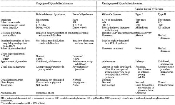
|
|
Table 8-4. Differential Diagnosis of
Hereditary Jaundice with Normal Liver
Chemistries and No Signs or Symptoms of Liver Disease
|
P.216
Type
II
- Patients have partial
deficiency of glucuronyl transferase (autosomal dominant with incomplete
penetrance). Not related to type I syndrome; may be homozygous form of
Gilbert's disease. Patient may not become jaundiced until adolescence.
Neurologic complications are rare.
- Serum indirect bilirubin
= 625 mg/dL. Increases with fasting or removal of lipid from diet. May
decrease to <5 mg/dL with phenobarbital treatment.
Dubin-Johnson
Syndrome (Sprinz-Nelson Syndrome)
- (Autosomal
recessive disease due to inability to transport bilirubin-glucuronide
through hepatocytes into canaliculi, but conjugation of
bilirubin-glucuronide is normal. Characterized by mild chronic, recurrent
jaundice; hepatomegaly and right upper quadrant abdominal pain may be
present. Usually is compensated except in periods of stress. Jaundice
[innocuous and reversible] may be produced by estrogens, birth control
pills, or last trimester of pregnancy. May resemble mild viral hepatitis.)
- See Table
8-4.
- Serum bilirubin is increased (310 mg/dL; rarely ≤ 30 mg/dL);
significant amount is direct.
- Urine contains bile and
urobilinogen.
- BSP excretion is impaired with late (1.5- to 2-hr) increase; virtually pathognomonic.
- Other liver function tests are normal.
- Urine total coproporphyrin is usually normal but ~80% is
coproporphyrin I (normally 75% is coproporphyrin III); diagnostic of
Dubin-Johnson syndrome. Not useful to detect individual heterozygotes.
- Liver biopsy shows large amounts of yellow-brown or slate-black
pigment in centrolobular hepatic cells (lysosomes) and small amounts in Kupffer's
cells.
Fatty
Liver
- Laboratory findings are
due to underlying conditions (most commonly alcoholism; nonalcoholic fatty
liver is commonly associated with noninsulin dependent diabetes mellitus
[≤ 75%], obesity [69100%], hyperlipidemia [2081%]; malnutrition,
toxic chemical exposure)
- Biopsy of liver establishes the diagnosis
- Nonalcoholic fatty liver
is distinguished by negligible history of alcohol consumption and negative
random blood alcohol assays.
- Liver function tests
- Most commonly, serum AST
and ALT are increased 23×; usually ALT >AST.
- Serum ALP is normal or
slightly increased in <50% of patients.
- Increased serum ferritin
(≤ 5×) and transferrin saturation in ~60% of cases.
- Other liver function
tests are usually normal.
- Serologic tests for
viral hepatitis are negative.
- Cirrhosis occurs in
≤ 50% of alcoholic and ≤ 17% of nonalcoholic cases.
- Biochemically different
form occurs in acute fatty liver of pregnancy, Reye's syndrome,
tetracycline administration.
- Fatty
liver may be the only postmortem finding in cases of sudden, unexpected
death
Fatty
Liver Of Pregnancy, Acute
- (Incidence
of 1 per 13,328 deliveries; usually occurs after 35th week of pregnancy.
Medical emergency because of high maternal and fetal mortality, which is
markedly improved by termination of pregnancy.)
- Often associated with
toxemia
- Increased AST and ALT to
~300 U (rarely >500 U) are used for early screening in suspicious
cases; ratio is not helpful in differential diagnosis.
- Increased WBC in >80%
of cases (often >15,000/cu mm)
- Evidence of DIC in
>75% of patients
P.217
- Serum uric acid is
increased disproportionately to BUN and creatinine, which may also be
increased.
- Serum bilirubin may be normal
early but will rise unless pregnancy terminates.
- Blood ammonia is usually
increased.
- Blood glucose is often
decreased, sometimes markedly.
- Neonatal liver function
tests are usually normal, but hypoglycemia may occur.
- Biopsy of liver confirms the diagnosis
Gallbladder
and Bile Duct Cancer
- Laboratory findings
reflect varying location and extent of tumor infiltration that may cause
partial intrahepatic duct obstruction or obstruction of hepatic or common
bile duct, metastases in liver, or associated cholangitis; 50% of patients
have jaundice at the time of hospitalization.
- Laboratory findings of
duct obstruction are of progressively increasing severity in contrast to
the intermittent or fluctuating changes due to duct obstruction caused by
stones. A papillary intraluminal duct carcinoma may undergo periods of
sloughing, producing the findings of intermittent duct obstruction.
- Anemia is present.
- Cytologic examination of aspirated duodenal fluid may demonstrate malignant cells.
- Silver-colored stool due to jaundice combined with GI bleeding may be seen in
carcinoma of duct or ampulla of Vater.
- Laboratory findings of
the preceding cholelithiasis are present (gallbladder cancer occurs in ~3%
of patients with gallstones).
Gilbert's
Disease
- (Chronic,
benign, intermittent, familial [autosomal dominant with incomplete
penetrance], nonhemolytic unconjugated hyperbilirubinemia with evanescent
increases of indirect serum bilirubin, which is usually discovered on
routine laboratory examinations; due to defective transport and
conjugation of unconjugated bilirubin. Jaundice is usually accentuated by
pregnancy, fever, exercise, and various drugs, including alcohol and birth
control pills. Rarely identified before puberty. May be mildly
symptomatic. 37% prevalence in total population.)
- See Table
8-4.
- Presumptive diagnostic
criteria
- Exclusion of other
diseases.
- Unconjugated
hyperbilirubinemia on several occasions.
- Liver chemistries and
hematologic parameters are normal.
- Indirect serum bilirubin is increased
transiently and has been previously normal at least once in ≤ 33% of
patients. It may rise to 18 mg/dL but usually is <4 mg/dL. Considerable
daily and seasonal fluctuation. Fasting (<400 calories/day) for 72 hrs
causes elevated indirect bilirubin to increase >100% in Gilbert's
disease but not in healthy persons (increase <0.5 mg/dL) or those with
liver disease or hemolytic anemia. Fasting bilirubin returns to baseline
1224 hrs after resumption of normal diet. Combination of basal total
bilirubin >1.2 mg/dL and fasting increase of unconjugated bilirubin
>1 mg/dL has sensitivity of 84%, specificity of 78%, positive
predictive value of 85%, negative predictive value of 76%. Provocative
tests are rarely needed. Direct serum bilirubin is normal but may give
elevated results by liquid diazo methods but not by dry methods or
chromatography. Enzyme inducers (e.g., phenobarbital) normalize
unconjugated bilirubin in 12 wks. Prednisone administration reduces
bilirubin concentration.
- Liver function tests are
usually normal.
- Fecal urobilinogen
usually normal but may be decreased.
- Urine shows no increased
bilirubin.
- Liver biopsy is normal.
Heart
Failure (Congestive), Liver Function Abnormalities
- Pattern of abnormal liver
function tests is variable depending on severity of heart failure; the
mildest cases show only slightly increased ALP and slightly decreased
serum
P.218
albumin; moderately severe cases also show slightly increased serum bilirubin
and GGT; one-fourth to three-fourths of the most severe cases also show
increased AST and ALT (≤ 200 U/L) and LD (≤ 400 U/L). All return to
normal when heart failure responds to treatment. Serum ALP is usually the last
to become normal, and this may occur weeks to months later.
- Serum bilirubin is
frequently increased (indirect more than direct); usually 15 mg/dL. It
usually represents combined right- and left-sided failure with hepatic
engorgement and pulmonary infarcts. Serum bilirubin may suddenly rise
rapidly if superimposed myocardial infarction occurs.
- AST and ALT are
disproportionately increased compared with other liver function tests in
left-sided heart failure.
- PT may be slightly
increased, with increased sensitivity to anticoagulant drugs.
- Serum cholesterol and
esters may be decreased.
- Urine urobilinogen is
increased. Urine bilirubin is increased in the presence of jaundice.
- These
findings may occur with marked liver congestion due to other conditions
(e.g., Chiari's syndrome [occlusion of hepatic veins] and constrictive
pericarditis)
Hemochromatosis
See Fig. 8-4.
Due To
- Hereditary
hemochromatosis is an autosomal recessive defect in the ability of the
duodenum to regulate iron absorption; abnormal gene present in 10% of
white Americans; frequency of homozygosity >3 in 1000. 13% of
heterozygotes develop iron overload; may be due to coincidental condition
with altered iron absorption or metabolism.
- Other primary causes of
iron overload (may have one hemochromatosis allele)
- Neonatal hemochromatosis
- Juvenile hemochromatosis
- African iron overload
- Aceruloplasminemia
- Secondary
- Increased intake (e.g.,
excessive medicinal iron ingestion, long-term frequent transfusions,
Bantu siderosis)
- Anemias with increased
erythropoiesis (especially thalassemia major; also thalassemia minor,
some other hemoglobinopathies, paroxysmal nocturnal hemoglobinuria,
sideroblastic anemias, refractory anemias with hypercellular bone marrow,
pyruvate kinase deficiency, pyridoxine-responsive anemia, X-linked
iron-loading anemia, etc.)
- Chronic hemodialysis
- Porphyria cutanea tarda
(minor)
- Alcoholic liver disease
(minor; deposited in Kupffer's cells, not hepatocytes)
- After portal-systemic
shunt
- Congenital
atransferrinemia
- Increased transferrin saturation (= serum iron ÷ total iron-binding capacity ×
100); usually >70% and frequently approaches 100%; repeat fasting
transferrin saturation >60% in men and >50% in women without other
known causes probably represents hemochromatosis; 5062% usually indicates
heterozygous state but occasionally found in homozygous persons. Most
heterozygotes have no detectable changes unless a secondary cause (e.g.,
thalassemia) is present. If value is increased, patient should be retested
(fasting) twice at weekly intervals. Screening discovers hemochromatosis
in 23 of 1000 persons; should be sought especially in patients with
diabetes mellitus, congestive heart failure, idiopathic cardiomyopathy,
arthritis, alcoholic cirrhosis, bronze skin, hypogonadism.
- Increased serum ferritin (usually >1000 µg/L); increased in approximately
two-thirds of patients with hemochromatosis. Is good index of total body
iron but has limited
P.219
value for screening because may be increased in acute
inflammatory conditions and less sensitive than transferrin saturation in early
cases. May not be increased in patients who have not yet
accumulated excess amounts of iron (e.g., children, young adults, premenopausal
women). >5000 µg/L indicates tissue damage (e.g., liver degeneration)
with release of ferritin into circulation. >350 µg/L in fasting men and
>250 µg/L in women is recommended for screening. Critical threshold
associated with cirrhosis is unknown. Liver biopsy is probably not indicated if
serum ferritin is normal.
|
|
|
Fig. 8-4. Sequence of tests for
hemochromatosis screening and treatment.
|
- Serum iron is increased (usually >200 µg/dL in women and >300 µg/dL in
men and typically >1000 µg/dL) but should not be only screening test
because of many other conditions in which it occurs. Confirm by measuring
repeat fasting sample at least two more times. Serum
iron levels may show marked diurnal variation, with lowest values in
evening and highest between 7 a.m. and noon.
P.220
- TIBC is decreased (~200 µg/dL; often approaches zero; generally higher in secondary than
primary type).
- Liver biopsy is needed to confirm or refute diagnosis, grade amount of iron, and
assess tissue damage (presence of fibrosis/cirrhosis, other liver
diseases). Is indicated when repeat fasting serum ferritin (>750 mg/L)
and transferrin saturation are increased after 46 wks of abstinence from
alcohol. Histologic examination confirms increased stainable iron (special
stain) in perilobular hepatocytes and biliary epithelium in hereditary
hemochromatosis with little in Kupffer's cells (in contrast to secondary
iron overload) or bone marrow, with or without inactive cirrhosis. In
later stages, liver biopsy alone does not distinguish hereditary
hemochromatosis from secondary hemochromatosis. Liver iron is increased
(normal 2002000 µg/gm in men and 2001600 µg/gm in women). >1000
µg/100 mg of dry liver is consistent with homozygous state but level may
reach 5000. Some heterozygotes may reach 1000 µg/100 mg but do not
progress beyond this level. Fibrosis or cirrhosis usually does not occur
at levels <2000 µg/100 mg dry liver unless
alcoholism is also present. For chemical analysis of iron, use acid-washed
needle and place specimen in iron-free container. Liver iron and serum
ferritin may also be increased in alcoholic cirrhosis but levels are not
as abnormal (<2× normal) as in hemochromatosis. Liver iron must be
related to patient age: hepatic iron index (micrograms/gram divided by
55.8 × age) in homozygotes is ≥1.9; in heterozygotes usually ≤
1.5. False negative may be due to phlebotomy treatment; false positive may
be due to secondary hemosiderosis. Another calculation is liver iron
(micromoles/gram dry weight) divided by patient age; value >2 in
homozygotes; <2 in heterozygotes, healthy persons, patients with
alcoholic liver disease.
- Other tests to assess
iron stores (when liver biopsy is not possible)
- Chelating agent (0.5 gm
IM deferoxamine mesylate) causes urinary excretion >5 mg/24 hrs in
hereditary hemochromatosis but <2 mg/24 hrs in normal persons.
Measures only chelatable iron rather than total iron stores so may
underdiagnose hereditary hemochromatosis; not a useful diagnostic test.
- Weekly phlebotomy for
510 wks causes iron deficiency in alcoholic liver disease but >50
weekly phlebotomies are required in hereditary hemochromatosis.
- Presence of excess iron in other tissue biopsy sites (e.g.,
synovia, GI tract) should arouse suspicion of hereditary hemochromatosis;
iron stains should be done.
- Bone marrow biopsy
stained for iron is not useful for diagnosis of hereditary
hemochromatosis.
- Liver function tests
depend on presence and degree of liver damage (e.g., cirrhosis).
- On average, women have
serum ferritin concentrations 1000 µg/L less than men; men have twice the
incidence of cirrhosis (25%) and diabetes (15%) compared with women.
- Laboratory findings due to
involvement of various organs
- Insulin-dependent
diabetes mellitus in 4075% of cases; glucose intolerance
- Osteoarthritis and
chondrocalcinosis (pseudogout) in 50% of cases
- Cardiomyopathy in 33% of
cases (congestive heart failure)
- Hypogonadism/pituitary dysfunction
in ~50% of cases
- Skin pigmentation
- Underlying diseases
- Laboratory findings due to complications and sequelae
- Increased susceptibility
to severe bacterial infection, especially Yersinia
sepsis (also occurs in other iron overload conditions).
- Cirrhosis in 69% of
cases. Does not resolve with phlebotomy. Increased risk of hepatocellular
carcinoma. Associated alcoholism.
- Hepatocellular carcinoma
develops in ≤ 30% of cases and has become the chief cause of death
in hereditary hemochromatosis.
- Portal hypertension.
- When diagnosis of hereditary
hemochromatosis is established, other family members should be screened;
one-fourth of siblings have the disease; 5% of patients' children are
homozygous for hemochromatosis gene. Relatives with negative results should be
rescreened every 5 yrs.
- Genotyping is not used for screening to discover sporadic cases
but useful to identify patient's siblings at risk because HLA-identical
sibs almost always are also homozygous for hemochromatosis gene and at
high risk for developing clinical disease. May be useful to distinguish
patients with primary hereditary hemochromatosis from cirrhotic patients
with secondary iron overload and siderosis.
P.221
- DNA test for hereditary
hemochromatosis gene is available, but diagnostic role is being evaluated.
C282Y or H63D present in 6997% of affected patients; would not identify
≤ 31% of clinically affected patients. May ultimately replace HLA
typing.
- Adequate treatment with
phlebotomy (13 U/wk) sufficient to maintain a mild anemia is determined
by Hct (3739%) before each phlebotomy. If >40%, an additional
treatment may be scheduled. Serum iron and ferritin are used only when anemia become refractory to establish whether iron
stores are exhausted. Maintenance phlebotomy (46 U/yr) can be monitored
with serum ferritin to indicate normal amount of storage iron. Insulin
requirement decreases in more than one-third of diabetics; liver function
tests often improve; arthritis, impotence, and sterility usually do not
improve. Removal of 450500 mL of blood causes loss of 200250 mg of iron.
Hemochromatosis,
Neonatal
- (Severe
iron overload disorder with onset in utero. Death
usually occurs soon after birth.)
- Oligohydramnios or less
commonly polyhydramnios may indicate intrauterine growth retardation or
fetal hydrops.
- Fulminant liver failure including hyperbilirubinemia, decreased
transaminases, glucose, and albumin. Increased AFP. Variable fibrinogen
consumption, thrombocytopenia, anemia, acanthocytosis.
- Marked hepatic and extrahepatic (e.g., heart, pancreas, adrenal; not spleen) siderosis
with relative lack in RE cells.
- Liver iron analysis not
useful because high in healthy newborn.
Hepatic
Encephalopathy
- (Neurologic
and mental abnormalities in some patients with liver failure)
- Blood ammonia is increased in 90% of patients but does not reflect
the degree of coma. Normal level in comatose patient suggests another
cause of coma. Not reliable for diagnosis but may be useful to follow individual
patients. May be increased by tight tourniquet or vigorously clenched
fist; thus arterial specimen may be preferable.
- Respiratory alkalosis due
to hyperventilation is frequent.
- Hyponatremia and
iatrogenic hypernatremia are frequent complications and are associated
with a higher mortality rate.
- Hypokalemic metabolic
alkalosis may occur due to diuretic excess.
- Serum amino acid profile
is abnormal. All serum amino acids are markedly increased in coma due to
acute liver failure.
- CSF is normal except for
increased glutamine level.
- Diagnosis is clinical; characteristic laboratory findings are supportive but not
specific.
Hepatic
Failure, Acute
Due To
- Infection
- Viral hepatitis (e.g.,
hepatitis A, B, C, D, E; HSV 1, 2, 6; EBV, CMV).
- Acute liver failure
related to HSV is usually associated with immunosuppressive therapy.
- Develops in ~13% of
adults with acute icteric type B hepatitis with resultant death.
- Other causes rare (e.g.,
amebic abscesses, disseminated TB).
- Drugs (e.g.,
acetaminophen, methyltestosterone, isoniazid, halothane, idiosyncratic
reaction)
- Toxins (e.g., phosphorus,
death-cap mushroom [Amanita phalloides])
- Acute fatty liver
- Pregnancy
- Reye's syndrome
- Drugs (e.g.,
tetracycline)
- Ischemic liver necrosis
- Shock
- Budd-Chiari syndrome
(acute)
- Wilson's disease with
intravascular hemolysis
P.222
- Congestive heart failure
- Extracorporeal
circulation during open heart surgery
- Marked infiltration by
tumor
- Acute leukemia
- Lymphoma
- Hodgkin's disease
- Non-Hodgkin's lymphoma
- Burkitt's lymphoma
- Malignant histiocytosis
- Serum bilirubin progressively increases; may become very high.
- Increased serum AST, ALT,
may fall abruptly terminally; serum ALP and GGT may be increased.
- Serum cholesterol and
esters are markedly decreased.
- Decreased albumin and
total protein
- Electrolyte
abnormalities, e.g.,
- Hypokalemia (early)
- Metabolic alkalosis due
to hypokalemia
- Respiratory alkalosis
- Lactic acidosis
- Hyponatremia,
hypophosphatemia
- Hypoglycemia in ~5% of
patients
- Laboratory findings associated with
- Hepatic encephalopathy
- Hepato-renal syndrome
- Coagulopathy
- Decreased factors II,
V, VII, IX, X cause prolonged PT and aPTT (PT is never normal in acute
hepatic failure).
- Decreased antithrombin
III.
- Platelet count
<100,000 in two-thirds of patients.
- Hemorrhage, especially
in GI tract
- Bacterial and fungal
infections, especially streptococci and S. aureus
- Ascites
- As patient deteriorates,
titers of HBsAg, and HBeAg may often fall and
disappear.
Hepatitis,
Acute Viral
- See Table
8-5 and Fig. 8-5.
- Different types of viral
hepatitis cannot be distinguished by clinical features or routine
chemistries; serologic tests are needed.
Prodromal
Period
- Serologic markers appear in serum (Table 8-6
- Bilirubinuria occurs
before serum bilirubin increases.
- Increase in urinary
urobilinogen and total serum bilirubin just before clinical jaundice
occurs.
- Serum AST and ALT both rise during the preicteric phase and show
very high peaks (>500 U) by the time jaundice appears.
- ESR is normal.
- Leukopenia (lymphopenia
and neutropenia) is noted with onset of fever, followed by relative
lymphocytosis and monocytosis; may find plasma cells and <10% atypical
lymphocytes (in infectious mononucleosis level is >10%).
Asymptomatic
Hepatitis
Biochemical evidence of acute hepatitis is
scant and often absent.
Acute
Icteric Period
- (Tests
show parenchymal cell damage.)
- Serum bilirubin is 5075%
direct in the early stage; later, indirect bilirubin is proportionately
more.
P.223
- Serum AST and ALT fall
rapidly in the several days after jaundice appears and become normal 25
wks later.
- In
hepatitis associated with infectious mononucleosis, peak levels are
usually <200 U and peak occurs 23 wks after onset, becoming normal by
the fifth week.
- In
toxic hepatitis, levels depend on severity; slight elevations may be associated
with therapy with anticoagulants, anovulatory drugs, etc.; poisoning
(e.g., carbon tetrachloride) may cause levels ≤ 300 U.
- In
severe toxic hepatitis (especially carbon tetrachloride poisoning), serum enzymes may be
1020× higher than in acute hepatitis and show a different pattern, i.e.,
increase in LD > AST > ALT.
- In
acute hepatitis, ALT > AST > LD.
- Other liver function
tests are often abnormal, depending on severity of the
diseasebilirubinuria, abnormal serum protein electrophoresis, ALP, etc.
- Serum cholesterol/ester ratio
is usually depressed early; total serum cholesterol is decreased only in
severe disease.
- Serum phospholipids are
increased in mild but decreased in severe hepatitis. Plasma vitamin A is
decreased in severe hepatitis.
- Urine urobilinogen is
increased in the early icteric period; at peak of the disease it
disappears for days or weeks; urobilinogen simultaneously disappears from
stool.
- ESR is increased; falls
during convalescence.
- Serum iron is often
increased.
- Urine: Cylindruria is
common; albuminuria occurs occasionally; concentrating ability is
sometimes decreased.
Defervescent
Period
- Diuresis occurs at onset
of convalescence.
- Bilirubinuria disappears,
whereas serum bilirubin is still increased.
- Urine urobilinogen
increases.
- Serum bilirubin becomes
normal after 36 wks.
- ESR falls.
Anicteric
Hepatitis
Laboratory findings are the same as in the
icteric type, but abnormalities are usually less marked and serum bilirubin
shows slight or no increase.
Acute
Fulminant Hepatitis with Hepatic Failure
Cholangiolitic
Hepatitis
Same as acute hepatitis, but evidence of
obstruction is more prominent (e.g., increased serum ALP and direct serum
bilirubin), and tests of parenchymal damage are less marked (e.g., AST increase
may be 36× normal).
Chronic
Hepatitis
- See Table
8-7.
- Occurs in 510% of adults
with acute HBV.
- HBV hepatitis is
generally divided into three stages:
- Stage of acute
hepatitis: Usually lasts 16 mos with mild or no symptoms.
- AST and ALT are
increased >10×.
- Serum bilirubin is
usually normal or only slightly increased.
- HBsAg gradually rises
to high titers and persists; HBeAg also appears.
- Gradually merges with
next stage.
- Stage of chronic
hepatitis: Transaminases increased >50% for >6 mos duration; may
last only 1 yr or for several decades with mild or severe symptoms; most
cases resolve, but some develop cirrhosis and liver failure.
- AST and ALT fall to
210× normal range.
- HBsAg usually remains
high, and HBeAg remains present.
- Chronic carrier stage:
Patients are usually, but not always, healthy and asymptomatic.
- AST and ALT fall to
normal or <2× normal.
P.224
|
|
|
Table 8-5. Comparison of Different Types
of Viral Hepatitis
|
P.225
P.226
|
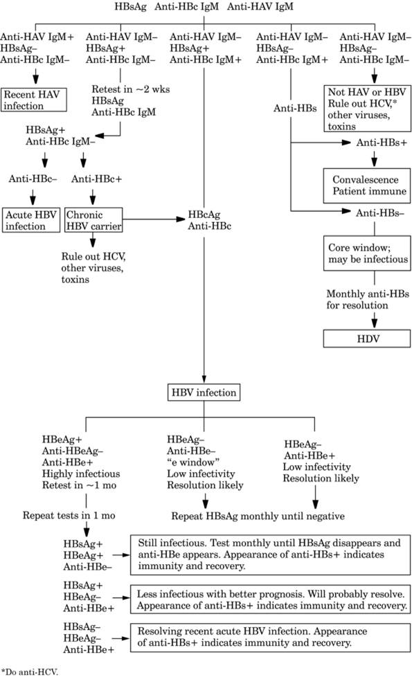
|
|
Fig. 8-5. Algorithm illustrating use of
serologic tests for diagnosis of acute hepatitis.
|
P.227
|
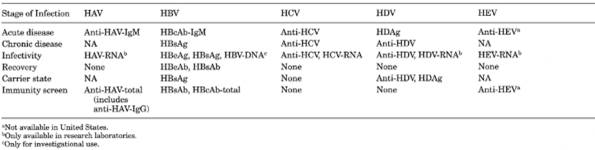
|
|
Table 8-6. Serologic Markers of Viral
Hepatitis
|
P.228
|
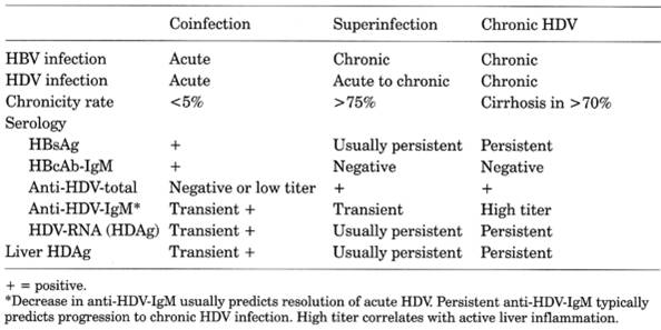
|
|
Table 8-7. Comparison of Types of
Hepatitis D Virus (HDV) Infections
|
- HBeAg disappears, and
anti-HBe appears.
- HBsAg titer falls
although may still be detectable; anti-HBs subsequently develops,
marking the end of carrier stage.
- Anti-HBc is usually
present in high titer (>1:512).
- Laboratory findings due
to sequelae, e.g.,
- GN or nephrotic syndrome
due to deposition of HBeAg or HBcAg in glomeruli, which often progresses
to chronic renal failure.
Hepatitis,
Alcoholic
- Diagnosis is established by liver biopsy and history of alcohol intake. Liver
biopsy should be performed for any alcoholic patient with enlarged liver
as the only way to make definite diagnosis of alcoholic hepatitis. Many
alcoholics have normal liver biopsies.
- Increased
serum GGT and MCV >100 together or separately are useful clues for
occult alcoholism.
- Ratio of desialylated
transferrin to total transferrin >0.013 has been reported to have 81%
sensitivity and 98% specificity for ongoing alcohol consumption.
- Serum AST is increased
(rarely >300 U/L), but ALT is normal or only slightly elevated.
- AST and ALT are more
specific but less sensitive than GGT. Levels of AST and ALT do not
correlate with severity of liver disease. AST/ALT ratio >1 associated
with AST <300 U/L will identify 90% of patients with alcoholic liver
disease; is particularly useful for differentiation from viral hepatitis,
in which increase of AST and ALT are about the same.
- Cholestasis in ≤
35% of patients.
- In acute alcoholic
hepatitis, GGT level is usually higher than AST level. GGT is often
abnormal in alcoholics even with normal liver histology. Is more useful as
index of occult alcoholism or to indicate that elevated serum ALP is of
bone or liver origin than to follow course of patient, for which AST and
ALT are most useful.
- Serum ALP may be normal
or moderately increased in 50% of patients and is not useful as a
diagnostic test.
- Serum bilirubin may be
mildly increased except with cholestasis; is not useful as a diagnostic
test. However, if bilirubin continues to increase during a week of therapy
in the hospital, a poor prognosis is indicated.
- Decreased serum albumin
and increased polyclonal globulin with disproportionately increased IgA
are frequent. Decreased albumin means long-standing or relatively severe
disease.
- Increased PT that is not
corrected by parenteral administration of 10 mg/day of vitamin K for 3
days is best indicator of poor prognosis.
- Discriminant function to
assess severity of alcoholic hepatitis = 4.6 × (PT [secs] control PT) +
serum bilirubin. Discriminant function >32 is equated with severe
disease.
P.229
- Increased WBC
(>15,000) in up to one-third of patients with shift to left (WBC is
decreased in viral hepatitis); normal WBC may indicate folic acid
depletion.
- Anemia in >50% of
patients may be macrocytic (folic acid or vitamin B deficiency), microcytic (iron or pyridoxine deficiency), mixed, or
hemolytic.
- Metabolic alkalosis may
occur due to K loss with pH normal or
increased, but pH <7.2 often indicates that
disease is becoming terminal.
- In terminal stage of
chronic alcoholic liver disease (last week before death), there is often
decrease of serum sodium and albumin and increase of PT and serum bilirubin;
AST and LD decrease from previously elevated levels.
- Indocyanine green (50
mg/kg) is abnormal in 90% of patients.
- Compared to nonalcoholic
patients, alcoholic patients as a group show an increase in a number of
blood components (e.g., AST, phosphorus, ALP, GGT, MCV, MCH, Hb, WBC) and
a decrease in others (e.g., total protein, BUN); however, these variations
usually remain within the reference range. These changes may last for
>6 wks after abstaining from alcohol.
- Laboratory findings due
to sequelae or complications
- Fatty liver
- Cirrhosis
- Portal hypertension
- Infections (e.g., GU
tract, pneumonia, peritonitis)
- DIC
- Hepatorenal syndrome
- Encephalopathy
Hepatitis,
Autoimmune Chronic Active
- Criteria
for diagnosis (all must be present for definite diagnosis)
|
|
Probable
|
Definite
|
|
Increased serum AST or ALT
concentrations
|
X
|
X
|
|
Increased serum ALP <3× normal
concentration
|
|
X
|
|
Increased serum total or gamma globulin
or IgG
|
|
|
|
>1.5× upper
limit of normal
|
|
X
|
|
1.01.5× upper
limit of normal
|
X
|
|
|
Antibody titers to nucleus, smooth
muscle or liver/kidney
microsome type 1 >1:80 (adults) or >1:20 (children)
|
|
X
|
|
Lower titers or presence of other
antibodies
|
X
|
|
|
Absence of markers for viral hepatitis
(HAV, HBV, HCV, CMV, EBV)
|
X
|
X
|
|
Absence of excess alcohol consumption
|
|
|
|
<25 gm/day
(women) or <35 gm/day (men)
|
|
X
|
|
<40 gm/day
(women) or <50 gm/day (men)
|
X
|
|
|
Exposure to blood products
|
|
|
|
No
|
|
X
|
|
Yes, but
unrelated to disease
|
X
|
|
|
Exposure to hepatotoxic drugs
|
|
|
|
No
|
|
X
|
|
Yes, but
unrelated to disease
|
X
|
|
|
Compatible histologic findings and
absence of biliary
lesions, copper deposits or other changes suggestive
of other causes of lobular hepatitis
|
X
|
X
|
|
Hepatitis,
Chronic Active
(Inflammatory liver disease present >6 mos.)
Due To
- Viruses
- HBV (with or without
HDV)
- HCV (with or without
hepatitis G virus [HGV])
P.230
- Metabolic disorders
- Wilson's disease
- Alpha -antitrypsin deficiency
- Hemochromatosis
- Primary biliary
cirrhosis
- Sclerosing cholangitis
- Drugs, e.g.,
- Methyldopa
- Nitrofurantoin
- Isoniazid
- Oxyphenacetin
- Nonalcoholic fatty liver
- Alcoholic hepatitis
- Autoimmune causes
- Type I (lupoid)
(antismooth muscle; antiactin)
- Type II
(antikidney-liver-microsomal)
- Type III (antisoluble
liver antigen)
Hepatitis
A
- Serum bilirubin usually
510× normal. Jaundice lasts a few days to 12 wks. Usually not infectious
after onset of jaundice.
- Serum AST and ALT
increased to hundreds for 13 wks.
- Relative lymphocytosis is
frequent.
Serologic
Tests for Viral Hepatitis A (HAV)7
- See Tables
8-5 and , and Figs. 8-3,
, and .
- Anti-HAV
IgM appears at the same time as symptoms in >99% of cases, peaks within
first month, becomes nondetectable in 12 mos (usually 6 mos). Presence
confirms diagnosis of recent acute infection.
- Anti-HAVtotal is predominantly IgG except immediately after acute HAV infection,
when it is mostly IgM and IgA. Almost always positive at onset of acute
hepatitis and is usually detectable for life; found in 45% of adult
population; indicates previous exposure to HAV, recovery, and immunity to
type A hepatitis. Negative anti-HAVtotal effectively excludes acute HAV.
Positive anti-HAVtotal does not distinguish recent from past infection,
for which anti-HAV IgM test is needed. Test for anti-HAVtotal is
relatively insensitive (minimum detection amount = 100 mU/mL) and may not
detect protective antibody response after one dose of inactivated HAV
vaccine (minimum protective antibody is <10 mU/mL).
- Serial testing is usually
not indicated.
- Tests for anti-HAVtotal
and anti-HAV IgM are not influenced by normal doses of immune globulin.
- HAV antigen and HAV RNA
are available only as research tools.
Hepatitis
B
See Tables 8-5 and , and Figs. 8-5 and .
Serologic
Tests for Viral Hepatitis B (HBV)
See Table 8-6 and Tables 8-8, , , ,
and .
Use
- Differential diagnosis of
hepatitis
- Screening of blood and
organ donors
- Determination of immune
status for possible vaccination
P.231
|
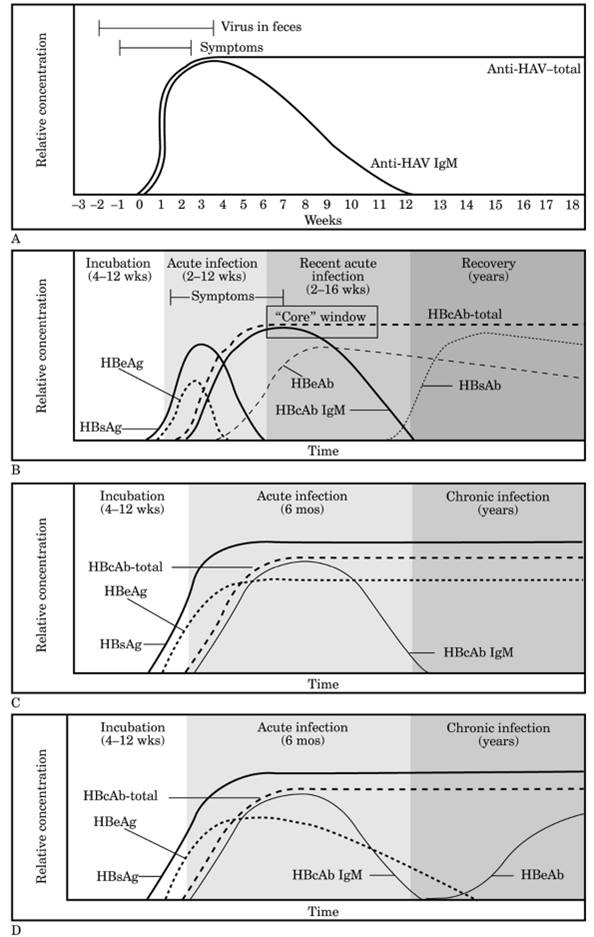
|
|
Fig. 8-6. Hepatitis serologic profiles. A: Antibody response to hepatitis A. B:
Hepatitis B core window identification. C, D:
Hepatitis B chronic carrier profiles: no seroconversion (C);
late seroconversion (D). (Reproduced with
permission of Hepatitis Information Center,
Abbott Laboratories, Abbott Park, IL.)
|
P.232
|
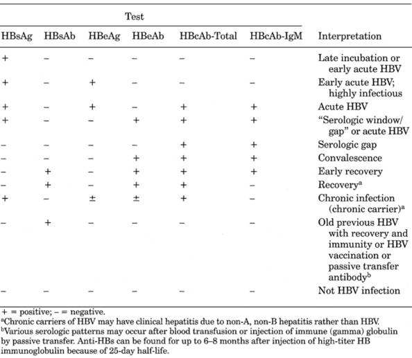
|
|
Table 8-8. Serologic Tests for Hepatitis
B Virus Infections
|
Hepatitis
B Surface Antigen (HBsAg)
Earliest indicator of HBV infection. Usually appears in
2741
days (as early as 14 days). Appears 726 days before
biochemical abnormalities. Peaks as ALT rises. Persists during the acute illness. Usually disappears 1220
wks after onset of symptoms or laboratory abnormalities in 90% of cases. Is the
most reliable serologic marker of HBV infection.
Persistence >6 mos defines carrier state. May also be
found in chronic infection. Hepatitis B vaccination does not cause a
positive HBsAg. Titers are not of clinical value. Present sensitive assays
detect <1.0 ng/mL of circulating antigen, which is the level needed to find
1015% of reactive blood donors who carry antigen but express only low levels.
Is never detected in some patients, and diagnosis is based on presence of HBc
IgM.
HBsAg
and Blood Transfusions
- Transfusion of blood
containing HBsAg causes hepatitis or appearance of HBsAg in blood in
>70% of recipients; needle stick with such blood causes hepatitis in
45% of cases. Transfusion of blood not containing HBsAg causes anicteric
hepatitis in 16% of recipients and icteric hepatitis in 2%.
- Screening out of blood
donors with HBsAg reduces posttransfusion hepatitis by 2540%.
- When HBsAg carriers are
discovered (e.g., in screening program), 6080% show some evidence of
hepatic damage.
- Persons with a positive
test for HBsAg should never be permitted to donate blood or plasma.
- HBsAg is found in
|
Chronic persistent hepatitis
|
|
|
Chronic active hepatitis
|
|
|
Cirrhosis
|
P.233
|
|
Patients undergoing multiple transfusions
|
|
|
Drug addicts
|
|
|
Blood donor population
|
<0.1%
|
|
Prevalence in United States
|
|
|
|
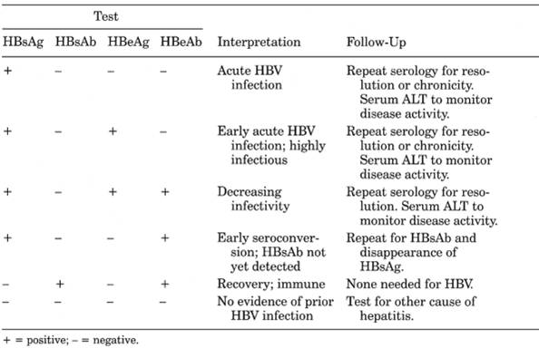
|
|
Table 8-9. Serologic Tests for Hepatitis
B Virus Infection Follow-Up
|
| |
Antibody
to HBsAg (Anti-HBsAg)
- Presence of antibody (titer ≥10 mU/mL); (without detectable HBsAg) indicates
recovery from HBV infection, absence of infectivity, and immunity from
future HBV infection; patient does not need gamma globulin administration
if exposed to infection; this blood can be transfused.
|
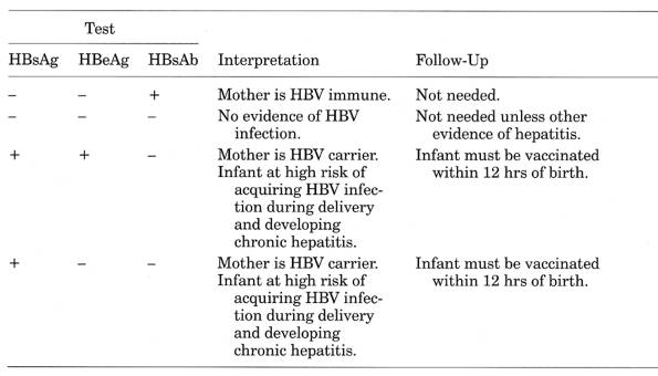
|
|
Table 8-10. Serologic Tests for Prenatal
Screening for Hepatitis B Virus
|
|
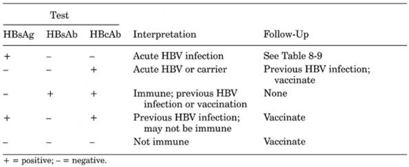
|
|
Table 8-11. Serologic Tests for Candidate
for Hepatitis B Virus Vaccination
|
- May also occur after
transfusion by passive transfer.
- Found in 80% of patients
after clinical cure. Appearance may take several weeks or months after
HBsAg has disappeared and after ALT has returned to normal, causing a
serologic gap during which time (usually 2- to 6-wk window) only
IgManti-HBsAg can identify patients who are recovering but may still be
infectious.
- Presence can be used to
show efficiency of immunization program. Appears in ~90% of healthy
adults after three-dose deltoid muscle immunization; 3050% of these lose
antibodies in 7 yrs and require boosters. Revaccination of nonresponders
produces adequate antibody in <50% after three additional doses.
- A few persons acquire
HBV infection after developing high titers of anti-HBsAg due to a mutant
HBV virus.
- In fulminant
hepatitisantibody is produced early and may coexist with low antigen
titer.
- In chronic carriersno
IgM antibody is present but antigen titers are very high.
Hepatitis
Be Antigen (HBeAg)
- Indicates highly infectious state. Appears within 1 wk after HBsAg; in acute cases
disappears before disappearance of HBsAg; is found only when HBsAg is
found. Occurs early in disease before biochemical changes and disappears
after serum ALT peak. Usually lasts 36 wks. Is a marker of active HBV
replication in liver; with few exceptions, is present only in persons with
circulating HBV DNA and is used as alternative to HBV DNA assay.
- Is useful to determine
resolution of infection. Persistence >20 wks suggests progression to
chronic carrier state and possible chronic hepatitis. Presence in
HBsAg-positive mothers indicates 90% chance that infant will acquire HBV
infection.
- Absence of HBeAg is not
indicator of benign nonprogressive disease.
- May be HBeAg negative and
HBV DNA positive in patients infected with an HBV mutant who do not
synthesize HBeAg.
Antibody
to HBe (Anti-HBe)
Appears after HBeAg disappears and remains detectable
for years. Indicates decreasing infectivity, suggests good
prognosis for resolution of acute infection. Association with anti-HBc in
absence of HBsAg and anti-HBs confirms recent acute infection (216 wks).
|
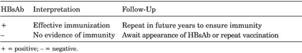
|
|
Table 8-12. Serologic Tests for Hepatitis
B Virus Vaccination FollowUp
|
P.235
Antibody
to Hepatitis B Core AntigenTotal (Anti-HBcTotal)
- Occurs early in acute infection, 410 wks after appearance of HBsAg, at same time as
clinical illness; persists for years or for lifetime. Anti-HBctotal and
HBsAg are always present and anti-HBsAg is absent in chronic HBV
infection.
- Anti-HBc
IgM is the earliest specific antibody; usually occurs 2 wks after
HBsAg. Is found in high titer for a short time during the acute disease
stage that covers the serologic window and then declines to low levels
during recovery (see Fig. 8-6); may be detectable
≤ 6 mos. May be the only serologic marker present after HBsAg and
HBeAg have subsided but before these antibodies have appeared (serologic
gap or window). Because this is the only test unique to recent infection,
it can differentiate acute from chronic HBV. It is the only serologic test
that can differentiate recent and remote infection with one specimen.
However, because some patients with chronic hepatitis B infection become
positive for anti-HBc IgM during flares, it is not an absolutely reliable
marker of acute illness. Before anti-HBc IgM disappears, anti-HBc IgG
appears and lasts indefinitely.
- Anti-HBc detects
virtually all persons who have been previously infected with HBV and can
therefore serve as surrogate test for other infectious agents (e.g., HCV).
Exclusion of anti-HBcpositive donors reduces the incidence of
posttransfusion hepatitis and possibly of other virus infections (e.g.,
AIDS) due to the frequency of dual infection. Present without other
serologic markers and with normal AST in ~2% of routine blood donors; 70%
of cases are due to recovery from subclinical HBV (and individual may be
infectious) and the rest are considered false-positives. False-positive
anti-HBc can be confirmed by immune response pattern to hepatitis B
vaccination. Anti-HBc is not protective (unlike anti-HBsAg) and therefore
cannot be used to distinguish acute from chronic infection.
- HBV
DNA (by PCR) is the most sensitive and specific assay for early
evaluation of HBV and may be detected when all other markers are negative
(e.g., in immunocompromised patients). May become negative before HBeAg
becomes negative. Measures HBV replication even when HBeAg is not
detectable. Marked decrease in patients who respond to therapy; those with
concentrations <200 ng/L are more likely to respond to therapy.
Other
Laboratory Findings
- Very high serum ALT and bilirubin are not reliable indicators of patient's
clinical course, but prolonged PT, especially >20 secs, indicates the
likely development of acute hepatic insufficiency; therefore the PT should
be performed when patient is first seen.
- Acute fulminant
hepatitis may be indicated by triad of prolonged PT, increased PMNs, and
nonpalpable liver with likely development of coma.
- Acute viral hepatitis B
completely resolves in 90% of patients within 12 wks with disappearance
of HBsAg and development of anti-HBs.
- Relapse, usually within
1 yr, has been recognized in 20% of patients by some elevation of ALT and
changes in liver biopsy.
- Chronic hepatitis
(disease for >6 mos and ALT >50% above normal): 70% of these
patients have benign chronic persistent hepatitis and 30% have chronic
active hepatitis that can progress to cirrhosis and liver failure.
- Effective treatment of
chronic HBV hepatitis causes ALT, HBeAg, and HBV DNA to become normal.
- Chronic carriers have
also been defined as those who are either HBsAg positive on two occasions
>6 mos apart or have one specimen that is HBsAg positive and anti-HBc
IgM negative but antiHBc-positive.
- 10% of adults and 90% of
children ≤ 4 yrs old become chronic carriers; 25% of these develop
cirrhosis and high risk of hepatoma. HBV carriers should be screened
periodically with serum AFP and ultrasonography or CT scan of liver for
hepatoma.
- Laboratory indicators for
favorable response to interferon:
- Pretreatment serum ALT
>100 U/L (high ALT may indicate better host immune response to HBV)
- HBV DNA <200 ng/L
(pg/mL)
- Absence of HIV
- Also duration <4 yrs
and acquisition of infection after 6 yrs of age
- Laboratory effects of
interferon treatment:
- Serum ALT may increase
to >1000 U/L.
P.236
|
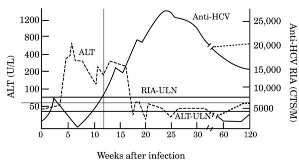
|
|
Fig. 8-7. Comparison of serum ALT and
antihepatitis C virus findings in acute hepatitis C. Chronic infection is
indicated by broken lines. (CTS/M = counts/minute; RIA = radioimmunoassay;
ULN = upper limit of normal.)
|
- 10% of patients show
sustained disappearance of HBV DNA and clearance of HBeAg.
- If serum ALT is
persistently increased despite lack of HBeAg, presence of an
HBeAg-negative mutant that may have emerged during treatment is
suggested.
- 510% of patients with
seroconversion due to therapy will have reactivation in next 10 yrs; this
is usually transitory.
- Laboratory
contraindications to interferon therapy for chronic hepatitis B:
- Liver decompensation
- Serum albumin <3.0
gm/L
- Serum bilirubin >3.0
mg/dL
- PT increased >3×
- Portal hypertension
(e.g., ascites, bleeding esophageal varices, encephalopathy)
- Hypersplenism
- WBC <2000/cu mm
- Platelet count
<70,000
- Autoimmune disease
(e.g., RA, polyarteritis nodosa)
- Major system impairment
- Other (e.g., pregnancy,
current IV drug abuse, psychiatric)
Hepatitis
C (Formerly Non-A, Non-B Hepatitis)8,
- See Fig.
8-7 and Tables 8-5 and .
- Can remain infectious for
years.
- ~85% of acute cases
become chronic with viremia.
- Of chronic carriers with
or without abnormal ALT values,
- 15% experience
resolution.
- 70% develop chronic
hepatitis (average time = 10 yrs).
- 1020% develop
cirrhosis despite normal liver function tests (average time = 20 yrs).
- ~50% die of
consequences of HCV infection.
P.237
- Fulminant hepatitis is
rare.
- Hepatocellular
carcinoma may occur in ~20% of cirrhosis patients (average time = 30 yrs)
and 15% of those with HCV infections.
- 5075% of all liver
cancers are HCV associated.
- ~40% of liver
transplantations in United
States are performed to treat chronic
hepatitis C with cirrhosis.
- Routine screening for HCV
should be performed and HCV should be ruled out in hepatitis in persons
who
- Ever injected illegal
drugs.
- Received clotting factor
concentrates produced before 1987 (7090% of severe hemophiliacs are
infected with HCV).
- Ever were on long-term
hemodialysis.
- Ever received blood from
donor who later tested positive for HCV (27% of blood donors in United States
are asymptomatic carriers).
- Ever received blood or
components or organ transplant before July 1992.
- Have persistently
abnormal serum ALT.
- Causes ≤ 25% of
sporadic cases of acute viral hepatitis in adults, 90% of cases of
posttransfusion hepatitis.
- Source of infection:
injected drug use = 42%; occupational exposure = ~5%; transfusion =
<1%; dialysis = 0.6%; household contact = 3%; heterosexual transmission
= 6% (cumulative risk may be 18%); unidentified = 42%.
- Perinatal infection at
time of birth in 5% of infants of HCV-infected mothers.
- Biochemical and
histologic evidence of abnormality occurs in 7% of sporadic cases, ≤
60% of posttransfusion cases, and ≤ 80% of immunosuppressed
patients.
- Occult HBV infection is
present in approximately one-third of patients with chronic HCV liver
disease by HBV DNA analysis of liver biopsy.10
- May be associated with mixed cryoglobulinemia with vasculitis (see Chapter 11), thyroiditis, Sjögren's syndrome,
membranoproliferative GN, and porphyria cutanea tarda, which should be
ruled out in cases of hepatitis C, and HCV infection should be ruled out
in patients with those disorders. Patients with alcoholic liver disease
have more rapidly progressive disease with higher ALT values and more
severe histologic changes.
Increased
Serum Transaminases
- Levels characteristically show unpredictable waxing and waning
pattern, returning to almost normal levels (formerly called acute relapsing
hepatitis); pattern is highly suggestive but only occurs in 25% of cases.
- May be extreme (>10×
normal).
- Patients with monophasic
ALT response usually recover completely with no biopsy evidence of
residual disease.
- ALT is usually <800 U.
ALT cannot be relied on to determine whether to perform liver biopsy in
chronic hepatitis C; biopsy is needed to define severity.
- ALT is primary marker to
monitor therapy. In chronic HCV, AST/ALT ratio >1 has specificity and
positive predictive value of 100% for cirrhosis although sensitivity is
52%. Ratio does not correlate with serum ALP, bilirubin, albumin, or PT.
- Anicteric patients with
ALT >300 U/L are at high risk for progressing to chronic hepatitis.
Liver
Biopsy
- Use
- Diagnose chronic active
hepatitis
- Assess disease
progression and indication for antiviral therapy
- No consistent correlation
between serum ALT and severity of liver pathology; significant liver
damage can occur with normal ALT.
- Exclude coexisting or
alternative (e.g., alcohol-related) diseases
P.238
Antibody to Hepatitis C Virus
(anti-HCV) (by EIA
- Use
- Screening of populations
with low and high prevalence, including blood donors
- Initial evaluation of
patients with liver disease, including those with increased serum ALT
- Positive results should
be verified by a supplemental assay (i.e., recombinant immunoblot assay
[RIBA]) showing reactivity with ≥2 viral antigens; indeterminate in
≤ 10% of cases.
- Interpretation
- Indicates past or present
infection but does not differentiate between acute, chronic, and resolved
infection.
- Sensitivity ≥97%;
only ~80% in chronic carriers. Low positive predictive value in
low-prevalence population.
- Seroconversion: average
time after exposure = 23 wks with EIA-3. Detected in 80% of patients
within 15 wks, in >90% within 5 mos, in >97% by 6 mos after exposure
or 23 mos after increase in ALT. Therefore serial assay of anti-HCV and
ALT for up to 1 yr after suspected acute hepatitis may be needed for
diagnosis. Negative EIA rules out HCV infection in low-risk group.
- Present in 7085% of
cases of chronic posttransfusion NANB hepatitis but is relatively
infrequent in acute cases. Present in 70% of IV drug abusers, 20% of
hemodialysis patients, and only 8% of homosexual men positive for HIV.
- Prevalence in normal
blood donors is 0.52.0%. In routine blood donor screening, estimates are
that 4070% of initial reactors prove not to be true positives. Surrogate
markers fail to detect one-third to one-half of blood units positive for
anti-HCV. Found in 710% of transfusion recipients. Only one-third of
anti-HCV donors had increased ALT and 54% were positive for anti-HBc.
- In one study, anti-HCV
was positive in 75% of patients with hepatocellular carcinoma, 56% of
patients with cirrhosis, and 7% of controls.
- Present in various
quality assurance and calibration sera; overall rate = 43% with much
higher rates in some proficiency samples.
- Because resolves slowly,
is considered chronic only with evidence of activity >12 mos.
- Interferences
- False-positive
- Autoimmune diseases
(≤ 80% of cases of autoimmune chronic active hepatitis).
- EIA and RIBA are also
found in polyarteritis nodosa (~10%) and SLE (~2%).
- RF.
- Hypergammaglobulinemia.
- Paraproteinemia.
- Passive antibody
transfer.
- Anti-idiotypes.
- Antisuperoxide
dismutase (a human enzyme used in the cloning process).
- Repeated freezing and
thawing or prolonged storage of blood specimens.
- False-negative
- Early acute infection
- Immunosuppression
- Immunoincompetence
- Repeated freezing and
thawing or prolonged storage of blood specimens
RIBA
- Positive EIA should be evaluated
with RIBA-2; negative RIBA indicates false-positive EIA.
- Positive RIBA indicates
past or previous exposure.
- Confirms positive EIA in
>50% of cases; in high-risk population RIBA confirms diagnosis in
>88% of cases.
- Increasingly replaced by
HCV RNA.
HCV
RNA Assay
(By reverse transcriptase PCR [RT-PCR])
P.239
Qualitative
tests
- Use
- Diagnose acute HCV
infection before seroconversion; can detect virus as early as 12 wks
after exposure.
- Detection may be
intermittent; one negative RT-PCR is not conclusive.
- Monitor patients on
antiviral therapy
- Evaluate indeterminate
RIBA results
- False-positive and
false-negative results may occur.
Quantitative
Tests
- (RT-PCR and branched DNA;
not presently approved by U.S. Food and Drug Administration.)
- Quantitative tests from
different manufacturers do not yield identical results.
- Determines concentration
of HCV RNA.
- Large spontaneous
fluctuations in RNA level; therefore should measure two times or more to
evaluate changes due to therapy.
- RT-PCR yields positive
results for 7585% of persons positive for anti-HCV and >95% of persons
with acute or chronic HCV hepatitis.
- Use
- May be used to assess
likelihood of response to antiviral therapy. Patients with pretreatment
level <2 million copies/mL (by PCR or quantitative branched DNA) are
most likely to respond to interferon therapy. Positive test after 12 wks
of interferon therapy predicts failed response; negative test has ~30%
predictive value for sustained response.
- Less sensitive than
qualitative test RT-PCR.
- Earliest marker for
diagnosis of fulminant hepatitis C. Negative test in patient with
fulminant hepatitis rules out HCV infection.
- Confirm persistent HCV
infection after liver transplantation when anti-HCV is positive and serum
ALT is normal.
- Diagnose chronic
hepatitis patients with
- Negative anti-HCV
- False-positive serologic
tests due to autoantibodies
- Not used to exclude
diagnosis of HCV infection.
- Not used to determine
treatment end point.
HCV
Genotyping
- Presently a research tool
with no clinical utility. At least six genotypes and >90 subtypes. A
correlation may exist between genotype and disease. Mixed infections often
occur.
|
HCV Genotype
|
Occurrence (%)
|
|
1a
|
|
Higher rate of chronic hepatitis;
poorer response to interferon therapy and higher likelihood of relapse
|
|
1b
|
|
More severe liver disease; higher risk
of hepatocellular carcinoma
|
|
- Other genotypes have
various geographic distributions.
- Antiviral therapy is
recommended for patients with greatest risk of progression to cirrhosis
- Positive anti-HCV with
- Persistently increased
ALT
- Detectable HCV RNA
- Liver biopsy showing at
least moderate inflammation and necrosis or fibrosis
- Indicators of response to
antiviral therapy
- ~50% show normal serum
ALT.
- 33% lose detectable HCV
RNA in serum; loss associated with remission. Presence after sustained
response to interferon indicates late relapse.
- 50% relapse after
therapy ends.
- Decreased interferon
response occurs in <15% of patients; indicated by
- Higher serum HCV RNA
titers
- HCV genotype 1
P.240
- Laboratory
contraindications to interferon therapy
- Persistently increased
serum ALT
- Cytopenias
- Hyperthyroidism
- Renal transplantation
- Evidence of autoimmune
disease
- No tests are routinely
available for other HCV viruses.
Hepatitis
D (Delta)
- See Tables
8-5, and .
- Hepatitis D is due to a
transmissible virus that depends on HBV for expression and replication. It
may be found for 714 days in the serum during acute infection. Delta
agent can be an important cause of acute or chronic hepatitis. The course
depends on the presence of HBV infection. HDV hepatitis is often severe
with relatively high mortality in acute disease and frequent development
of cirrhosis in chronic disease. Chronic HDV infection is more severe and
has higher mortality than other types of viral hepatitis. Prevalence in
United States is 110% of HBsAg carriers, principally in high-risk groups
of IV drug abusers and multiply transfused patients but uncommon in other
groups at risk for HBV infection (e.g., health care workers, male
homosexuals).
Serologic
Tests for HDV
- See Tables
8-5, , and .
- Serum HDAg and HDV-RNA
appear during incubation period after HBsAg and before rise in AST, which
often shows a biphasic elevation. HBsAg and HDAg are transient; HDAg
resolves with clearance of HBsAg. Anti-HDV appears soon after clinical
symptoms but titer is often low and short-lived. Anti-HDVtotal test is
commercially available; HDAg and anti-HDV-IgM testing is available only in
research laboratories.
- Coinfection means
simultaneous acute HBV and acute HDV infection; usually causes acute
limited illness with additive liver damage due to each virus, followed by
recovery. Usually is self-limited; <5% of cases become chronic. ~3%
have fulminant course.
- Superinfection means
acute HDV infection in a chronic HBV carrier. Mortality = 220%; >80%
develop chronic hepatitis. Serum anti-HDV appears and rises to high
sustained titers indicating continuing replication of HDV; intrahepatic
HDAg is present. HDV-RNA persists in low titers.
- Diagnosis of HDV hepatitis is made by presence of anti-HDV in patient with
HBsAg-positive hepatitis. Anti-HDV assay should not be performed unless
diagnosis of HBV is confirmed.
- Acute coinfection is distinguished from
superinfection by presence of serum HBsAg and anti-HBc-IgM, which indicate acute
HBV.
- Chronic HDV infection occurs in ≤ 80% of acute cases; shows presence of HBsAg and
high titer of anti-HDV (RIA titer >1:100 suggests chronic HDV
hepatitis) and absence of anti-HBc-IgM in serum. Confirm by liver biopsy
showing HDAg by immunofluorescence or immunoperoxidase.
- Serum anti-HDV-IgM documents acute HDV infection; low levels remain in persistent
infection.
- Western blot can
demonstrate serum HDV-Ag when RIA is negative. Persistence correlates
with development of chronic HDV hepatitis and viral antigen in liver
biopsy.
- DNA probe for HDV-RNA in
serum to monitor HDV replication.
- Serum anti-HDV may be
sought in patients with HBsAg-positive chronic or acute hepatitis in
high-risk group or with severe disease or with biphasic acute hepatitis or
acute onset in chronic hepatitis.
Hepatitis
E
- See Table
8-5 and Table 8-6.
- Recent travel to certain
areas (e.g., Mexico, India, Africa, Burma,
Russia)
- Serologic markers for hepatitis A, B, and C and other causes of acute hepatitis (e.g.,
EMB, CMV) are absent.
- Antibody to hepatitis E can be detected by fluorescent antibody blocking assay and by
Western blot; not commercially available.
P.241
|
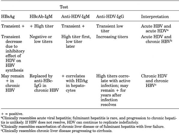
|
|
Table 8-13. Serologic Diagnosis of Hepatitis
B Virus (HBV) and Hepatitis D Virus (HDV)
|
Hepatitis
G
- (Due
to single-stranded RNA virus of Flaviviridae family. HGV RNA found in
~12% of American blood donors; higher in multiply transfused persons,
those with hepatitis B or C, drug addicts. Benign course; more studies
needed to determine if causes acute or chronic hepatitis.)
- Infection tends to
persist for many years.
- Serum ALT is persistently
normal; increase is due to concomitant HCV infection.
- Serologic assays under
development.
- Detected by RT-PCR.
- Of hemodialysis patients
- ≤ 5% are HGV
positive.
- ~25% have anti-HCV and
~15% are PCR positive for HCV.
- ~5% are HBsAg positive.
- >50% had anti-HBs or
anti-HBc (representing resolved HBV infection).
Hepatitis,
Neonatal
Infectious
Causes
- Adenovirus
- Coxsackievirus B
- CMV
- HAV and HBV
- HSV
- Listeria
- Rubella
- Syphilis
- Toxoplasmosis
- Varicella
- Unknown agent
P.242
Metabolic
Causes
- Alpha -antitrypsin
deficiencycauses 2035% of cases of neonatal liver disease.
- Cystic fibrosis rarely
presents as prolonged neonatal jaundice.
- Dubin-Johnson syndrome
- Fructosemia
- Galactosemia
- Gaucher's disease
- Glycogen storage disease
type IV
- Histiocytosis X
- Hypothyroidism
- Hypopituitarism
- Leprechaunism
- Niemann-Pick disease
- Tyrosinemia
- Zellweger syndrome
- Jaundice in infants
receiving parenteral alimentationmany are premature and have various
complications (e.g., RDS, septicemia, acidosis, congenital heart disease).
- Increased AST, ALT, ALP
- Serum proteins normal
- Increased serum bile
acids
- Increased serum ammonia
- Abnormal plasma amino
acid patterns (increased threonine, serine, methionine)
Associated
with Hemolytic Disease of Newborn
- Occurred in 10% of cases
(inspissated bile syndrome) before modern prevention of Rh disease.
- Cord blood direct
bilirubin ≥2 mg/dL indicates that syndrome will develop.
- Jaundice may persist for
34 wks.
- Most cases have required
exchange transfusion.
Clinical
and Laboratory Findings
- Jaundice at birth, or
days or weeks later. Both direct and indirect bilirubin levels are
increased in variable proportions.
- Mild hemolytic anemia is
usual.
- Increased AST, ALT, etc.;
may be marked and usually greater than in biliary atresia, but increases
are not useful for differentiating the two conditions.
- Laboratory findings as in
acute viral hepatitis.
- Liver biopsy to
differentiate from biliary atresia and to avoid unnecessary surgery is
useful in ~65% of patients but it may be misleading.
- I-rose bengal excretion test indicates
complete biliary obstruction if <10% of the radioactivity is excreted
in stools during 4872 hrs and incomplete obstruction if >10%. Complete
obstruction is found in all infants with biliary atresia and in ~20% with
neonatal hepatitis and severe cholestasis. Administration of phenobarbital
and cholestyramine increases the I-rose bengal
excretion in neonatal hepatitis but not in extrahepatic atresia. Some
authors have suggested a repeat test in 34 wks before exploratory surgery
if rose bengal test indicates complete
obstruction.
- Laboratory tests for
various causal agents.
- Laboratory findings of
chronic liver disease, which develops in 3050% of these infants.
- Whenever mother has
hepatitis during pregnancy or is HBsAg positive, test cord blood and baby's blood every 6 mos. If baby develops HBsAg or
anti-HBs, measure liver chemistries at periodic intervals. Infants who
acquire hepatitis in utero or at time of birth may develop clinical acute
hepatitis with abnormal liver chemistries, benign course, or development
of HBsAb. Infants who are asymptomatic but develop HBsAg often become
chronic carriers with biochemical and liver biopsy evidence of chronic
hepatitis and increased likelihood of hepatoma. (See Serologic
Tests for HBV.
Hepatocellular
Carcinoma (Hepatoma)
- Serum AFP present in 50% of white and 7590% of nonwhite patients; may be present
for up to 18 mos before symptoms; is sensitive indicator of recurrence in
treated patients but a normal postoperative level does not ensure absence
of metastases. Levels >500 ng/dL in adults strongly suggest primary
carcinoma of liver.
P.243
- Serum GGT hepatoma-specific band (HSBs I′, II, II′) by electrophoresis activity
>5.5 U/L has sensitivity of 85%, specificity of 97%, accuracy of 92%.
Does not correlate with AFP or tumor size.14
- Laboratory findings
associated with underlying disease (>60% occur with preexisting
cirrhosis).
- Hemochromatosis (≤
20% of patients die of hepatoma).
- HBV, HCV.
- More frequent in
postnecrotic than in alcoholic cirrhosis.
- Cirrhosis associated
with alpha -antitrypsin deficiency
and other inborn errors of metabolism, e.g., tyrosinemia.
- Clonorchis
sinensis infection is associated with cholangiosarcoma.
- Relative absence of
hepatoma associated with cirrhosis of Wilson's disease.
- Sudden progressive
worsening of laboratory findings of underlying disease (e.g., increased
serum ALP, LD, AST, bilirubin).
- Hemoperitoneumascites in ~50% of patients but tumor cells found irregularly.
- Laboratory findings due
to obstruction of hepatic veins (Budd-Chiari syndrome), portal veins, or
inferior vena cava may occur.
- Occasional marked
hypoglycemia unresponsive to epinephrine injection; occasional
hypercalcemia.
- ESR and WBC sometimes
increased.
- Anemia is common;
polycythemia occurs occasionally.
- Serologic markers of HBV
frequently present.
- CEA in bile is increased
in patients with cholangiocarcinoma and intrahepatic stones but not in
patients with benign stricture, choledochal cysts, sclerosing
cholangitis. Increases with progression of disease and declines with tumor
resection. Does not correlate with serum bilirubin or ALP.
- Serum CEA is usually
normal.
Hyperbilirubinemia,
Neonatal
Due To
Unconjugated
- Increased destruction of
RBCs
- Isoimmunization (e.g.,
incompatibility of Rh, ABO, other blood groups)
- Biochemical defects of
RBCs (e.g., G-6-PD deficiency, pyruvate deficiency, hexokinase
deficiency, congenital erythropoietic porphyria, alpha and gamma
thalassemias)
- Structural defects of
RBCs (e.g., hereditary spherocytosis, hereditary elliptocytosis,
infantile pyknocytosis)
- Infection
- Viral (e.g., rubella)
- Bacterial (e.g.,
syphilis)
- Protozoal (e.g.,
toxoplasmosis)
- Extravascular blood
(e.g., subdural hematoma, ecchymoses, hemangiomas)
- Erythrocytosis (e.g.,
maternal-to-fetal or twin-to-twin transfusion, delayed clamping of
umbilical cord)
- Increased enterohepatic
circulation
- Any cause of delayed
bowel motility
- Pyloric stenosisunconjugated
hyperbilirubinemia >12 mg/dL develops in 1025% of infants, usually
during second or third week, at which time vomiting begins; jaundice is
due to decreased hepatic glucuronyl transferase activity of unknown
mechanism.
- Duodenal and jejunal
obstruction may also be associated with exaggerated unconjugated
hyperbilirubinemia; level becomes normal 23 days after surgical relief.
- In Hirschsprung's
disease, unconjugated hyperbilirubinemia is usually more
mild.
- Meconium ileus,
meconium plug syndrome.
- Hypoperistalsis (e.g.,
induced by drugs, fasting)
P.244
- Endocrine and metabolic
- Neonatal
hypothyroidismassociated with prolonged and exaggerated unconjugated
hyperbilirubinemia in 10% of cases and is promptly alleviated by thyroid
hormone therapy.
- Always
rule out congenital hypothyroidism in cases of unexplained persistent or
excessive unconjugated hyperbilirubinemia; may be the only manifestation
of hypothyroidism
- Infants of diabetic
mothersassociated with higher incidence of prolonged and exaggerated
unconjugated hyperbilirubinemia of unknown mechanism; not related to
severity or duration of diabetes.
- Drugs and hormones
(e.g., breast-milk jaundice, Lucey-Driscoll syndrome, novobiocin).
- Galactosemia.
- Tyrosinosis.
- Hypermethionemia.
- Heart failure.
- Hereditary
glucuronyl-transferase deficiency.
- Gilbert's syndrome.
- Interference of albumin
binding of bilirubin
- Drugs (e.g., aspirin,
sulfonamides)
- Severe acidosis
- Hematin
- Free fatty acids (e.g.,
periods of stress, inadequate caloric intake)
- Prematurity (serum albumin
may be 12 gm/dL less than in full-term infants)
- Neonatal physiologic
hyperbilirubinemia
Conjugated
- Premature
infants with these conditions have more severe hyperbilirubinemia than
full-term infants
- Biliary
obstructionusually due to extrahepatic biliary atresia but may be due to
choledochal cyst, obstructive inspissated bile plugs, or bile ascites
- Neonatal hepatitis
- Sepsis, especially E. coli pyelonephritis (moderate azotemia, acidosis,
increased serum bilirubin, slight hemolysis, normal or slightly increased
AST)
- Hereditary diseases
(e.g., galactosemia, alpha -antitrypsin deficiency,
cystic fibrosis, hereditary fructose intolerance, tyrosinemia, infantile
Gaucher's disease, familial intrahepatic cholestasis [Byler's disease])
- In the course of hemolytic
disease of the newborndue to liver damage of unknown cause.
Differential Diagnosis
- Unconjugated
hyperbilirubinemia is serum level >1.5 mg/dL. Conjugated
hyperbilirubinemia is direct-reacting serum level >1.5 mg/dL when this
fraction is >10% of total serum bilirubin (because in newborn with
marked elevation of unconjugated bilirubin level, ≤ 10% of the
unconjugated bilirubin will act as direct reacting in the van den Bergh
reaction).
- Mixed hyperbilirubinemia
shows conjugated bilirubin as 2070% of total and usually represents
disorder of hepatic cell excretion or bile transport.
- Visible icterus before 36
hrs of age indicates hemolytic disorder.
- Diagnostic studies should
be performed whenever serum bilirubin is >12 mg/dL.
- After hemolytic disease
and hepatitis, the most frequent cause of hyperbilirubinemia is
enterohepatic circulation of bilirubin.
- Visible icterus
persisting after seventh day is usually due to impaired hepatic excretion,
most commonly due to breast-milk feeding or congenital hypothyroidism.
- Increase in direct
bilirubin usually indicates infection or inflammation of liver, but can
also be seen in galactosemia and tyrosinosis.
Hyperbilirubinemia,
Neonatal Nonphysiologic
- See Fig.
8-8.
- Cause should be sought
for underlying pathologic jaundice if:
- Total serum bilirubin is
>7 mg/dL during first 24 hrs or increases by >5 mg/dL/day or
visible jaundice.
- Peak total serum
bilirubin is >12.5 mg/dL in white or black full-term infants or >15
mg/dL in Hispanic or premature infants.
P.245
- Direct serum bilirubin
is >1.5 mg/dL.
- Clinical jaundice lasts
longer than 7 days in full-term or 14 days in premature infants or occurs
before age 36 hrs or with dark urine (containing bile).
- Initial tests in
unconjugated hyperbilirubinemia:
- Serial determinations of
total and direct bilirubin
- CBC including RBC
morphology, platelet count, normoblast and reticulocyte counts
- Blood type, mother and
infant
- Direct Coombs' test
- Maternal blood for
antibodies and hemolysins
- Blood cultures
- Urine microscopy and
culture
- Serologic tests for
infection
- Serum thyroxine (T ) and thyroid-stimulating hormone (TSH)
- Urine for nonglucose
reducing substances
Hyperbilirubinemia,
Neonatal Physiologic
- (Transient
unconjugated hyperbilirubinemia [physiologic jaundice] that occurs in
almost all newborns)
- In normal full-term
neonate, average maximum serum bilirubin is 6 mg/dL (up to 12 mg/dL is in
physiologic range), which occurs during the second to fourth days and then
rapidly falls to ~2.0 mg/dL by fifth day (phase I physiologic jaundice).
Declines slowly to <1.0 mg/dL during fifth to tenth days, but may take
1 mo to fall to <2 mg/dL (phase II physiologic jaundice). Phase I due
to deficiency of hepatic bilirubin glucuronyl transferase activity and
sixfold increase in bilirubin load presented to liver. In Asian and American
Indian newborns, the average maximum serum levels are approximately double
(1014 mg/dL) the levels in non-Asians, and kernicterus is more frequent.
Serum bilirubin >5 mg/dL during first 24 hrs of life is indication for
further workup because of risk of kernicterus.
- In
older children (and adults) icterus is apparent clinically when serum
bilirubin is >2 mg/dL, but in newborns clinical icterus is not apparent
until serum bilirubin is >57 mg/dL; therefore only half of full-term
newborns show clinical jaundice during first 3 days of life
- In premature
infantsaverage maximum serum bilirubin is 1012 mg/dL and occurs during
the fifth to seventh days. Serum bilirubin may not fall to normal until
30th day. Further workup is indicated in all premature infants with
clinical jaundice because of risk of kernicterus in some low-birth-weight
infants with serum levels of 1012 mg/dL.
- In postmature infants and
half of small-for-date infantsserum bilirubin is <2.5 mg/dL and
physiologic jaundice is not seen. When mothers have received phenobarbital
or used heroin, physiologic jaundice is also less severe.
- When a
pregnant woman has unconjugated hyperbilirubinemia, similar levels occur
in cord blood, but when the mother has conjugated hyperbilirubinemia
(e.g., hepatitis), similar levels are not present
in cord blood
Hyperbilirubinemia;
Neonatal, Transient Familial (Lucey-Driscoll Syndrome)
- Syndrome is due to
progestational steroid in mother's serum only during last trimester of
pregnancy, which inhibits glucuronyl transferase activity; disappears ~2
wks postpartum.
- Newborn infants have
severe nonhemolytic unconjugated hyperbilirubinemia, usually up to 20
mg/dL during first 48 hrs, and a high risk of kernicterus.
Hyperbilirubinemia
In Older Children
Due To
- All
cases of conjugated hyperbilirubinemia also show some increase of
unconjugated serum bilirubin
Unconjugated
- Gilbert's disease
- Administration of drugs
(e.g., novobiocin)
- Occasionally other
conditions (e.g., thyrotoxicosis, after portacaval shunt in cirrhosis)
P.246
|
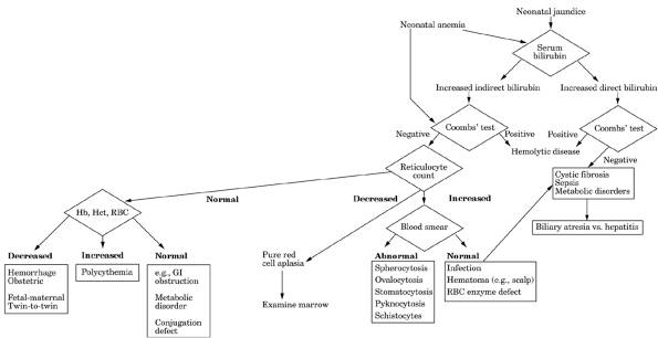
|
|
Fig. 8-8. Algorithm for workup of
neonatal jaundice and anemia.
|
P.247
Conjugated
- Dubin-Johnson syndrome
- Rotor's syndrome
- Acute viral hepatitis
causes most cases in children.
- Cholestasis due to
chemicals and drugs or associated with other diseases (e.g., Hodgkin's
disease, sickle cell disease)
Jaundice
(Cholestatic and Hepatocellular), Comparison
|
|
Hepatocellular
|
Cholestasis
|
Infiltration
|
|
Disease example
|
Acute viral hepatitis
|
Common duct stone
|
Metastatic tumor
|
|
Serum bilirubin (mg/dL)
|
|
|
Usually <4, often normal
|
|
AST, ALT (U/mL)
|
Markedly increased, often 5001000
|
May be slightly increased, <200
|
May be slightly increased, <100
|
|
Serum ALP
|
12× normal
|
35× normal
|
24× normal
|
|
PT
|
Increased in severe disease
|
Increased in chronic cases
|
Normal
|
|
Response to
parenteral
vitamin K
|
No
|
Yes
|
|
|
*Serum bilirubin >10 mg/dL is rarely
seen with common duct stone and usually indicates carcinoma.
|
|
Increased serum ALP <3× normal in 15% of patients
with extrahepatic biliary obstruction, especially if obstruction is
incomplete or due to benign conditions.
|
|
- Occasionally AST and LD
are markedly increased in biliary obstruction or liver cancer.
Metabolism,
Inborn Errors, Causing Liver Disorder
Inborn
Errors of Carbohydrate Metabolism
- Glycogen storage diseases
- Galactosemia
- Fructose intolerance
Inborn
Errors of Protein Metabolism
- Tyrosinemia
- Urea cycle enzyme defects
Inborn
Errors of Lipid Metabolism
- Gaucher's disease
- Gangliosidosis
- Cholesterol ester storage
disease
- Niemann-Pick disease
- Lipodystrophy
- Wolman's disease
Others
- Mucopolysaccharidoses
- Wilson's disease
- Hepatic porphyria
- Alpha -antitrypsin
deficiency
- Byler's disease
- Cystic fibrosis
Pylephlebitis,
Septic
- Increased WBCs and PMNs
in >90% of patients; usually >20,000/cu mm
- Anemia of varying
severity
- Moderate increase in
serum bilirubin in ~33% of patients
P.248
- Other liver function
tests positive in ~25% of patients
- Needle biopsy of liver
not helpful; contraindicated
- Blood culture sometimes
positive
- Laboratory findings due
to preceding disease (e.g., acute appendicitis, diverticulitis, ulcerative
colitis)
- Laboratory findings due
to complications (e.g., portal vein occlusion)
Rotor's
Syndrome
- (Autosomal
recessive, familial, asymptomatic, benign defective uptake and storage of
conjugated bilirubin and possibly in transfer of bilirubin from liver to
bile or in intrahepatic binding; usually detected in adolescents or
adults. Jaundice may be produced or accentuated by pregnancy, birth
control pills, alcohol, infection, surgery.)
- See Table
8-4.
- Mild chronic fluctuating nonhemolytic conjugated
hyperbilirubinemia (usually <10 mg/dL).
- BSP excretion is
impaired.
- Other liver function test are normal.
- Liver biopsy is normal;
no pigment is present.
- Urine coproporphyrins are markedly increased especially
coproporphyrin I (increased more than III).
Space-Occupying
Lesions
Due To
- Neoplasms (e.g., primary
hepatocellular carcinoma, metastasis)
- Cysts
- Echinococcus
- ≤ 40% of patients
with autosomal dominant polycystic renal disease
- Abscesses (amebic,
pyogenic)
- Granulomas
- Sarcoidosis
- Infections (e.g., TB,
cat-scratch bacillus, Q fever, Lyme disease, secondary syphilis)
- Drugs (e.g., gold,
quinidine, diltiazem, hydralazine, methimazole, tocainide)
- Increased serum ALP is the most useful index of partial
obstruction of the biliary tree in which serum bilirubin is usually normal
and urine bilirubin is increased.
- Increased in 80% of
patients with metastatic carcinoma.
- Increased in 50% of
patients with TB.
- Increased in 40% of patients
with sarcoidosis.
- Increased frequently in
patients with amyloidosis.
- Increase in serum LAP
parallels that in ALP but is not affected by bone disease.
- Whenever the ALP is
increased, a simultaneous increase of 5′-NT establishes biliary
disease as the cause of the elevated ALP.
- AST is increased in 50%
of patients (≤ 300 U).
- ALT is increased less
frequently (≤ 150 U).
- Detection of metastases by panel of blood tests (ALP, LD,
transaminase, bilirubin) has sensitivity of 85%. ALP or GGT alone has
sensitivity of 2533% and specificity of ≤ 75%. Serum LD is often
increased in cancer even without liver metastases.
- Radioactive scanning of
the liver has 65% sensitivity.
- Blind needle biopsy of the liver is positive in 6575% of patients.
- Laboratory findings due to primary disease (e.g., increased serum
CEA in colon carcinoma, carcinoid syndrome, pyogenic liver abscess)
Transplantation
Of Liver
Indications
- Liver Failure
Due To
- Arterial thrombosis
- Autoimmune liver disease
- Biliary atresia (infants)
- Budd-Chiari syndrome
- Cirrhosis
P.249
- Alcoholic
- Postnecrotic
- Primary biliary
- Secondary biliary
- Hepatitis
- Inborn errors of
metabolism
- Alpha -antitrypsin deficiency
- Protein C deficiency
- Crigler-Najjar syndrome
type I
- Cystic fibrosis
- Erythropoietic
protoporphyria
- Glycogen storage
diseases type I and IV
- Hemophilias A and B
- Homozygous type II
hyperlipoproteinemia
- Hyperoxaluria type I
- Niemann-Pick disease
- Tyrosinemia
- Urea cycle enzyme
deficiencies
- Wilson's disease
- Laboratory indications,
e.g.,
- Portal hypertension with
intractable ascites
- Hypersplenism and/or
bleeding esophageal varices
- Poor synthesis function
(e.g., decreased albumin, fibrinogen, prolonged PT)
- Progressive hyperbilirubinemia
- Liver trauma
- Polycystic liver disease
- Rejection of liver
transplant (causes 20% of retransplants)
- Reye's syndrome
- Sclerosing cholangitis
- Unresectable liver
neoplasms confined to liver
- Venoocclusive disease
Contraindications
- Extrahepatic neoplasms
- Positive serology for
HBsAg, HBcAb, HIV
- Sepsis other than of
hepatobiliary system
- Stage 4 hepatic coma
- Unrelated failure of
other organ systems
Postoperative
Complications
|
Early
|
Reported Incidence
|
|
Primary nonfunction due to graft
ischemia
|
|
|
Portal vein thrombosis
|
|
|
Hepatic artery thrombosis
|
|
|
Hyperacute rejection
|
|
|
Early acute rejection
|
|
|
Immunosuppressant therapy toxicity
|
|
|
Hepatorenal syndrome
|
|
|
Hepatopulmonary syndrome
|
|
|
Infection/sepsis
|
|
|
- Later
- Acute and chronic
rejection
- Side effects of
immunosuppressant therapy
- Biliary stenosis
- Recurrence of disease
(especially hepatitis B, hepatitis C, EBV-associated lymphoproliferative
disorders)
- Vanishing bile duct
syndrome
Rejection
- Most episodes occur
within first 3 mos; patients are usually asymptomatic.
P.250
- Electrolytes must be
monitored rapidly to treat cardiac arrest due to sudden release of large
amounts of potassium from perfused liver and to monitor IV fluid
replacement. Ionized calcium is lost due to chelation by citrate in
transfused blood; left ventricular dysfunction may occur when serum level
is <1.2 mEq/L. Serum sodium is monitored to avoid postoperative
neurologic complications due to rapid increase during transplant and
postoperative periods (e.g., central pontine myelinolysis). Normalization
of serum HCO
and anion gap signifies early function of liver transplant and of kidneys.
- Serum
GGT is the most sensitive marker for rejection; rises early during
rejection before serum ALP and bilirubin. Is more specific than other
markers because other complications (e.g., CMV infection) cause relatively
low levels compared with AST and ALT.
- Serum ALP lags behind serum GGT and bilirubin indicators of
rejection.
- In uncomplicated cases,
serum ALP and GGT remain within reference range.
- AST and ALT rise after reperfusion of the allograft; increase to
>45× upper limit of reference range even in uncomplicated cases.
Persistent or late increases may be due to rejection or to other causes
such as viral infections (e.g., CMV, HSV, adenovirus),
occlusion of hepatic artery, liver abscess.
- Serum total and direct bilirubin are monitored with enzymes and are useful to help
differentiate between biliary obstruction (suggesting rejection) and
hepato-cellular disease. Increase may be early sign of rejection but less
useful than enzymes.
- Monitoring of serum
cyclosporine is important because it is metabolized in the liver and
proportion of cyclosporine and its metabolites may be altered when
postoperative liver function is not maintained.
- PT and aPTT monitor
synthesis of coagulation factors; specific factor measurements are not
needed.
- Cultures from appropriate
sites are performed for evidence of infection.
- Liver biopsy is gold standard for diagnosis
- Distinguish causes of
rejection that have no specific biochemical pattern (e.g., acute
rejection, chronic rejection, opportunistic viral infection, recurrence
of HBV infection, CMV, changes in hepatic blood perfusion, unrecognized
disease in donor liver).
- Differentiate from
cholangitis, hepatitis, ischemic injury, which may mimic rejection.
- Substantial number of false-positives occur.
- Laboratory findings due
to immunosuppression therapy
- Nephrotoxicity
- Liver toxicity (e.g.,
serum cyclosporine concentration >1200 ng/dL)
- Infection (e.g.,
bacterial, fungal, HBV, CMV, HSV, EBV)
- Cancer (e.g.,
non-Hodgkin's lymphoma, Kaposi's sarcoma, carcinomas of cervix, perineum,
lip)
- Complications of
hypertension
- In rare cases, genetic
defects (e.g., factor XI deficiency) can be transmitted to the recipient
and cause postoperative complications.
Trauma
- Serum LD is frequently
increased (>1400 U) 812 hrs after major injury. Shock due to any
injury may also increase LD.
- Other serum enzymes and
liver function tests are not generally helpful.
- Findings of abdominal
paracentesis
- Bloody fluid (in ~75% of
patients) confirming traumatic hemoperitoneum and indicating exploratory
laparotomy.
- Nonbloody fluid
(especially if injury occurred >24 hrs earlier).
- Microscopicsome red
and white blood cells.
- Determine amylase,
protein, pH, presence of bile.
Wilson's Disease
- (Autosomal
recessive defect impairs copper excretion by liver, causing copper
accumulation in liver.)
- Heterozygous
gene for Wilson's disease occurs in 1 of 200 in the general population; 10%
of these have decreased serum ceruloplasmin; liver copper is not increased
(<250
P.251
µg/gm of dry liver). Serum copper and ceruloplasmin
and urine copper levels are inadequate to detect heterozygous state.
- Homozygous
gene (clinical Wilson's disease) occurs in 1 of 200,000 in the general
population. Screening with DNA probes may become useful to detect
homozygous infants.
Serum
Ceruloplasmin
- Decreased
(<20 mg/dl) In
- Wilson's disease. (It is normal in 25% of patients with overt Wilson's disease.)
May not be decreased in Wilson's disease with acute or fulminant liver
involvement (ceruloplasmin is an acute-phase reactant).
- Healthy infants
(therefore cannot use test for Wilson's disease in first year of life)
- 1020% of persons
heterozygous for Wilson's disease
- Renal protein loss (e.g.,
nephrosis)
- Malabsorption (e.g.,
sprue)
- Malnutrition
- Inherited ceruloplasmin
deficiency (rare)
- Serum
ceruloplasmin (<20 mg/dL) with increased hepatic copper (>250 µg/gm)
occurs only in Wilson's disease or normal infants aged <6 mos.
- Increased
In
- Pregnancy
- Use of estrogen or birth
control pills
- Thyrotoxicosis
- Cirrhosis
- Cancer
- Acute inflammatory
reactions (e.g., infection, RA)
- (May cause green color of
plasma.)
- Total serum copper is
decreased and generally parallels serum ceruloplasmin. Not a good
indicator because changes during course of disease.
- Free
(nonceruloplasmin) copper in serum is increased and causes excess
copper deposition in tissues and excretion in urine. Calculated from
difference between total serum copper and ceruloplasmin-bound copper. Free
copper (µg/dL) = total serum copper (µg/dL) ceruloplasmin (mg/dL) × 3.
Is virtually 100% sensitive and specific.
- Urinary copper is
increased (>100 µg/24 hrs; normal <50 µg/24 hrs); may be normal in
presymptomatic patients and increased in other types of cirrhosis.
- Liver biopsy shows high copper concentration (>250 µg/gm of dry
liver; normal = 2045) and should be used to confirm the diagnosis.
(Special copper-free needle should be used.) Copper concentrations may
vary between nodules; thus extensive sampling may be necessary to confirm
diagnosis. May also be elevated in cholestatic syndromes (e.g., primary
biliary cirrhosis, primary sclerosing cholangitis, extrahepatic biliary
cirrhosis, Indian childhood cirrhosis), which are easily differentiated
from Wilson's disease by increased serum ceruloplasmin.
- Histochemical staining of paraffin-embedded liver specimens for
copper and copper-associated protein is diagnostic in appropriate clinical
context but may be negative in Wilson's disease and present in other
hepatic disorders.
- Liver biopsy may show no
abnormalities, moderate to marked fatty changes with or without fibrosis,
or active or inactive cirrhosis.
- Findings of liver function tests may not be abnormal, depending on
the type and severity of disease. In patients presenting
with acute fulminant hepatitis, Wilson's disease is suggested if a
disproportionately low serum ALP and relatively mild increase in AST and
ALT are seen. Should also be ruled out in any patient <30 yrs with
hepatitis (with negative serology for viral hepatitis), Coombs'-negative
hemolysis, or neurologic symptoms to allow early diagnosis and treatment
of Wilson's disease
- Radiocopper loading test: Cu is administered IV or
by mouth and serum concentration is plotted against time. Serum Cu disappears within 46 hrs and then reappears in persons without
Wilson's disease; secondary reappearance is absent in Wilson's disease
because incorporation of Cu into ceruloplasmin is
decreased. Useful test in patients with normal ceruloplasmin levels or
increased hepatic copper due to other forms of liver disease, or
heterozygous carriers of Wilson's disease gene, or when liver biopsy is
contraindicated.
- Aminoaciduria (especially
cystine and threonine), glucosuria, hyperphosphaturia, hypercalciuria,
uricosuria, and decreased serum uric acid and phosphorus may
P.252
occur due to renal proximal tubular dysfunction;
distal renal tubular acidosis is less common.
- Coombs'-negative
nonspherocytic hemolytic anemia may occur.
- Other tests that have
been used in diagnosis of heterozygotes may not be available locally:
- D-penicillamine
administration induces increased urinary copper excretion.
- Excretion of radioactive
copper.
- Conversion of ionic
radioactive copper to radioactive ceruloplasmin.
- Copper content of
cultured fibroblasts.
- DNA markers have been
used for detection of homozygous and heterozygous patients.
- Laboratory findings due
to complications
- Cirrhosis and sequelae
(e.g., ascites, esophageal varices, liver failure).
- Hypersplenism (e.g.,
anemia, leukopenia, thrombocytopenia).
- Acute liver failure
characterized by very high serum bilirubin (often >30 mg/dL) and
decreased ALP; ALP/bilirubin ratio <2.0 is said to distinguish this
from other causes of liver failure.
- Laboratory findings due
to effects of therapeutic agents
- Long-term treatment with
copper-depleting agents may sometimes cause a mild sideroblastic anemia
and leukopenia due to copper deficiency.
- Penicillamine toxicity
(e.g., nephrotic syndrome, thrombocytopenia, etc.).
- All transplant recipients
have complete reversal of underlying defects in copper metabolism.
Tests
for Pancreatic Disease
Amylase,
Serum
- See Fig.
8-9.
- (Composed
of pancreatic and salivary types of isoamylases; distinguished by various
methodologies; nonpancreatic etiologies are almost always salivary; both
types may be increased in renal insufficiency.)
Increased
In
- Acute pancreatitis. Urine
levels reflect serum changes by a time lag of 610 hrs.
- Acute exacerbation of
chronic pancreatitis
- Drug-induced acute
pancreatitis (e.g., aminosalicylic acid, azathioprine, corticosteroids,
dexamethasone, ethacrynic acid, ethanol, furosemide, thiazides,
mercaptopurine, phenformin, triamcinolone)
- Obstruction of pancreatic
duct by
- Stone or carcinoma
- Drug-induced spasm of
Oddi's sphincter (e.g., opiates, codeine, methyl choline, cholinergics,
chlorothiazide) to levels 215× normal
- Partial obstruction plus
drug stimulation (see discussion of cholecystokinin-secretin
test)
- Biliary tract disease
- Common bile duct
obstruction
- Acute cholecystitis
- Complications of
pancreatitis (pseudocyst, ascites, abscess)
- Pancreatic trauma
(abdominal injury; after ERCP)
- Altered GI tract
permeability
- Ischemic bowel disease
or frank perforation
- Esophageal rupture
- Perforated or
penetrating peptic ulcer
- Postoperative upper
abdominal surgery, especially partial gastrectomy (up to 2× normal in
one-third of patients)
- Acute alcohol ingestion
or poisoning
- Salivary gland disease
(mumps, suppurative inflammation, duct obstruction due to calculus,
radiation)
- Malignant tumors
(especially pancreas, lung, ovary, esophagus; also breast, colon); usually
>25× ULN, which is rarely seen in pancreatitis
- Advanced renal
insufficiency. Often increased even without pancreatitis.
- Macroamylasemia
P.253
|
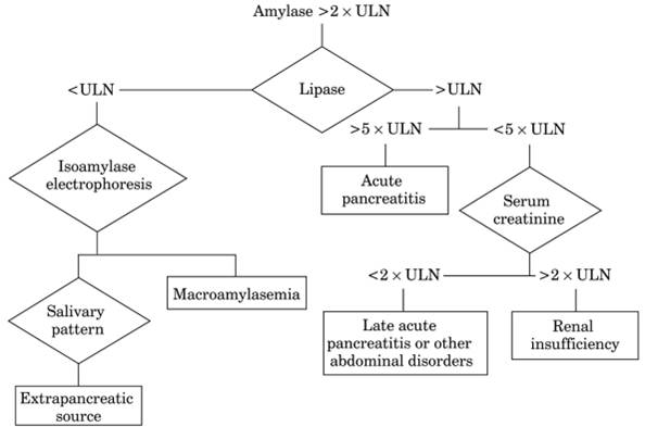
|
|
Fig. 8-9. Algorithm for increased serum
amylase and lipase. (ULN = upper limit of normal.)
|
- Others, e.g., chronic
liver disease (e.g., cirrhosis; ≤ 2× normal), burns, pregnancy
(including ruptured tubal pregnancy), ovarian cyst, diabetic ketoacidosis,
recent thoracic surgery, myoglobinuria, presence of myeloma proteins, some
cases of intracranial bleeding (unknown mechanism), splenic rupture,
dissecting aneurysm
- It has been suggested
that a level >1000 U/L is usually due to surgically correctable lesions
(most frequently stones in biliary tree), the pancreas being negative or
showing only edema; but 200500 U is usually associated with pancreatic
lesions that are not surgically correctable (e.g., hemorrhagic pancreatitis,
necrosis or pancreas).
- Increased
serum amylase with low urine amylase may be seen in renal insufficiency and
macroamylasemia. Serum amylase ≤ 4× normal in renal disease only
when creatinine clearance is <50 mL/min due to pancreatic or salivary
isoamylase but rarely >4× normal in absence of acute pancreatitis.
Decreased
In
- Extensive marked
destruction of pancreas (e.g., acute fulminant pancreatitis, advanced
chronic pancreatitis, advanced cystic fibrosis). Decreased levels are
clinically significant only in occasional cases of fulminant pancreatitis.
- Severe liver damage (e.g.,
hepatitis, poisoning, toxemia of pregnancy, severe thyrotoxicosis, severe
burns)
- Methodologic interference
by drugs (e.g., citrate and oxalate decrease activity by binding calcium
ions)
- Amylasecreatinine
clearance ratio = (urine amylase concentration ÷ serum amylase
concentration) × (serum creatinine concentration ÷ urine creatinine
concentration) × 100
- Normal
- Macroamylasemia: <1%;
very useful for this diagnosis.
- Acute pancreatitis:
>5%; use is presently discouraged for this diagnosis.
May Be
Normal In
- Patients with relapsing
chronic pancreatitis
- Patients with
hypertriglyceridemia (technical interference with test)
P.254
- Frequently normal in
patients with acute alcoholic pancreatitis.
Lipase,
Serum
- (Method
should always include colipase in reagent.)
- See Fig.
8-9.
Increased
In
- Acute pancreatitis
- Perforated or penetrating
peptic ulcer, especially with involvement of pancreas
- Obstruction of pancreatic
duct by
- Stone
- Drug-induced spasm of
Oddi's sphincter (e.g., codeine, morphine, meperidine, methacholine,
cholinergics), to levels 215× normal
- Partial obstruction plus
drug stimulation
- Chronic pancreatitis
- Acute cholecystitis
- Small bowel obstruction
- Intestinal infarction
- Acute and chronic renal
failure (increased 23× in 80% of patients and 5× in 5% of patients)
- Organ transplant (kidney,
liver, heart), especially with complications (e.g., organ rejection, CMV
infection, cyclosporin toxicity)
- Alcoholism
- Diabetic ketoacidosis
- After ERCP
- Some cases of
intracranial bleeding (unknown mechanism)
- Macro forms in lymphoma,
cirrhosis
- Drugs
- Induced acute
pancreatitis (see preceding section on serum amylase)
- Cholestatic effect
(e.g., indomethacin)
- Methodologic
interference (e.g., pancreozymin [contains lipase], deoxycholate,
glycocholate, taurocholate [prevent inactivation of enzyme], bilirubin
[turbidimetric methods])
- Chronic liver disease
(e.g., cirrhosis) (usually ≤ 2× normal)
Decreased
In
Methodologic interference (e.g., presence
of hemoglobin, calcium ions)
Usually
Normal In
- Mumps
- Values are lower in
neonates.
- Macroamylasemia
Diseases
of the Pancreas
Cystic
Fibrosis Of Pancreas
- (Autosomal
recessive disorder with abnormal ion transport due to failure of chloride
regulation; incidence of 1 in 1500 to 1 in 2000 in whites with a carrier
frequency of 1 in 20; 1 in 17,000 in American blacks; marked heterogeneity
among patients.)
Quantitative
Pilocarpine Iontophoresis Sweat Test (Properly Performed)
- Striking increase in sweat sodium (>60 mEq/L) and chloride (>60 mEq/L)
and, to a lesser extent, potassium is present in virtually all homozygous
patients; value is 35×
P.255
higher than in healthy persons or in those with other
diseases. Is consistently present throughout life from time of birth, and
degree of abnormality is not related to severity of disease or organ
involvement. Sensitivity = 98%, specificity = 83%, positive predictive value =
93%. Sweat chloride is somewhat more reliable than sodium
for diagnostic purposes.
- In children, chloride
>60mEq/L is considered positive for cystic fibrosis.
- 4060 mEq/L is
considered borderline and requires further investigation.
- <40
mEq/L is considered normal.
- ≤ 80 mEq/L may be
normal for adults.
- On occasion 12% of
cystic fibrosis patients have normal, borderline, or variable values.
- Rare patients with
borderline values have only mild disease.
- Sweat potassium is not
diagnostically valuable because of overlap with normal controls.
- Increased sweat sodium
and chloride are not useful for detection of heterozygotes (who have
normal values) or for genetic counseling.
- Sweat chloride ≥80 mEq/L on repeated occasions with characteristic clinical
manifestations or family history confirm diagnosis of cystic fibrosis.
- A broad range of sweat
values is seen in patients with this disease and in normal persons but
overlap is minimal.
- Sweat values (mEq/L)
|
|
Chloride
|
Sodium
|
Potassium
|
|
|
|
|
|
Mean
|
Range
|
Mean
|
Range
|
Mean
|
Range
|
|
Cystic fibrosis
|
|
|
|
|
|
|
|
Normal
|
|
|
|
|
|
|
|
- Note that one instrument
(Wescor; Logan, Utah) measures sweat conductivity, not
sweat chloride, which is not equivalent. Sweat conductivity measurement is
considered a screening test; patients with values ≥50 mEq/L should
have quantitative sweat chloride test.
Interferences
- Sweat
testing is fraught with problems and technical and laboratory errors are
very frequent; should be performed in duplicate and repeated at least once
on separate days on samples >100 mg of sweat
- Values may be increased
to cystic fibrosis range in healthy persons when sweat rate is rapid
(e.g., exercise, high temperature), but pilocarpine test does not increase
sweating rate.
- Mineralocorticoids
decrease sodium concentration in sweat by ~50% in normal subjects and
1020% in cystic fibrosis patients, whose final sodium concentration
remains abnormally high.
Increased
In
- Endocrine disorders
- Untreated adrenal
insufficiency (Addison's disease)
- Hypothyroidism
- Vasopressin-resistant
diabetes insipidus
- Familial
hypoparathyroidism
- Pseudohypoaldosteronism
- Metabolic disorders
- Malnutrition
- Glycogen storage disease
type I (von Gierke's disease)
- Mucopolysaccharidosis IH
and IS
- Fucosidosis
- Genitourinary disorders
- Klinefelter's syndrome
- Nephrosis
- Allergic/immunologic
disorders
- Hypogammaglobulinemia
- Prolonged infusion with
prostaglandin E
- Atopic dermatitis
P.256
- Neuropsychologic
disorders
- Anorexia nervosa
- Autonomic dysfunction
- Others
- Ectodermal dysplasia
- G-6-PD deficiency
- Serum chloride, sodium,
potassium, calcium, and phosphorus are normal unless complications occur
(e.g., chronic pulmonary disease with accumulation of CO ;
massive salt loss due to sweating causing hyponatremia). Urine
electrolytes are normal. Submaxillary saliva has slightly increased
chloride and sodium but not potassium; considerable overlap with results
for normal persons prevents diagnostic use.
- Submaxillary saliva is
more turbid, with increased calcium, total protein, and amylase. These
changes are not generally found in parotid saliva.
- Serum protein
electrophoresis shows increasing IgG and IgA with progressive pulmonary
disease; IgM and IgD are not appreciably increased.
- Serum albumin is often
decreased (because of hemodilution due to cor pulmonale; may be found
before cardiac involvement is clinically apparent).
- Laboratory changes secondary to complications that should
also suggest diagnosis of cystic fibrosis
- Salt-loss syndromes
- Hypochloremic metabolic
alkalosis and hypokalemia due to excessive loss of electrolytes in sweat
and stool
- Acute salt depletion
- Respiratory
abnormalities
- Chronic lung disease
(especially upper lobes) with laboratory changes of decreased pO , accumulation of CO , metabolic alkalosis,
severe recurrent infection, secondary cor pulmonale, etc. Nasal polyps,
pansinusitis; normal sinus radiographs are strong evidence against
cystic fibrosis.
- BAL usually shows
increased PMNs (>50% in cystic fibrosis; ~3% in normal persons) with
high absolute neutrophil count; is strong evidence of cystic fibrosis
even in absence of pathogens.
- Bacteriology: Special
culture techniques should be used in these patients. Before 1 yr of age,
S. aureus is found in 25% and Pseudomonas in 20% of respiratory tract cultures; in
adults Pseudomonas grows in 80% and S. aureus in 20%. Haemophilus
influenzae is found in 3.4% of cultures. Pseudomonas
aeruginosa is found increasingly often after treatment of
staphylococcal infection, and special identification and susceptibility
tests should be performed on P. aeruginosa. Pseudomonas cepacia is becoming more important in
older children. Increasing serum antibodies against P.
aeruginosa can document probable infection when cultures are
negative.
- Gastrointestinal
abnormalities
- Chronic/recurrent
pancreatitis.
- Pancreatic enzyme
activity is lost in 80% of patients, decreased in 10%, and normal in 10%
of patients. Protein-calorie malnutrition, hypoproteinemia, fat-soluble
vitamin deficiency (see Malabsorption). Stool and
duodenal fluid show lack of trypsin digestion of radiographic film
gelatin; useful screening test up to age 4; decreased chymotrypsin
production (see bentiromide test). Impaired
glucose intolerance in ~40% of patients, with glycosuria and
hyperglycemia in 8%, precedes diabetes mellitus.
- Cirrhosis (in >25%
of patients at autopsy), especially before age 30 years; hypersplenism;
cholelithiasis.
- Meconium ileus during
early infancy; causes 2030% of cases of neonatal intestinal
obstruction; present at birth in 8% of these children. Almost all of
them will develop the clinical picture of cystic fibrosis.
- GU tract abnormalities
- Aspermia in 98% due to
obstructive changes in vas deferens and epididymis, confirmed by
testicular biopsy.
- Neonatal screening using dried filter paper blood test that measures immunoreactive
trypsin has been used. Normal
in ≤ 10% of cystic fibrosis infants. Increased false-negative rate
in meconium ileus.
- DNA genotyping
(using blood; can use buccal scrapings) to confirm diagnosis based on two
mutations is highly specific but not very sensitive. Supports diagnosis of
cystic
P.257
fibrosis, but failure to detect gene mutations does
not exclude cystic fibrosis because of large number of alleles. Substantial
number of cystic fibrosis patients have unidentified gene mutation. Helpful
when sweat test is borderline or negative. Can also be used
for prenatal diagnosis and carrier screening. Identical genotypes can be
associated with different degrees of disease severity.
- Nasal electrical potential-difference measurements may be more reliable than sweat
tests but are much more complex to perform.
Macroamylasemia
- Serum amylase persistently increased (often 14×
normal) without apparent cause. Serum lipase is normal. Normal pancreatic
to salivary amylase ratio.
- Urine amylase normal or
low.
- Amylasecreatinine clearance ratio <1% with normal renal
function is very useful for this diagnosis; should make the clinician suspect
this diagnosis.
- Macroamylase is identified in serum by special gel filtration or ultracentrifugation
technique.
- May be found in ~1% of
randomly selected patients and 2.5% of persons with increased serum
amylase. Same findings may also occur in patients with
normal-molecular-weight hyperamylasemia in which excess amylase is
principally salivary gland isoamylase types 2 and 3.
- When associated with
pancreatic disease, serum lipase may be elevated.
Pancreatic
Carcinoma
Body
or Tail
- Laboratory tests are
often normal.
- Serum markers for tumor CA 19-9, CEA, etc. (see Chapter
16
- In carcinoma of
pancreas, CA 19-9 has sensitivity of 70%, specificity of 87%, positive
predictive value of 59%, negative predictive
value of 92%. No difference in sensitivity between local disease and
metastatic disease. Often normal in early stages, therefore not useful
for screening. Increased value may help differentiate benign disease from
cancer. Declines to normal in 36 mos if cancer is completely removed, so
may be useful for prognosis and follow-up. Detects tumor recurrence 220
wks before clinical evidence appears. Not specific for pancreas because
high levels may also occur in other GI cancers, especially those
affecting colon and bile duct.
- Testosterone/dihydrotestosterone
ratio is <5 (normal = ~10) in >70% of men with pancreatic cancer
(due to increased conversion by tumor). Less sensitive but more specific
than CA 19-9; present in higher proportion of stage I tumors.
- The most useful diagnostic test is
ultrasonography or CAT scanning followed by ERCP (at which time fluid is also obtained
for cytologic and pancreatic function studies). This combination correctly
diagnoses or rules out cancer of pancreas in ≥90% of cases. ERCP
with brush cytology has sensitivity ≤ 25% and specificity ≤ 100%.
- CEA level in bile
(obtained by percutaneous transhepatic drainage) was reported increased in
76% of a small group of cases.
- Serum amylase and lipase
may be slightly increased in early stages (<10% of cases); with later
destruction of pancreas, they are normal or decreased. They may increase
after secretin-pancreozymin stimulation before destruction is extensive;
therefore, the increase is less marked with a diabetic glucose tolerance
curve. Serum amylase response is less reliable.
- Glucose tolerance curve is of the diabetic type with overt
diabetes in 20% of patients with pancreatic cancer. Flat blood sugar curve
with IV
tolbutamide tolerance test indicates destruction of islet cell tissue. Unstable, insulin-sensitive diabetes that develops in an older
man should arouse suspicion of carcinoma of the pancreas.
- Secretin-cholecystokinin stimulation evidences duct obstruction when duodenal
intubation shows decreased volume of duodenal contents (<10 mL/10-min
collection period) with usually normal bicarbonate and enzyme levels in
duodenal contents. Acinar destruction (as in pancreatitis) shows normal
volume (2030 mL/10-min collection period), but bicarbonate and enzyme
levels may be decreased. Abnormal
P.258
volume, bicarbonate, or both is found in 6080% of
patients with pancreatitis or cancer. In carcinoma, the test result depends on
the relative extent and combination of acinar destruction and of duct
obstruction. Cytologic examination of duodenal contents shows malignant cells
in 40% of patients. Malignant cells may be found in up to 80% of patients with
periampullary cancer.
- Serum LAP is increased
(>300 U) in 60% of patients with carcinoma of pancreas due to liver
metastases or biliary tract obstruction. It may also be
increased in chronic liver disease.
- Triolein I test demonstrates
pancreatic duct obstruction with absence of lipase in the intestine
causing flat blood curves and increased stool excretion.
- Radioisotope scanning of
pancreas may be done (selenium 75) for lesions >2 cm.
- Needle biopsy has reported sensitivity of 5796%; false-positives are rare.
Head
- The abnormal pancreatic
function tests and increased tumor markers that occur with carcinoma of
the body of the pancreas may be evident.
- Laboratory findings due to complete obstruction of common bile duct
- Serum bilirubin
increased (1225 mg/dL), mostly direct (increase
persistent and nonfluctuating).
- Serum ALP increased.
- Urine and stool
urobilinogen absent.
- Increased PT; normal
after IV vitamin K administration.
- Increased serum
cholesterol (usually >300 mg/dL) with esters not decreased.
- Other liver function
tests are usually normal.
Pancreatitis,
Acute
- Serum lipase increases within 36 hrs with peak at 24 hrs and usually returns to
normal over a period of 814 days. Is superior to amylase; increases to a
greater extent and may remain elevated for up to 14 days after amylase
returns to normal. In patients with signs of acute pancreatitis,
pancreatitis is highly likely (clinical specificity = 85%) when lipase
≥5× ULN, if values change significantly with time, and if amylase
and lipase changes are concordant. (Lipase should
always be determined whenever amylase is determined.) New
methodology improves clinical utility. Urinary lipase is not clinically
useful. It has been suggested that a lipase/amylase ratio >3 (and
especially >5) indicates alcoholic rather than nonalcoholic
pancreatitis. Acute pancreatitis or organ rejection is highly likely if
lipase is ≥5× ULN, but unlikely if <3× ULN. (See Fig.
8-9)
- Serum amylase increase begins in 36 hrs, rises rapidly within 8 hrs in 75% of patients,
reaches maximum in 2030 hrs, and may persist for 4872 hrs. >95% sensitivity
during first 1224 hrs. The increase may be ≤ 40× normal, but the
height of the increase and rate of fall do not correlate with the severity
of the disease, prognosis, or rate of resolution; however, an increase of
>710 days suggests an associated cancer of pancreas or pseudocyst,
pancreatic ascites, or nonpancreatic cause. Similar
high values may occur in obstruction of pancreatic duct; they tend to fall
after several days. >10% of patients with acute pancreatitis
(especially when seen more than 2 days after onset of symptoms) may have
normal values, even when dying of acute pancreatitis. May also be
normal in patients with relapsing chronic pancreatitis and patients with
hypertriglyceridemia (technical interference with test). Frequently normal
in acute alcoholic pancreatitis. Acute abdomen due to GI infarction or
perforation rather than acute pancreatitis is suggested by only moderate
increase in serum amylase and lipase (<3× ULN), evidence of bacteremia.
1040% of patients with acute alcoholic intoxication have elevated serum
amylase (about half of amylases are salivary type); patients often present
with abdominal pain, but increased serum amylase is usually <3× ULN.
Levels >25× ULN indicate metastatic tumor rather than pancreatitis.
- Serum
pancreatic isoamylase can distinguish elevations due to salivary amylase,
which may account for 25% of all elevated values. (In healthy persons, 40%
of total serum amylase is pancreatic type and 60% is salivary type.)
P.259
- Only slight increase in
serum amylase and lipase values suggests a different diagnosis than acute
pancreatitis. Many drugs increase both amylase and
lipase in serum.
- Serum calcium is
decreased in severe cases 19 days after onset (due to binding to soaps in
fat necrosis). The decrease usually occurs after amylase and lipase levels
have become normal. Tetany may occur. (Rule out
hyperparathyroidism if serum calcium is high or fails to fall in
hyperamylasemia of acute pancreatitis.)
- Increased urinary amylase
tends to reflect serum changes by a time lag of 610 hrs, but sometimes
increased urine levels are higher and of longer duration than serum
levels. The 24-hr level may be normal even when some of the 1-hr specimens
show increased values. Measurement of amylase levels in hourly samples of
urine may be useful. Ratio of amylase clearance to creatinine clearance is
increased (>5%) and its use avoids the problem of timed urine
specimens; also increased in any condition that decreases tubular
reabsorption of amylase (e.g., severe burns, diabetic ketoacidosis,
chronic renal insufficiency, multiple myeloma, acute duodenal
perforation). Considered not specific; its use now discouraged by some but
still recommended by others.
- Serum bilirubin may be
increased when pancreatitis is of biliary tract origin but is usually
normal in alcoholic pancreatitis. Serum ALP, ALT, and AST may increase and
parallel serum bilirubin rather than amylase, lipase, or calcium levels.
Marked amylase increase (e.g., >2000 U/L) also favors biliary tract
origin. Fluctuation >50% in 24 hrs of serum bilirubin, ALP, ALT, and
AST suggests intermittent biliary obstruction.
- Serum trypsin (by RIA) is increased. High sensitivity makes a
normal value useful for excluding acute pancreatitis. But low specificity
(increased in large proportion of patients with hepatobiliary, bowel, and other
diseases and renal insufficiency; increased in 13% of patients with
chronic pancreatitis and 50% with pancreatic carcinoma) and RIA technology
limit utility.
- WBC is slightly to moderately increased (10,00020,000/cu mm).
- Hemoconcentration occurs
(increased Hct). Hct may be decreased in severe hemorrhagic pancreatitis.
- Glycosuria appears in 25%
of patients.
- Methemalbumin may be
increased in serum and ascitic fluid in hemorrhagic (severe) but not
edematous (mild) pancreatitis; may distinguish these two conditions but
not useful in diagnosis of acute pancreatitis.
- Hypokalemia, metabolic
alkalosis, or lactic acidosis may occur.
- Laboratory findings due
to predisposing conditions (may be multiple)
- Alcohol abuse accounts
for ~36% of cases.
- Biliary tract disease
accounts for 17% of cases.
- Idiopathic origin
accounts for >36% of cases.
- Infections (especially
viral such as mumps, coxsackievirus and CMV infections, AIDS).
- Trauma and postoperative
condition account for >8% of cases.
- Drug effects (e.g.,
steroids, thiazides, azathioprine, estrogens, sulfonamides; valproic acid
in children) account for >5% of cases.
- Hypertriglyceridemia
(hyperlipidemia types V, I, IV) accounts for 7% of cases.
- Hypercalcemia from any
cause.
- Tumors (pancreas,
ampulla).
- Anatomic abnormalities
of ampullary region causing obstruction (e.g., annular pancreas, Crohn's
disease, duodenal diverticulum).
- Hereditary.
- Renal failure; renal
transplantation.
- Miscellaneous (e.g.,
collagen vascular disease, pregnancy, ischemia, scorpion bites, parasites
obstructing pancreatic duct [Ascaris, fluke],
Reye's syndrome, fulminant hepatitis, severe hypotension, cholesterol
embolization).
- Laboratory findings due
to complications
- Pseudocysts of pancreas.
- Pancreatic abscess.
- Polyserositis
(peritoneal, pleural, pericardial, synovial surfaces). Ascites may
develop, cloudy or bloody or prune juice fluid, 0.52.0 L in volume,
containing increased amylase with a level higher than that of serum
amylase. No bile is evident (unlike in perforated ulcer). Gram stain
shows no bacteria (unlike in infarct of intestine). Protein >3 gm/dL
and marked increase in amylase.
- ARDS (with pleural
effusion, alveolar exudate, or both) may occur in ~40% of patients;
arterial hypoxemia is present.
P.260
- DIC.
- Hypovolemic shock.
- Others.
- Prognostic laboratory
findings
- On admission
- WBC >16,000/cu mm
- Blood glucose >200
mg/dL
- Serum LD >350 U/L
- Serum AST >250 U/L
- Age >55 yrs
- Within 48 hrs
- Serum calcium <8.0
mg/dL
- Decrease in Hct >10
points
- Increase in BUN >5
mg/dL
- Arterial pO <60 mm Hg
- Metabolic acidosis with
base deficit >4 mEq/L
- Mortality
- 1% if three signs are
positive
- 15% if three or four
signs are positive
- 40% if five or six
signs are positive
- 100% if seven or more
signs are positive
- Degree of amylase
elevation has no prognostic significance.
Pancreatitis,
Chronic
- See
also Malabsorption.
- Cholecystokinin-secretin
test measures the effect of IV administration of cholecystokinin and
secretin on volume, bicarbonate concentration, and amylase output of
duodenal contents and increase in serum lipase and amylase. This is the
most sensitive and reliable test (gold standard) for chronic pancreatitis,
especially in the early stages. Is technically difficult and is often not
performed accurately; gastric contamination must be avoided. Some
abnormality occurs in >85% of patients with chronic pancreatitis.
Amylase output is the most frequent abnormality. When all three are
abnormal, there is a greater frequency of abnormality in the tests listed
below.
- Normal duodenal contents
- Volume: 95235 mL/hr
- Bicarbonate
concentration: 74121 mEq/L
- Amylase output:
87,000267,000 mg
- Serum amylase and lipase
increase after administration of cholecystokinin and secretin in ~20% of
patients with chronic pancreatitis. They are more often abnormal when
duodenal contents are normal. Normally serum lipase and amylase do not
rise above normal limits.
- Fasting serum amylase and
lipase are increased in 10% of patients with chronic pancreatitis.
- Serum pancreolauryl test
- Fluorescein dilaurate
taken with breakfast is acted on by a pancreas-specific cholesterol ester
hydrolase, releasing fluorescein, which is absorbed from gut and measured
in serum; preceded by administration of secretin and followed by
administration of metoclopramide. Reported sensitivity = 82%, specificity
91%.20
- Diabetic OGTT results in
65% of patients with chronic pancreatitis and frank diabetes in >10% of
patients with chronic relapsing pancreatitis. When GTT is normal in the
presence of steatorrhea, the cause should be sought elsewhere than in the
pancreas.
- Laboratory findings due to malabsorption (occurs when >90% of exocrine
function is lost) and steatorrhea.
- Bentiromide test is
usually abnormal with moderate to severe pancreatic insufficiency.
- Schilling test may show
mild malabsorption of Vitamin B
- Xylose tolerance test
and small bowel biopsy are not usually done but are normal.
P.261
- Chemical analysis of
fecal fat demonstrates steatorrhea. It is more sensitive than tests using
triolein I.
- Triolein I testing is abnormal in one-third of patients with chronic
pancreatitis.
- Starch tolerance test is
abnormal in 25% of patients with chronic pancreatitis.
- Laboratory findings due
to causes of chronic pancreatitis and pancreatic exocrine insufficiency
- Alcoholism in 6070%
- Idiopathic in 3040%
- Obstruction of
pancreatic duct (e.g., trauma, pseudocyst, pancreas divisum, cancer or
obstruction of duct or ampulla)
- Other causes
occasionally (e.g., cystic fibrosis, primary hyperparathyroidism,
heredity, protein caloric malnutrition, miscellaneous [Z-E syndrome, Shwachman
syndrome, alpha -antitrypsin deficiency,
trypsinogen deficiency, enterokinase deficiency, hemochromatosis,
parenteral hyperalimentation])
- Radioactive selenium scanning of pancreas
yields variable findings in different clinics.
- CT, ultrasonography, ERCP
are most accurate for diagnosing and staging chronic pancreatitis.
Pseudocyst
Of Pancreas
- Serum direct bilirubin is
increased (>2 mg/dL) in 10% of patients.
- Serum ALP is increased in
10% of patients.
- Fasting blood sugar is
increased in <10% of patients.
- Duodenal contents after
secretin-pancreozymin stimulation usually show decreased bicarbonate
content (<70 mEq/L) but normal volume and normal content of amylase,
lipase, and trypsin.
- Findings of pancreatic cyst aspiration
- Best when panel of tests
is used.
- High fluid viscosity and
CEA indicate mucinous differentiation and exclude pseudocyst, serous
cystadenoma, other nonmucinous cysts or cystic tumors.
- Increased CA 72-4, CA
15-3, and tissue polypeptide antigen are markers of malignancy; if all
are low, pseudocyst or serous cystadenoma is most likely.
- CA 125 is increased in
serous cystadenoma.
- Pancreatic enzymes,
leukocyte esterase, and NB/70K are increased in pseudocysts.
- Cytologic examination.
- Laboratory findings of
preceding acute pancreatitis (this is mild and unrecognized in one-third
of patients). Persistent increase of serum amylase and lipase after an
episode of acute pancreatitis may indicate formation of a pseudocyst.
- Laboratory findings due
to conditions preceding acute pancreatitis (e.g., alcoholism, trauma,
duodenal ulcer, cholelithiasis).
- Laboratory findings due
to complications
- Infection
- Perforation
- Hemorrhage by erosion of
blood vessel or into a viscus
REFERENCES
1. Sheth SG, Gordon FD,
Chopra S. Nonalcoholic Steatohepatitis. Ann Intern
Med .
2. Mendlein J, et al. Iron
overload, public health and genetics. Ann Intern Med .
3. Edwards CQ, Kushner JP.
Screen for hemochromatosis. N Engl J Med .
4.
Press RD,
et al. Hepatic iron overload. Am J Clin Pathol .
5. Sheth SG, Gordon FD,
Chopra S. Nonalcoholic steatohepatitis. Ann Intern
Med .
6. Czaja AJ. The variant
forms of autoimmune hepatitis. Ann Intern Med .
7. Lemon SM. Type A viral hepatitis: epidemiology, diagnosis, and prevention. Clin Chem
8. MMWR Recommendations
for prevention and control of hepatitis C virus infection and HCV-related
chronic disease. US Department of
Health and Human Services, Centers for Disease Control and Prevention; 1998 Oct
16; 47/no RR-19.
9. Fairfax MR, Merline
JR, Podzorski RP. Am Soc Clin
Pathol Check Sample
Microbiology No. 97-1.
10. Cacciola I, et al.
Occult hepatitis B virus infections in patients with chronic hepatitis C liver
disease. N Engl J Med .
11. Masuko K, et al.
Infection with hepatitis GB virus C in patients on maintenance hemodialysis. N Engl J Med .
12. Alter HJ. The cloning and clinical implications of HGV and HGBV-C. N Engl J Med .
13. De Lamballerie X,
Charrel RN, Dussol B. Hepatitis GB virus C in patients on hemodialysis. N Engl J Med .
14. Yao DF, et al.
Diagnosis of hepatocellular carcinoma by quantitative detection of
hepatoma-specific bands of serum g-glutamyltransferase. Am J Clin Pathol .
15. Stremmel W, et al. Wilson disease: clinical
presentation, treatment, and survival. Ann Intern Med .
16. Tietz NW, Shuey DF.
Lipase in serumthe elusive enzyme: an overview. Clin
Chem .
17. Stern RC. The diagnosis of cystic fibrosis. N
Engl J Med .
18. Warshaw AL, Fernandez-del Castillo C. Pancreatic cancer. N Engl J Med .
19. Ranson JHC. Etiological
and prognostic factors in human acute pancreatitis: a review. Am J Gastroenterol .
20. Dominguez-Munoz JE,
Malfertheiner P. Optimized serum pancreolauryl test for differentiating
patients with and without chronic pancreatitis. Clin
Chem .
21. Centeno BA. Fine needle aspiration biopsy of the pancreas. Clin Lab Med .
